. 2013 Jun 26;5(191):191ra83.
doi: 10.1126/scitranslmed.3005576.

Surface-mediated bone tissue morphogenesis from tunable nanolayered implant coatings

Affiliations

Surface-mediated bone tissue morphogenesis from tunable nanolayered implant coatings

Nisarg J Shah et al. Sci Transl Med. .

Abstract

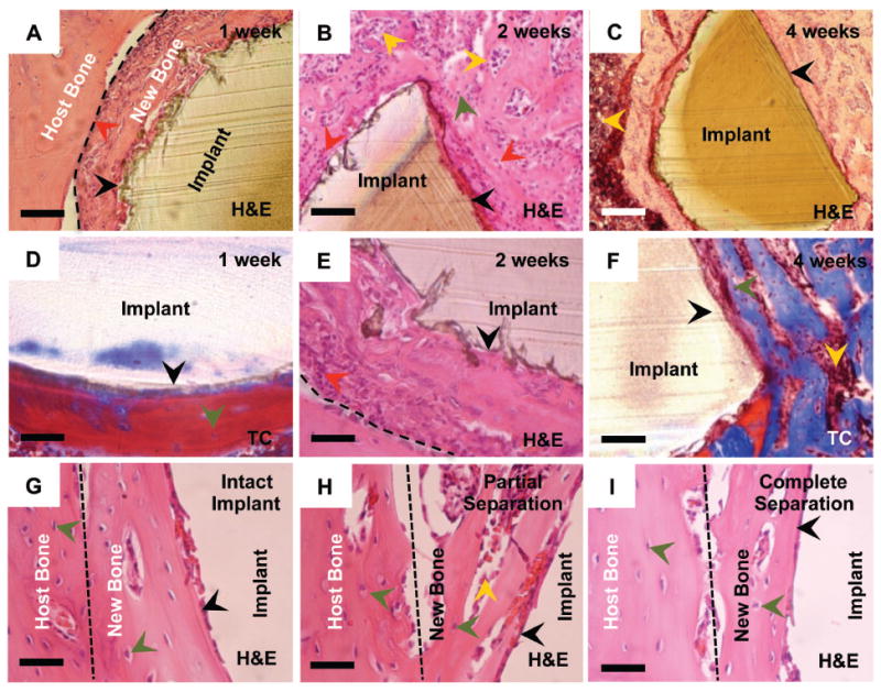
The functional success of a biomedical implant critically depends on its stable bonding with the host tissue. Aseptic implant loosening accounts for more than half of all joint replacement failures. Various materials, including metals and plastic, confer mechanical integrity to the device, but often these materials are not suitable for direct integration with the host tissue, which leads to implant loosening and patient morbidity. We describe a self-assembled, osteogenic, polymer-based conformal coating that promotes stable mechanical fixation of an implant in a surrogate rodent model. A single modular, polymer-based multilayered coating was deposited using a water-based layer-by-layer approach, by which each element was introduced on the surface in nanoscale layers. Osteoconductive hydroxyapatite (HAP) and osteoinductive bone morphogenetic protein-2 (BMP-2) contained within the nanostructured coating acted synergistically to induce osteoblastic differentiation of endogenous progenitor cells within the bone marrow, without indications of a foreign body response. The tuned release of BMP-2, controlled by a hydrolytically degradable poly(β-amino ester), was essential for tissue regeneration, and in the presence of HAP, the modular coating encouraged the direct deposition of highly cohesive trabecular bone on the implant surface. In vivo, the bone-implant interfacial tensile strength was significantly higher than standard bioactive bone cement, did not fracture at the interface, and had long-term stability. Collectively, these results suggest that the multilayered coating system promotes biological fixation of orthopedic and dental implants to improve surgical outcomes by preventing loosening and premature failure.


Conflict of interest statement

Competing interests: H.J.S. is a paid consultant for Pfizer Inc., and owns stock. N.J.S., M.N.H. and P.T.H. are co-inventors on a patent application for osteogenic LbL coatings (U.S. Serial Number: 13/746,902, International PCT Patent Application Number: PCT/US13/22430). The authors have no other competing financial interests.

Figures

Figure 1
Figure 1. Structured coatings for bone regeneration are made up of two composite multilayers
(A to C) The base coating contains chitosan (Chi; 75-85% deacytelated chitin, Mν ~ 100 kDa) and hydroxyapatite [HAP; Ca10(PO4)6(OH)2] with poly(acrylic acid) (PAA; Mν ~450 kDa) in a bilayer repeat unit. (D and E) The osteogenic factor coating contains a hydrolytically degradable poly(β-amino ester) (Poly2, Mν ~ 11 kDa) and rhBMP-2 that are alternated with PAA on top of the osteoconductive base coating. (F) Schematic of the two sets of multilayers: osteoconductive and osteoinductive. (G) Cumulative release profile of rhBMP-2 from drilled implants. Data are means ± SEM (n = 9 per coating). (H) rhBMP-2 loading has a dose-dependent effect on calcium deposition, quantified by alizarin red at 14 days. Data are means ± SEM (n = 6-9). * P < 0.05**P < 0.01, ANOVA with a Tukey post hoc test.
Figure 2
Figure 2. In vivo evaluation of rhBMP-2 release
rhBMP-2 was loaded into the multilayers that coated smooth and drilled PEEK rods and then implanted in the tibias of rats (n = 41-45 per group). (A) Controlled and burst release of fluorescently labeled rhBMP-2 was tracked in vivo over 30 and 3 days respectively. (B) Radiant efficiency at the implant site over time (n = 4-6 per group). (C) Bone marrow flushed out of excised tibiae was assayed for rhBMP-2 using ELISA for smooth and drilled implants. Data are means ± SEM (n = 5-6 per group).
Figure 3
Figure 3. Mesenchymal stem cells differentiate into osteoblasts
Five color flow cytometry was used to assess the percentage of osteoblasts in cells isolated from the tibia marrow around smooth and drilled implants. Each point represents individual implants. Means ± SEM (n = 5 per group). *P < 0.05, **P < 0.01, ANOVA with a Tukey post hoc test. FACS plots are provided in fig. S4.
Figure 4
Figure 4. Tensile force testing of implants from the rat tibia
Force data from individual implants are presented from smooth and drilled implants. Data are means ± SEM (n = 5 implants per group time point). *p < 0.05; **p < 0.01; ***p < 0.001, ANOVA with a Tukey post hoc test. Interfacial tensile strength data are provided (table S1 and S2).
Figure 5
Figure 5. Histology of implants with various coating formulations demonstrating bone tissue morphogenesis at the implant interface
(A to F) Implants coated with X20 + Y60 at 1, 2 and 4 weeks post-implantation demonstrating the process of implant integration with the parent bone tissue. Cement lines (broken black line) are observed on some sections. (G) The plane of fracture in implants with the X20 + Y60 coating is indicated by a broken black line at 4 weeks which depict an intact implant, partial separation from the host bone and complete separation from the host bone. The new bone-implant interface is intact. Sections were viewed under bright field microscopy. Scale bars: (A and C) are 200 μm; (B) and (D to G) are 50 μm. Arrows: black, bone/implant interface; red, active osteoblasts; dark green, osteocytes; yellow, marrow cells. H&E: hematoxylin & eosin stain, TC: Masson’s trichrome stain.
Figure 6
Figure 6. Bone deposition in the channels of drilled implants
Representative sections (n = 5-6 per group) of drilled implants after 4 weeks, which were coated with X20 + Y60. (A) Granulation tissue (broken black line) penetrated the channel and supplied progenitor cells. (B) Newly deposited bone (blue) matures (red) and (C) gradually filled up the channel at 4 weeks. (D) Bone (blue and red) is present throughout the channel of a drilled implant. Sections were viewed under brightfield microscopy. Scale bars in (A, B and D) are 100μm and in (C) is 400 μm. Arrows: black, bone/implant interface; red, active osteoblasts; dark green, osteocytes; yellow, marrow cells. H&E: hematoxylin & eosin stain, TC: Masson’s trichrome stain.
Figure 7
Figure 7. μCT imaging of bone formation on drilled PEEK implants
(A) Radiographs of bone formation around drilled implants with different coatings at 1, 2, and 4 weeks. Red arrows indicate location of the implant. (B and C) The images in (A) were used to quantify bone regeneration at 2 and at 4 weeks within (B) and outside the medullary canal (C) (using regions of interest marked by dotted red circles). Each point represents individual implants. Data are means ± SEM (n = 5-6 per group). *p < 0.05, **p < 0.01, ANOVA with Tukey post hoc test. Data for smooth implants are provided in fig. S11.

References

    1. Labek G, Thaler M, Janda W, Agreiter M, Stöckl B. Revision rates after total joint replacement: cumulative results from worldwide joint register datasets. J Bone Joint Surg Br. 2011;93:293–297. - PubMed
    1. Ulrich SD, Seyler TM, Bennett D, Delanois RE, Saleh KJ, Thongtrangan I, Kuskowski M, Cheng EY, Sharkey PF, Parvizi J. Total hip arthroplasties: What are the reasons for revision? Int Orthop. 2008;32:597–604. - PMC - PubMed
    1. Lewis G. Alternative acrylic bone cement formulations for cemented arthroplasties: Present status, key issues, and future prospects. J Biomed Mater Res Part B Appl Biomater. 2007;84:301–319. - PubMed
    1. Berger RA, Lyon JH, Jacobs JJ, Barden RM, Berkson EM, Sheinkop MB, Rosenberg AG, Galante JO. Problems with cementless total knee arthroplasty at 11 years followup. Clin Orthop Relat Res. 2001;392:196–207. - PubMed
    1. Sun L, Berndt CC, Gross KA, Kucuk A. Material fundamentals and clinical performance of plasma-sprayed hydroxyapatite coatings: a review. J Biomed Mater Res A. 2001;58:570–592. - PubMed

Publication types