. 2013 Jun 26;33(26):10616-24.
doi: 10.1523/JNEUROSCI.0404-13.2013.

Emergence of orientation selectivity in the Mammalian visual pathway

Affiliations

Emergence of orientation selectivity in the Mammalian visual pathway

Benjamin Scholl et al. J Neurosci. .

Abstract

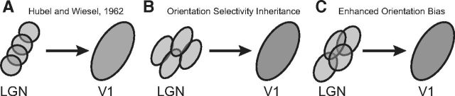
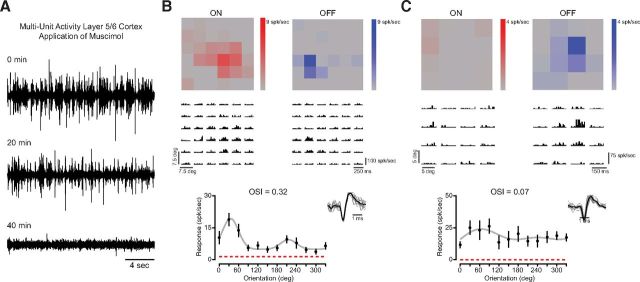
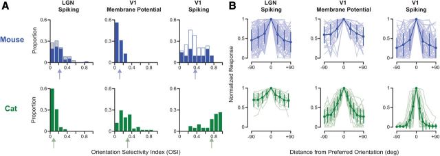
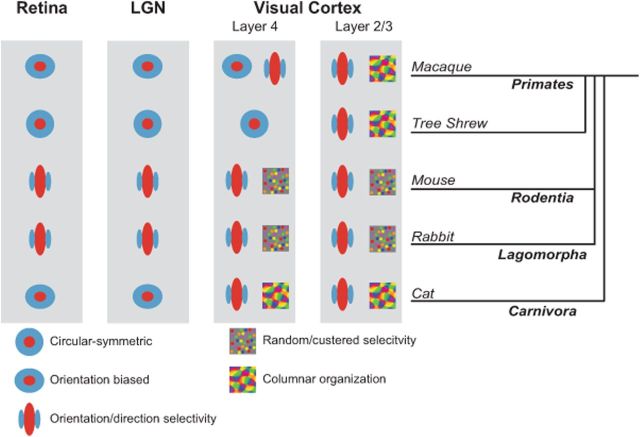
Orientation selectivity is a property of mammalian primary visual cortex (V1) neurons, yet its emergence along the visual pathway varies across species. In carnivores and primates, elongated receptive fields first appear in V1, whereas in lagomorphs such receptive fields emerge earlier, in the retina. Here we examine the mouse visual pathway and reveal the existence of orientation selectivity in lateral geniculate nucleus (LGN) relay cells. Cortical inactivation does not reduce this orientation selectivity, indicating that cortical feedback is not its source. Orientation selectivity is similar for LGN relay cells spiking and subthreshold input to V1 neurons, suggesting that cortical orientation selectivity is inherited from the LGN in mouse. In contrast, orientation selectivity of cat LGN relay cells is small relative to subthreshold inputs onto V1 simple cells. Together, these differences show that although orientation selectivity exists in visual neurons of both rodents and carnivores, its emergence along the visual pathway, and thus its underlying neuronal circuitry, is fundamentally different.


Figures

Figure 1.
Figure 1.
Models of the emergence of cortical orientation selectivity. A, Spatially offset LGN relay cells are combined to generate orientation selectivity in a Hubel and Wiesel framework (Hubel and Wiesel, 1962). B, Cortical orientation selectivity could be inherited from subcortical structures. C, Orientation bias of relay cells could generate orientation selectivity in combination with a Hubel and Wiesel framework.
Figure 2.
Figure 2.
Orientation selectivity in neurons of the mouse LGN. A, Example of an orientation-selective relay cell in mouse LGN. Mean cycled-averaged spiking responses to drifting gratings of each orientation (0–330°) are shown next to spontaneous activity during blank (mean-luminance) periods. The OSI was measured from peak responses (F1+F0), plotted for all orientations (black) with the mean spontaneous activity (red dashed line) and a Gaussian fit (gray). Sample waveforms for this isolated neuron are also shown. B, Example of a nonselective cell. C, Another example of a selective neuron which is direction selective, has high spontaneous firing rate, and lower spike rate for peak responses. D, Example of an orientation-biased cell with large spontaneous activity. E, Example of a neuron with oriented receptive field subregions matching the selectivity measured with drifting gratings. Orientation tuning and sample isolated waveforms (left) shown alongside the mean responses to white (ON) and black (OFF) patches of 2-D sparse noise stimulus (right).
Figure 3.
Figure 3.
Orientation selectivity of mouse LGN relay cells is unaffected by cortical inactivation. A, Inactivation of layer 5/6 in visual cortex by application of muscimol, a GABAA receptor agonist. Multiunit activity is reduced 20 min after application and completely abolished after 40 min. Visually evoked activity in visual cortex was abolished after 40 min of application. B, Example of an orientation-selective neuron and corresponding receptive field subregions recorded after cortical inactivation. ON and OFF subregions shown alongside recorded spiking activity to sparse noise stimuli. C, Example of a nonselective cell with a circular receptive field.
Figure 4.
Figure 4.
Comparison of orientation selectivity in mouse and cat. A, Distributions of OSI in mouse (blue) and cat (green) LGN spiking responses, V1 subthreshold membrane potential responses, and V1 spiking responses. Arrows indicate mean value for each distribution. V1 spiking OSI is based on the suprathreshold responses from intracellular records. Measurements made during cortical inactivation shown for mouse (gray). In mouse V1, some of the spiking OSI measurements were based on extracellular single-unit records (open blue). OSI distributions in mouse LGN and V1 subthreshold input are similar, whereas those in the cat show an enhancement of selectivity. In V1 of both mouse and cat, there is an enhancement of selectivity from subthreshold to spiking responses. B, Orientation tuning curves centered around preferred orientation (±90°) are shown across the visual pathway in mouse (blue) and cat (green). Each tuning curve was normalized by the peak response at the preferred orientation. Mean and SD are plotted over each population (light shading). Note that tuning curves shown for the cat LGN are from a subset of neurons (n = 13/35) for which we measured responses with small angle increments.
Figure 5.
Figure 5.
Comparison of orientation selectivity emergence across mammalian species. Orientation selectivity emerges in the retina of lagomorphs and rodents and is inherited by V1 neurons. In species of carnivores and primates, the transformation driving orientation selectivity occurs in visual cortex, although some selectivity in cat and primate is observed in the retina and LGN. Recordings along the visual pathway from macaque (Hubel and Wiesel, 1968; Essen and Zeki, 1978; Schall et al., 1986; Smith et al., 1990; Ts'o et al., 1990; Gur et al., 2005), tree shrew (Fitzpatrick, 1996; Bosking et al., 1997; Chisum et al., 2003), mouse (Dräger, 1975; Métin et al., 1988; Ohki et al., 2005; Weng et al., 2005; Ohki and Reid, 2007; Elstrott et al., 2008; Marshel et al., 2012; Piscopo et al., 2013), rabbit (Barlow et al., 1964; Levick, 1967; Levick et al., 1969; Stewart et al., 1971; Murphy and Berman, 1979; Taylor et al., 2000; Venkataramani and Taylor, 2010), and cat (Hubel and Wiesel, 1959, 1961, 1962, 1963; Boycott and Wässle, 1974; Cleland and Levick, 1974; Hammond, 1974; Levay and Gilbert, 1976; Levick and Thibos, 1980; Leventhal and Schall, 1983; Vidyasagar and Heide, 1984; Soodak et al., 1987; Shou and Leventhal, 1989; Thompson et al., 1994; Reid and Alonso, 1995; Shou et al., 1995; Ferster et al., 1996; Chung and Ferster, 1998; Usrey et al., 1999; Alonso et al., 2001; Kuhlmann and Vidyasagar, 2011; Viswanathan et al., 2011; Stanley et al., 2012). Note that the first emergence of orientation selectivity in the primate may depend on whether the thalamic inputs derive from the magnocellular or parvocellular pathway (Gur et al., 2005), and could either be located in layer 4Ca or layer 4Cb (Blasdel and Fitzpatrick, 1984; Ringach et al., 2002). Also note that although tree shrews are more closely related to primates than lagomorphs or rodents, they are not considered primates and the phylogenetic relationships remain unresolved (Cronin and Sarich, 1980; Luckett, 1980; MacPhee, 1993).

References

    1. Alonso JM, Usrey WM, Reid RC. Rules of connectivity between geniculate cells and simple cells in cat primary visual cortex. J Neurosci. 2001;21:4002–4015. - PMC - PubMed
    1. Anderson J, Lampl I, Reichova I, Carandini M, Ferster D. Stimulus dependence of two-state fluctuations of membrane potential in cat visual cortex. Nat Neurosci. 2000;3:617–621. doi: 10.1038/75797. - DOI - PubMed
    1. Barlow HB, Hill RM, Levick WR. Retinal ganglion cells responding selectively to direction and speed of image motion in the rabbit. J Physiol. 1964;173:377–407. - PMC - PubMed
    1. Blasdel GG, Fitzpatrick D. Physiological organization of layer 4 in macaque striate cortex. J Neurosci. 1984;4:880–895. - PMC - PubMed
    1. Bosking WH, Zhang Y, Schofield B, Fitzpatrick D. Orientation selectivity and the arrangement of horizontal connections in tree shrew striate cortex. J Neurosci. 1997;17:2112–2127. - PMC - PubMed

Publication types

MeSH terms

LinkOut - more resources