. 2013 Jun 26;33(26):10661-6.
doi: 10.1523/JNEUROSCI.1215-13.2013.

Concurrent maturation of inner hair cell synaptic Ca2+ influx and auditory nerve spontaneous activity around hearing onset in mice

Affiliations

Concurrent maturation of inner hair cell synaptic Ca2+ influx and auditory nerve spontaneous activity around hearing onset in mice

Aaron B Wong et al. J Neurosci. .

Abstract

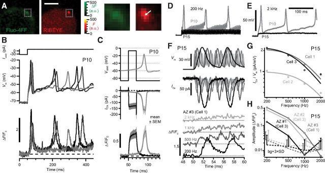
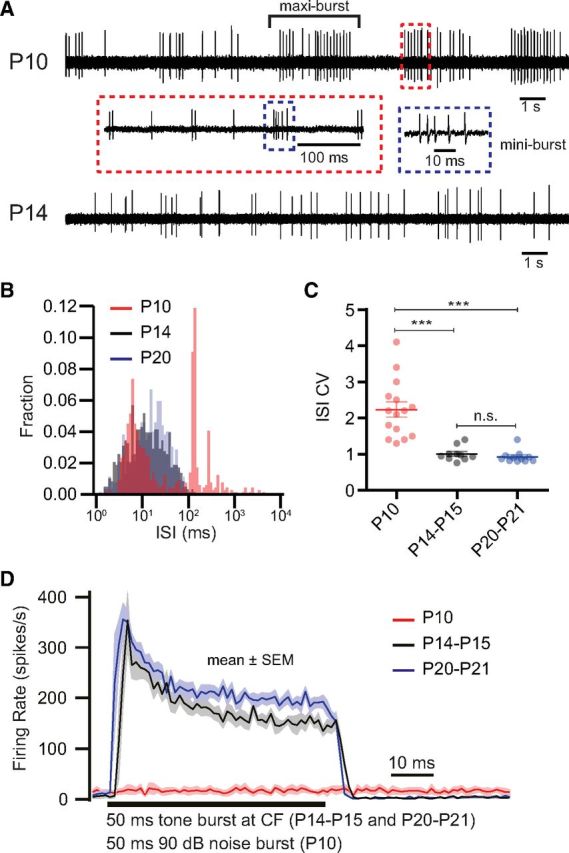
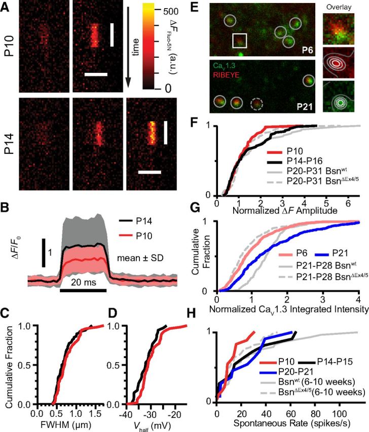
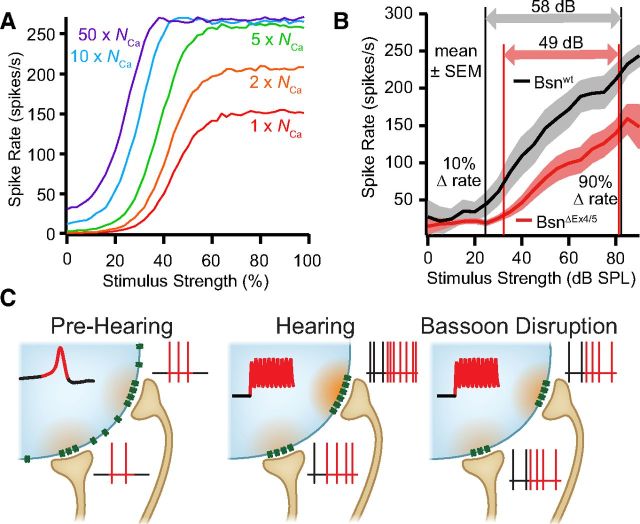
Hearing over a wide range of sound intensities is thought to require complementary coding by functionally diverse spiral ganglion neurons (SGNs), each changing activity only over a subrange. The foundations of SGN diversity are not well understood but likely include differences among their inputs: the presynaptic active zones (AZs) of inner hair cells (IHCs). Here we studied one candidate mechanism for causing SGN diversity-heterogeneity of Ca(2+) influx among the AZs of IHCs-during postnatal development of the mouse cochlea. Ca(2+) imaging revealed a change from regenerative to graded synaptic Ca(2+) signaling after the onset of hearing, when in vivo SGN spike timing changed from patterned to Poissonian. Furthermore, we detected the concurrent emergence of stronger synaptic Ca(2+) signals in IHCs and higher spontaneous spike rates in SGNs. The strengthening of Ca(2+) signaling at a subset of AZs primarily reflected a gain of Ca(2+) channels. We hypothesize that the number of Ca(2+) channels at each IHC AZ critically determines the firing properties of its corresponding SGN and propose that AZ heterogeneity enables IHCs to decompose auditory information into functionally diverse SGNs.


Figures

Figure 1.
Figure 1.
Naturalistic synaptic Ca2+ signal in IHCs before and after hearing onset. A, Change in fluorescence of the Ca2+ indicator Fluo-4FF (green) in a P10 IHC reveals a hotspot, colocalizing with the ribbon (red) marked by the fluorescent peptide (2 μm dimer). Scale bar, 3 μm. Boxes enlarged to the right. B, Small depolarizing current pulses (Icmd, top) applied to a prehearing IHC elicited APs (middle), overlay of three pulses. The normalized fluorescence change of Fluo-4FF (ΔF/F0) at a synaptic ribbon indicated transient Ca2+ influx (bottom) during each AP. C, Top, Immature IHCs were stimulated with a square depolarization or with the recorded waveform of a Ca2+ AP (Vcmd). The whole-cell Ca2+ current (ICa, middle) and ΔF/F0 (bottom) were similar for the two stimuli. ΔF/F0 traces are grand averages of 10 repetitions for each AZ (8 AZs in 4 IHCs). D, E, Differential responses of the membrane potential of a P10 (gray) or P15 (black) IHC in response to inwardly rectified sinusoidal current of 200 pA peak-to-peak amplitude at 200 Hz (D) and 2 kHz (E). F, The temporally compressed response of a P15 IHC to 200 Hz, 1 nA amplitude current was presented to IHCs in voltage clamp. Membrane potentials (Vm, top, corrected for attenuation as predicted by a resistor–capacitor circuit model), measured ICa (middle), and ΔF/F0 of Ca2+ indicator at an AZ (bottom) from a P15 IHC (AZ #3 in Cell 1, as in H) stimulated at four frequencies. Robust ΔF oscillations were observable up to 500 Hz. G, Amplitude of ICa (open circles) estimated as the fast Fourier transform magnitude at the stimulus frequency, normalized to Vm. Solid curves are fits to a filter function for three IHCs (see Materials and Methods). H, ΔF/F0 oscillation amplitudes of three AZs from two IHCs of similar ICa amplitude, estimated as the fast Fourier transform magnitude at the stimulus frequency. Dotted lines and error bars represent the mean and variation (3× SD) of background (bg) noise within ±100 Hz of the stimulus frequency, offset on x-axis for clarity. In these three AZs, the ΔF/F0 oscillation fell to noise level ≤2 kHz.
Figure 2.
Figure 2.
Developmental changes in SGN firing properties in vivo. A, A 20 s example trace of spontaneous spiking activity of single P10 (top) and P14 (bottom) SGNs. Note the alternation between silence and active periods in P10, whereby mini-bursts cluster in maxi-bursts. B, ISI histograms of representative SGNs in three age groups (red, P10; black, P14–P15; blue, P20–P21) during spontaneous activity. The strong peak for the P10 SGN at ∼120 ms shows the inter-mini-burst interval. C, CV of ISI for SGNs in three developmental age groups. A CV of ∼1 is typical for a stochastic Poisson process, whereas a higher CV is related to bursting activity. n.s., No significant difference; ***p < 0.001. D, Averaged PSTHs of SGNs in response to a 50 ms tone burst at characteristic frequency (CF; 30 dB above threshold, for P14–P15, n = 9 SGNs; P20–P21, n = 11 SGNs) or of a 50 ms noise burst (P10, n = 8 SGNs).
Figure 3.
Figure 3.
Developmental increase in heterogeneity of synaptic Ca2+ signals coincides with emergence of AZs with more Ca2+ channels and SGNs with higher spontaneous rates. A, Examples of Fluo-5N fluorescence change (ΔFFluo-5N) at synaptic sites during a 20 ms depolarization to −7 mV in prehearing (P10) and hearing (P14) IHCs, using line scans. All images were baseline subtracted (average of 50 ms period before depolarization) and have the same color lookup scale. Vertical bars indicate the duration of depolarization, and the horizontal bars indicate 2 μm. B, Average temporal profile of ΔF/F0 for prehearing (P10, red trace, n = 56) or hearing (P14–P16, black trace, n = 74). Shaded bands denote mean ± SD. C. Cumulative distributions of full-width at half-maximum (FWHM) of ΔFFluo-5N spatial profiles, estimated by Gaussian fit. No significant difference was found between the two age groups. D, Cumulative distributions of voltage of half-activation (Vhalf) of ΔFFluo-4FF amplitude for the two age groups, estimated by Boltzmann fit. E, Single confocal sections of immunolabeled tissue at P6 and P21 demonstrate an increase in Ca2+ channel (CaV1.3, green) accumulation around synaptic ribbons (RIBEYE, red). Right column, Two-dimensional Gaussian fit for intensity quantification (see Materials and Methods). F–H, Cumulative distributions show parallel increases over postnatal development for measurements of median-normalized ΔFFluo-5N (F), immunofluorescence of CaV1.3 puncta (G), and spontaneous rate of putative SGNs of wild-type mice (H). Compared with mature wild-type mice, mature but hearing-impaired bassoon mutant mice (BsnΔEx4/5) had generally smaller Ca2+ signals, less CaV1.3 immunofluorescence, and lower spike rates, similar to immature wild-type distributions. BsnΔEx4/5 and Bsnwt data were replotted from Frank et al. (2010) and Jing et al. (2013).
Figure 4.
Figure 4.
Ca2+ channel number at IHC AZs critically determines SGN firing and the combined dynamic range of SGN ensemble. A, Relationship between Ca2+ channel number (NCa) per AZ and SGN rate-level functions in a modified model (Meddis et al., 1990) of the IHC–SGN synapse. Changing NCa can account for diversity in spontaneous spike rate, sound sensitivity, and dynamic range. B, Average rate-level functions for mature wild-type and bassoon mutant mice (20 Bsnwt and 20 BsnΔEx4/5 SGNs). Arrows mark the dynamic range (10–90% between maximal and minimal rates) of the two neuron populations. C, Schematic of changes in IHC Ca2+ signaling and SGN spiking. Concurrent with hearing onset, AZs with stronger Ca2+ influx emerge at wild-type synapses (middle) but not at bassoon mutant synapses (right). We propose that these strong Ca2+ influx sites correspond to the SGNs that have high sensitivity and spontaneous rate (purple and blue traces in A).

Similar articles

Cited by

References

    1. Beutner D, Moser T. The presynaptic function of mouse cochlear inner hair cells during development of hearing. J Neurosci. 2001;21:4593–4595. - PMC - PubMed
    1. Beutner D, Voets T, Neher E, Moser T. Calcium dependence of exocytosis and endocytosis at the cochlear inner hair cell afferent synapse. Neuron. 2001;29:681–690. doi: 10.1016/S0896-6273(01)00243-4. - DOI - PubMed
    1. Blankenship AG, Feller MB. Mechanisms underlying spontaneous patterned activity in developing neural circuits. Nat Rev Neurosci. 2010;11:18–29. doi: 10.1038/nrn2759. - DOI - PMC - PubMed
    1. Brandt A, Striessnig J, Moser T. CaV1. 3 channels are essential for development and presynaptic activity of cochlear inner hair cells. J Neurosci. 2003;23:10832–10840. - PMC - PubMed
    1. Brandt A, Khimich D, Moser T. Few CaV1.3 channels regulate the exocytosis of a synaptic vesicle at the hair cell ribbon synapse. J Neurosci. 2005;25:11577–11585. doi: 10.1523/JNEUROSCI.3411-05.2005. - DOI - PMC - PubMed

Publication types

MeSH terms

LinkOut - more resources