. 2013 Jun 26;33(26):10741-9.
doi: 10.1523/JNEUROSCI.5419-12.2013.

Ionotropic glutamate receptors IR64a and IR8a form a functional odorant receptor complex in vivo in Drosophila

Affiliations

Ionotropic glutamate receptors IR64a and IR8a form a functional odorant receptor complex in vivo in Drosophila

Minrong Ai et al. J Neurosci. .

Abstract

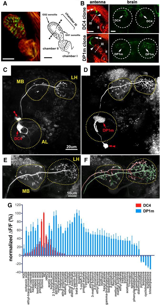
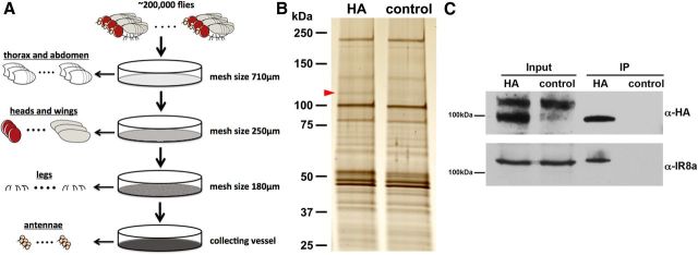
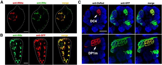
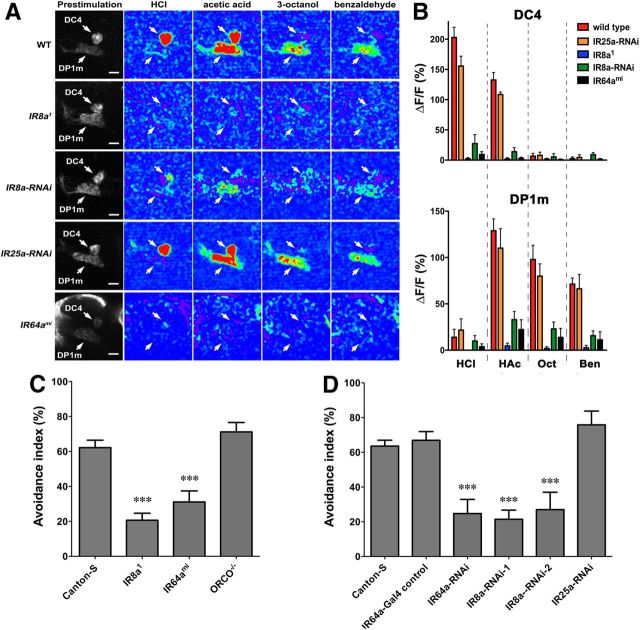
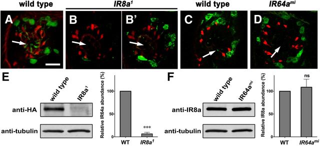
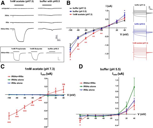
Drosophila olfactory sensory neurons express either odorant receptors or ionotropic glutamate receptors (IRs). The sensory neurons that express IR64a, a member of the IR family, send axonal projections to either the DC4 or DP1m glomeruli in the antennal lobe. DC4 neurons respond specifically to acids/protons, whereas DP1m neurons respond to a broad spectrum of odorants. The molecular composition of IR64a-containing receptor complexes in either DC4 or DP1m neurons is not known, however. Here, we immunoprecipitated the IR64a protein from lysates of fly antennal tissue and identified IR8a as a receptor subunit physically associated with IR64a by mass spectrometry. IR8a mutants and flies in which IR8a was knocked down by RNAi in IR64a+ neurons exhibited defects in acid-evoked physiological and behavioral responses. Furthermore, we found that the loss of IR8a caused a significant reduction in IR64a protein levels. When expressed in Xenopus oocytes, IR64a and IR8a formed a functional ion channel that allowed ligand-evoked cation currents. These findings provide direct evidence that IR8a is a subunit that forms a functional olfactory receptor with IR64a in vivo to mediate odor detection.


Figures

Figure 1.
Figure 1.
Anatomic separation and functional distinction of neurons innervating DC4 and DP1m. A, Left, Anti-HA (green) immunostaining of a whole-mount antenna from an IR64a-HA transgenic fly reveals the sensilla tips (green) of IR64a+ OSNs. Red autofluorescence depicts the outline of the sacculus. Right, Drawing of the sacculus. Double arrowheads point to a pair of thick cuticular flaps that separate dorsal and ventral compartments of Chamber III of the sacculus. B, Flip-out labeling of single cell clones (green) in flies carrying IR64a-GAL4, UAS-frt-Stop-frt-CD8GFP, and hs-Flip. Examples of single cells projecting bilaterally to either DC4 (top row) or DP1m (bottom row) are shown with images of the antennae on the left and the corresponding antennal lobes on the right. Arrowheads show sites of dendritic innervation in the sacculus; double arrowheads show cell bodies. The dotted line indicates cuticular flaps that separate dorsal and ventral compartments of Chamber III. The dotted circle indicates antennal lobe. Scale bars: A, B, 10 μm. C, D, PA-GFP labeling of a DC-PN (C) and a DP1m-PN (D) in the brains of nSyb-GAL4; UAS-C3PA flies. The outline of each neuropil was determined by the background fluorescence of PA-GFP. Red double arrowheads indicate PN cell bodies. E, F, PA-GFP labeling of a single DP1m-PN (E) followed by labeling of a single DC4-PN (F) in the same brain of a fly carrying nSyb-GAL4; UAS-C3PA. The axonal projections of the DP1m PN and DC4 PN were mapped in 3D space and labeled in green and red, respectively, by using the Vaa3D software (Peng et al., 2010). Note that axonal termini of the DP1m-PN (green) and DC4-PN (red) occupy largely nonoverlapping space within the LH. The double labeling was repeated three times with similar results. G, Odor tuning properties of DC4 (red) and DP1m (blue) glomeruli in flies carrying IR64a-GAL4; UAS-GCaMP3.0 were measured by calcium imaging. For each glomerulus, the GCaMP fluorescence intensity changes (ΔF/F) were normalized to the maximal ΔF/F response, which was defined as 100%.
Figure 2.
Figure 2.
IR8a binds to IR64a in vivo. A, Schematic drawing of large-scale isolation of antennal tissue for co-IP experiments. B, Silver stain of co-IP proteins from IR64a-HA transgenic flies (HA) and wild-type flies (control). The red arrowhead points to a silver stain positive band that is present in the HA sample but absent in control sample. C, Western blot analysis of independently prepared antennal tissue showing that IR8a is coimmunoprecipitated with IR64a-HA.
Figure 3.
Figure 3.
IR8a is expressed in IR64a+ neurons. A, Fluorescence micrographs of a cryosectioned wild-type antenna immunostained by anti-IR64a (red) and anti-IR8a (green) polyclonal antibodies. The dotted line outlines the antenna. B, A section of an antenna from IR8a-GAL4; UAS-GFP flies immunostained by anti-IR8a (green) and anti-GFP (red) showing that IR8a-GAL4 faithfully recapitulated endogenous IR8a expression. C, An antennal lobe from a fly carrying IR8a-GAL4; UAS-CD8GFP and PromoterIR64a-mCherry immunostained by anti-dsRed/mCherry (red; corresponding to IR64a promoter expression), anti-GFP (green; corresponding to IR8a promoter expression), and nc82 (blue). Note that red and green fluorescence represent the glomeruli labeled by IR64a and IR8a promoters, respectively. Top row, Focal plane showing the DC4 glomerulus. Bottom row: Focal plane showing the DP1m glomerulus of the same antennal lobe. Scale bars: 20 μm.
Figure 4.
Figure 4.
IR8a is required specifically in IR64a+ neurons for the physiological and behavioral responses to odorants. A, Calcium imaging of flies carrying IR64a-GAL4; UAS-GCaMP3.0 in wild-type (WT), IR8a mutant (IR8a1), IR8a-RNAi (IR64a-GAL4, UAS-dcr2, UAS-IR8aRNAi), IR25a-RNAi (IR64a-GAL4, UAS-dcr2, UAS-IR25aRNAi), or IR64a mutant (IR64ami) backgrounds. Arrows indicate DC4 and DP1m glomeruli. Scale bars, 10 μm. B, Fluorescence intensity changes (ΔF/F) of DC4 (top) and DP1m (bottom) in response to odorants were quantified. N = 5. HCl, Hydrochloric acid (3.6%); HAc, acetic acid (1%); Oct, 1-octanol (1%); Ben, benzaldehyde (1%). C, Avoidance to acetic acid in wild-type and different mutant flies in a T maze. N = 10∼16. D, Avoidance to acetic acid in flies carrying different UAS-RNAi transgenes driven by IR64a-GAL4. N = 8∼16. ***p < 0.01 (ANOVA with Tukey's test).
Figure 5.
Figure 5.
IR8a affects IR64a protein abundance. A–D, Cryosectioned antennae from wild-type, IR8a mutant, or IR64a mutant flies immunostained by anti-IR64a (A–B′, green) or anti-IR8a (C, D, green) and monoclonal antibody 21A6 (red). B′, An image of the same sacculus region as in B taken with increased laser power and more sensitive detector gain to overexpose the green fluorescence. Arrows point to the sensilla within the sacculus. Scale bar, 10 μm. E, F, Left, Western blot of dissected antennae (35 pairs of antennae per lane). Right, Relative protein abundance from Western blot was quantified by the gel analysis function of the ImageJ software. N = 3. ***p = 0.0015. ns, Not significantly different by Student's t test.
Figure 6.
Figure 6.
IR64a and IR8a form functional ion channels in Xenopus oocytes. A, Top left, Acetate (1 mM) induced an inward current in oocyte expressing both IR64a and IR8a. Top right, Acidic buffer induced a small and transient outward current and a small off response. Bottom, Responses to propionate, butyrate, and pH 8.5 buffer. Calibration: 100 nA, 20 s. B, I/V relationship in IR64a+IR8a-expressing oocytes in response to different stimuli. Insets, right, Examples of currents from an IR64a+IR8a-expressing oocyte clamped from −100 to 40 mV with 20 mV steps. *p < 0.05; **p < 0.01 [unpaired Student's t test compared to buffer (pH 7.3) control]. C, D, Relationship of odor-evoked currents (Iodor) and holding potentials. In each oocyte, evoked currents measured at each holding potential were normalized to the currents at the same holding potential in control buffer (pH 7.3) (see Materials and Methods). *p < 0.05; ***p < 0.01 (ANOVA with Tukey's test).

References

    1. Abuin L, Bargeton B, Ulbrich MH, Isacoff EY, Kellenberger S, Benton R. Functional architecture of olfactory ionotropic glutamate receptors. Neuron. 2011;69:44–60. doi: 10.1016/j.neuron.2010.11.042. - DOI - PMC - PubMed
    1. Ai M, Min S, Grosjean Y, Leblanc C, Bell R, Benton R, Suh GS. Acid sensing by the Drosophila olfactory system. Nature. 2010;468:691–695. doi: 10.1038/nature09537. - DOI - PMC - PubMed
    1. Basler K, Struhl G. Compartment boundaries and the control of Drosophila limb pattern by hedgehog protein. Nature. 1994;368:208–214. doi: 10.1038/368208a0. - DOI - PubMed
    1. Benton R, Vannice KS, Gomez-Diaz C, Vosshall LB. Variant ionotropic glutamate receptors as chemosensory receptors in Drosophila. Cell. 2009;136:149–162. doi: 10.1016/j.cell.2008.12.001. - DOI - PMC - PubMed
    1. Couto A, Alenius M, Dickson BJ. Molecular, anatomical, and functional organization of the Drosophila olfactory system. Curr Biol. 2005;15:1535–1547. doi: 10.1016/j.cub.2005.07.034. - DOI - PubMed

Publication types

MeSH terms

LinkOut - more resources