. 2013 Jun 26;33(26):10924-33.
doi: 10.1523/JNEUROSCI.0886-13.2013.

Estrogen mediates neuroprotection and anti-inflammatory effects during EAE through ERα signaling on astrocytes but not through ERβ signaling on astrocytes or neurons

Affiliations

Estrogen mediates neuroprotection and anti-inflammatory effects during EAE through ERα signaling on astrocytes but not through ERβ signaling on astrocytes or neurons

Rory D Spence et al. J Neurosci. .

Abstract

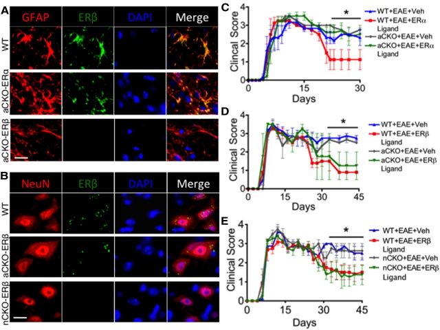
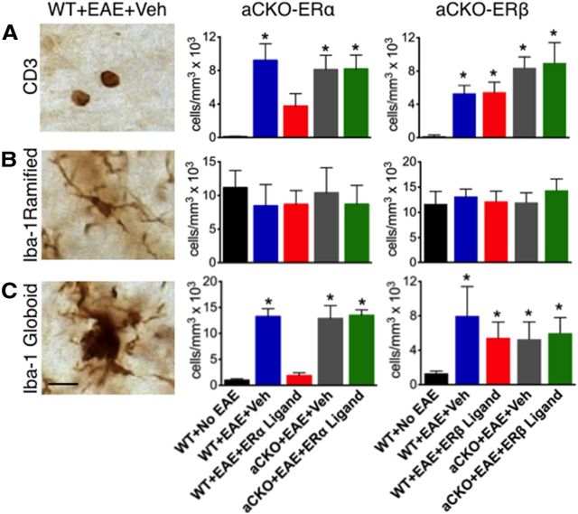
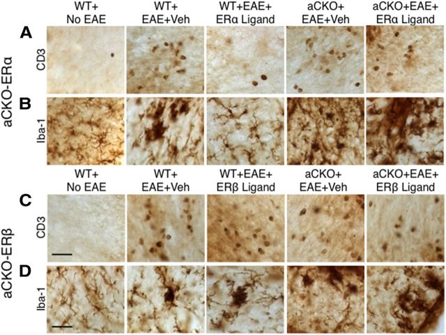
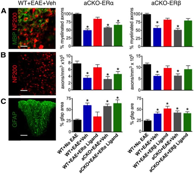
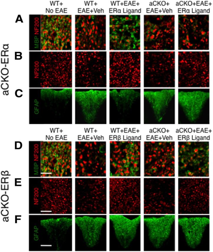
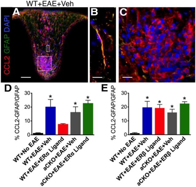
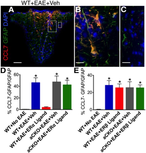
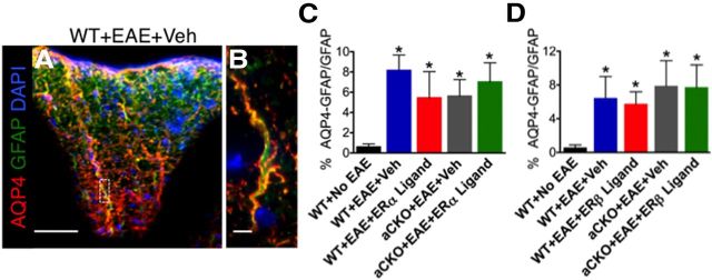
Estrogens can signal through either estrogen receptor α (ERα) or β (ERβ) to ameliorate experimental autoimmune encephalomyelitis (EAE), the most widely used mouse model of multiple sclerosis (MS). Cellular targets of estrogen-mediated neuroprotection are still being elucidated. Previously, we demonstrated that ERα on astrocytes, but not neurons, was critical for ERα ligand-mediated neuroprotection in EAE, including decreased T-cell and macrophage inflammation and decreased axonal loss. Here, we determined whether ERβ on astrocytes or neurons could mediate neuroprotection in EAE, by selectively removing ERβ from either of these cell types using Cre-loxP gene deletion. Our results demonstrated that, even though ERβ ligand treatment was neuroprotective in EAE, this neuroprotection was not mediated through ERβ on either astrocytes or neurons and did not involve a reduction in levels of CNS inflammation. Given the differential neuroprotective and anti-inflammatory effects mediated via ERα versus ERβ on astrocytes, we looked for molecules within astrocytes that were affected by signaling through ERα, but not ERβ. We found that ERα ligand treatment, but not ERβ ligand treatment, decreased expression of the chemokines CCL2 and CCL7 by astrocytes in EAE. Together, our data show that neuroprotection in EAE mediated via ERβ signaling does not require ERβ on either astrocytes or neurons, whereas neuroprotection in EAE mediated via ERα signaling requires ERα on astrocytes and reduces astrocyte expression of proinflammatory chemokines. These findings reveal important cellular differences in the neuroprotective mechanisms of estrogen signaling through ERα and ERβ in EAE.


Figures

Figure 1.
Figure 1.
Verification of gene deletion specificity in aCKO-ERα, aCKO-ERβ, and nCKO-ERβ mouse models and EAE disease severity scores showing protective effects of ERα in astrocytes, but not ERβ in astrocytes or neurons. A, Immunohistochemistry shows ERβ colocalized with GFAP in WT and aCKO-ERα, but not aCKO-ERβ mice with EAE. Scale bar, 12 μm. B, Immunohistochemistry shows ERβ colocalized with NeuN in WT and aCKO-ERβ, but not nCKO-ERβ mice with EAE. Scale bar, 11 μm. C, WT, but not aCKO-ERα mice, treated with ERα ligand had significantly better clinical scores compared with WT or aCKO-ERα mice with EAE treated with vehicle. *p < 0.05 versus WT + EAE + ERα ligand (repeated-measures ANOVA with post hoc Bonferroni pairwise analysis). D, WT and aCKO-ERβ mice treated with ERβ ligand had significantly better clinical scores compared with WT or aCKO-ERβ mice with EAE treated with vehicle. *p < 0.05 versus WT + EAE + ERα ligand and aCKO + EAE + ERα ligand (repeated-measures ANOVA with post hoc Bonferroni pairwise analysis). E, WT and nCKO-ERβ mice treated with ERβ ligand had significantly better clinical scores compared with WT or nCKO-ERβ mice with EAE treated with vehicle. *p < 0.05 versus WT + EAE + ERα ligand and nCKO + EAE + ERα ligand (repeated-measures ANOVA with post hoc Bonferroni pairwise analysis). n = 10 per group.
Figure 2.
Figure 2.
Quantification of how ERβ, unlike ERα, in astrocytes does not mediate reduction of CD3 T cells and Iba-1 globoid macrophages in EAE spinal cord. A, CD3 T cells were reduced in WT, but not aCKO-ERα, mice with EAE treated with ERα ligand. *p < 0.05 versus WT + No EAE and WT + EAE + ERα ligand (ANOVA with post hoc Bonferroni pairwise analysis). Treatment with ERβ ligand was unable to reduce CD3 T cells in WT or aCKO-ERβ with EAE. *p < 0.05 versus WT + No EAE (ANOVA with post hoc Bonferroni pairwise analysis). B, Iba-1 ramified microglia exhibited no significant difference in number across all experimental groups. C, Iba-1 globoid macrophages were significantly reduced in WT, but not aCKO-ERα, mice treated with EAE treated with ERα ligand. *p < 0.05 versus WT + No EAE and WT + EAE + ERα ligand (ANOVA with post hoc Bonferroni pairwise analysis). Treatment with ERβ ligand was unable to reduce CD3 T cells in WT or aCKO-ERβ mice with EAE. Scale bar, 12 μm. n = 6 per group. *p < 0.05 versus WT + No EAE (ANOVA with post hoc Bonferroni pairwise analysis).
Figure 3.
Figure 3.
Representative images of how ERβ, unlike ERα, in astrocytes does not mediate reduction of CD3 T cells and Iba-1 globoid macrophages in EAE spinal cord. A, B, CD3 T cells and Iba-1 globoid macrophages are increased in WT and aCKO-ERα mice with EAE compared with WT mice without EAE. Treatment with ERα ligand prevents this increase in WT, but not aCKO-ERα, mice with EAE. C, D, CD3 T cells and Iba-1 globoid macrophages are increased in WT and aCKO-ERβ mice with EAE compared with WT mice without EAE. Treatment with ERβ ligand did not prevent this increase in either WT or aCKO-ERβ mice. Scale bars: A, C, 41 μm; D, 27 μm.
Figure 4.
Figure 4.
Quantification of how ERβ, unlike ERα, in astrocytes does not protect against demyelination, axonal loss, and reactive astrogliosis. A, Myelinated NF200 axons fewer significantly reduced in WT mice with EAE, and treatment with ERα ligand prevented demyelination in WT, but not aCKO-ERα, mice. *p < 0.05 versus WT + No EAE and WT + EAE + ERα ligand (ANOVA with post hoc Bonferroni pairwise analysis). Treatment with ERβ ligand was able to reduce demyelination in WT and aCKO-ERβ mice. *p < 0.05 versus WT + No EAE, WT + EAE + ERβ ligand, aCKO + EAE + ERβ ligand (ANOVA with post hoc Bonferroni pairwise analysis). Scale bar, 15 μm. B, Numbers of NF200 axons were significantly reduced in WT mice with EAE, and treatment with ERα ligand prevented axonal loss in WT, but not aCKO-ERα, mice. *p < 0.05 versus WT + No EAE and WT + EAE + ERα ligand (ANOVA with post hoc Bonferroni pairwise analysis). Treatment with ERβ ligand was able to prevent axonal loss in WT and aCKO-ERβ mice. *p < 0.05 versus WT + No EAE, WT + EAE + ERβ ligand, aCKO + EAE + ERβ ligand (ANOVA with post hoc Bonferroni pairwise analysis). Scale bar, 40 μm. C, Reactive gliosis staining was significantly increased in WT mice with EAE, and treatment with ERα ligand prevented this increase in WT, but not aCKO-ERα, mice with EAE. *p < 0.05 versus WT + No EAE and WT + EAE + ERα ligand (ANOVA with post hoc Bonferroni pairwise analysis). Treatment with ERβ ligand was unable to prevent reactive gliosis in WT or aCKO-ERβ mice with EAE. *p < 0.05 versus WT + No EAE (ANOVA with post hoc Bonferroni pairwise analysis). Scale bar, 122 μm. n = 6 per group.
Figure 5.
Figure 5.
Representative images of how ERβ, unlike ERα, in astrocytes does not protect against demyelination, axonal loss, and gliosis in EAE spinal cord. A, D, Myelinated NF200 axons were decreased in WT mice with EAE. Treatment with ERα ligand reduced demyelination in WT, but not aCKO-ERα, mice with EAE. Treatment with ERβ ligand reduced demyelination in both WT and aCKO-ERβ mice with EAE. Scale bar, 22 μm. B, E, NF200 axons exhibited patchy reductions in numbers in WT mice with EAE. Treatment with ERα ligand reduced axonal loss in WT, but not aCKO-ERα, mice with EAE. Treatment with ERβ ligand reduced axonal loss in both WT and aCKO-ERβ mice with EAE. Scale bar, 50 μm. C, F, Reactive gliosis staining was increased in WT mice with EAE. Treatment with ERα ligand decreased reactive gliosis in WT, but not aCKO-ERα, mice with EAE. Treatment with ERβ ligand was unable to decrease reactive gliosis in WT and aCKO-ERβ mice with EAE. Scale bar, 130 μm.
Figure 6.
Figure 6.
ERα is required on astrocytes to reduce CCL2 expression within reactive astrocytes. A, CCL2 (red), GFAP (green), and DAPI (blue) are expressed in EAE spinal cord in a WT mouse with EAE. Scale bar, 120 μm. B, CCL2 is coexpressed with GFAP. Scale bar, 27 μm. C, CCL2 is coexpressed with infiltrating immune cells stained with DAPI. Scale bar, 27 μm. D, Treatment with ERα ligand was able to reduce coexpression of CCL2 with GFAP in WT, but not aCKO-ERα, mice with EAE. *p < 0.05 versus WT + No EAE and WT + EAE + ERα ligand (ANOVA with post hoc Bonferroni pairwise analysis). E, Treatment with ERβ ligand was not able to reduce coexpression of CCL2 with GFAP in WT or aCKO-ERβ mice with EAE. *p < 0.05 versus WT + No EAE (ANOVA with post hoc Bonferroni pairwise analysis). n = 5 per group.
Figure 7.
Figure 7.
ERα is required on astrocytes to reduce CCL7 expression within reactive astrocytes. A, CCL7 (red), GFAP (green), and DAPI (blue) are expressed in EAE spinal cord in a WT mouse with EAE. Scale bar, 120 μm. B, CCL7 is coexpressed with GFAP. Scale bar, 27 μm. C, CCL7 is not coexpressed in infiltrating immune cells (DAPI). Scale bar, 27 μm. D, Treatment with ERα ligand was able to reduce coexpression of CCL7 with GFAP in WT, but not aCKO-ERα, mice with EAE. *p < 0.05 versus WT + No EAE and WT + EAE + ERα ligand (ANOVA with post hoc Bonferroni pairwise analysis). E, Treatment with ERβ ligand was not able to reduce coexpression of CCL7 with GFAP in WT or aCKO-ERβ mice with EAE. *p < 0.05 versus WT + No EAE (ANOVA with post hoc Bonferroni pairwise analysis). n = 5 per group.
Figure 8.
Figure 8.
AQP4 expression within reactive astrocytes is increased in all groups with EAE, regardless of genotype or treatment. AQP4 (red), GFAP (green), and DAPI (blue) are expressed in EAE spinal cord in a WT mouse with EAE. Scale bar, 145 μm. B, AQP4 is coexpressed with GFAP. Scale bar, 8 μm. C, D, AQP4 coexpression with GFAP is increased in all groups with EAE, regardless of genotype or treatment. *p < 0.05 versus WT + No EAE (ANOVA with post hoc Bonferroni pairwise analysis). n = 5 per group.

Similar articles

Cited by

References

    1. Adzemovic MZ, Öckinger J, Zeitelhofer M, Hochmeister S, Beyeen AD, Paulson A, Gillett A, Thessen Hedreul M, Covacu R, Lassmann H, Olsson T, Jagodic M. Expression of Ccl11 associates with immune response modulation and protection against neuroinflammation in rats. PLoS One. 2012;7:e39794. doi: 10.1371/journal.pone.0039794. - DOI - PMC - PubMed
    1. Ajami B, Bennett JL, Krieger C, McNagny KM, Rossi FM. Infiltrating monocytes trigger EAE progression, but do not contribute to the resident microglia pool. Nat Neurosci. 2011;14:1142–1149. doi: 10.1038/nn.2887. - DOI - PubMed
    1. Arevalo MA, Santos-Galindo M, Bellini MJ, Azcoitia I, Garcia-Segura LM. Actions of estrogens on glial cells: implications for neuroprotection. Biochim Biophys Acta. 2010;1800:1106–1112. doi: 10.1016/j.bbagen.2009.10.002. - DOI - PubMed
    1. Argaw AT, Asp L, Zhang J, Navrazhina K, Pham T, Mariani JN, Mahase S, Dutta DJ, Seto J, Kramer EG, Ferrara N, Sofroniew MV, John GR. Astrocyte-derived VEGF-A drives blood–brain barrier disruption in CNS inflammatory disease. J Clin Invest. 2012;122:2454–2468. doi: 10.1172/JCI60842. - DOI - PMC - PubMed
    1. Barres BA. The mystery and magic of glia: a perspective on their roles in health and disease. Neuron. 2008;6:430–440. doi: 10.1016/j.neuron.2008.10.013. - DOI - PubMed

Publication types

MeSH terms

LinkOut - more resources