. 2013 Sep 1;305(5):F618-27.
doi: 10.1152/ajprenal.00309.2013. Epub 2013 Jun 26.

Proximal tubule PPARα attenuates renal fibrosis and inflammation caused by unilateral ureteral obstruction

Affiliations

Proximal tubule PPARα attenuates renal fibrosis and inflammation caused by unilateral ureteral obstruction

Shenyang Li et al. Am J Physiol Renal Physiol. .

Abstract

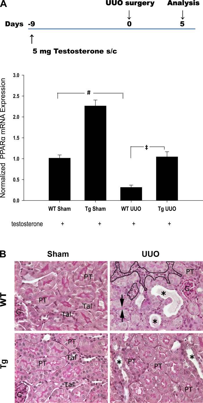
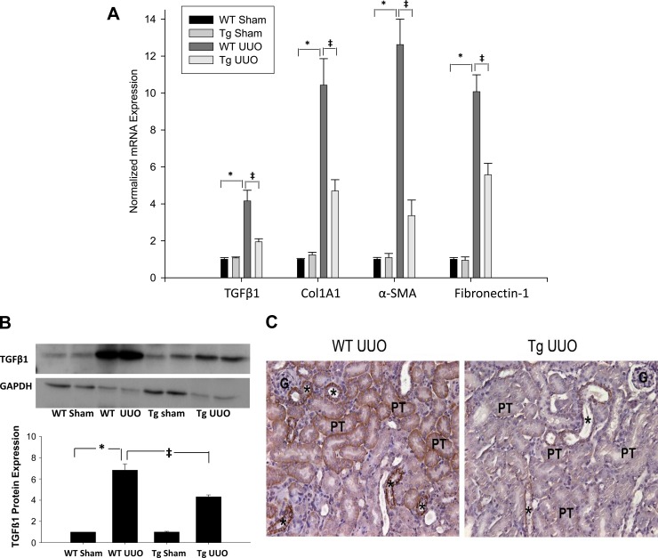
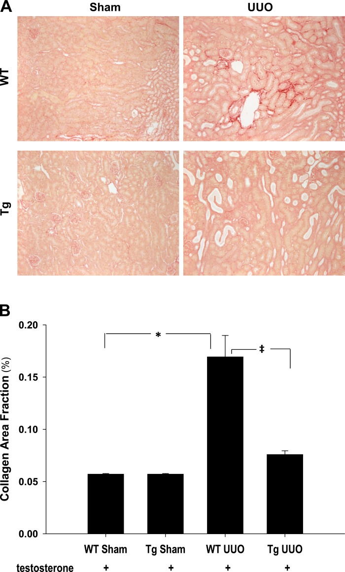
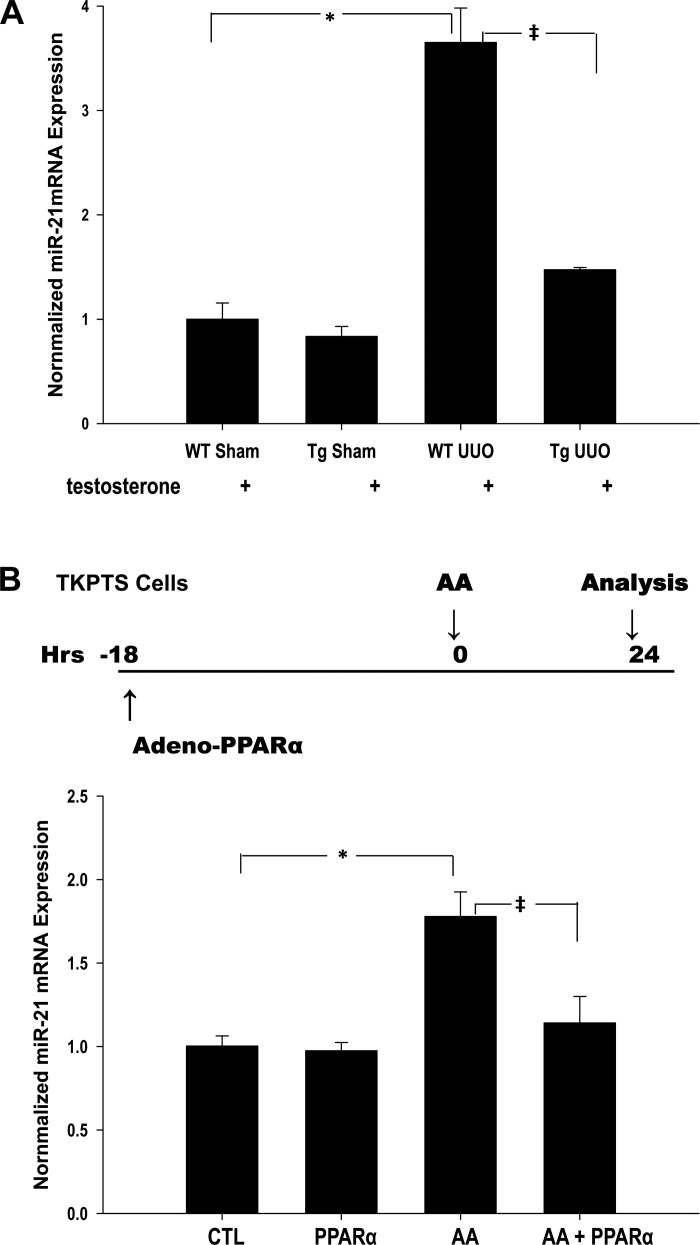
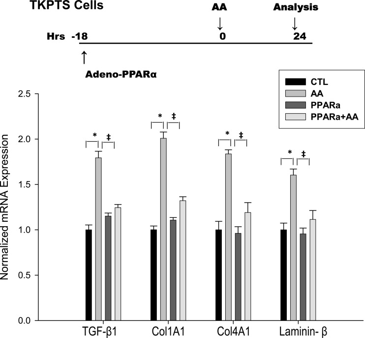
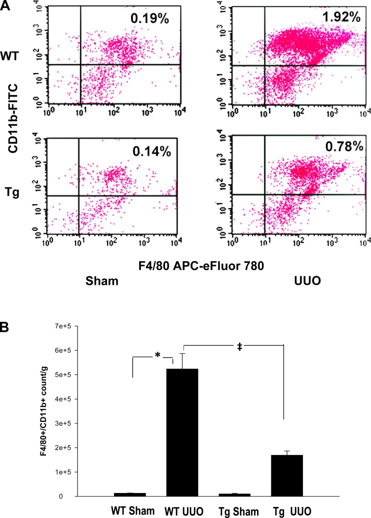
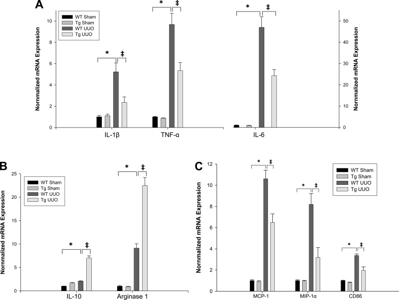
We examined the effects of increased expression of proximal tubule peroxisome proliferator-activated receptor (PPAR)α in a mouse model of renal fibrosis. After 5 days of unilateral ureteral obstruction (UUO), PPARα expression was significantly reduced in kidney tissue of wild-type mice but this downregulation was attenuated in proximal tubules of PPARα transgenic (Tg) mice. When compared with wild-type mice subjected to UUO, PPARα Tg mice had reduced mRNA and protein expression of proximal tubule transforming growth factor (TGF)-β1, with reduced production of extracellular matrix proteins including collagen 1, fibronectin, α-smooth muscle actin, and reduced tubulointerstitial fibrosis. UUO-mediated increased expression of microRNA 21 in kidney tissue was also reduced in PPARα Tg mice. Overexpression of PPARα in cultured proximal tubular cells by adenoviral transduction reduced aristolochic acid-mediated increased production of TGF-β, demonstrating PPARα signaling reduces epithelial TGF-β production. Flow cytometry studies of dissociated whole kidneys demonstrated reduced macrophage infiltration to kidney tissue in PPARα Tg mice after UUO. Increased expression of proinflammatory cytokines including IL-1β, IL-6, and TNF-α in wild-type mice was also significantly reduced in kidney tissue of PPARα Tg mice. In contrast, the expression of anti-inflammatory cytokines IL-10 and arginase-1 was significantly increased in kidney tissue of PPARα Tg mice when compared with wild-type mice subjected to UUO. Our studies demonstrate several mechanisms by which preserved expression of proximal tubule PPARα reduces tubulointerstitial fibrosis and inflammation associated with obstructive uropathy.

Keywords: interleukin-10; peroxisome proliferator-activated receptor; transforming growth factor-β.


Figures

Fig. 1.
Fig. 1.
A, top: schematic diagram showing the experimental time course. Animals received a subcutaneous pellet of 5 mg testosterone for 9 days before unilateral ureteral obstruction (UUO) surgery. Kidneys were harvested 5 days after UUO. A, bottom: effect of UUO on renal peroxisome proliferator-activated receptor (PPAR)α mRNA expression in both wild-type (WT) and transgenic (Tg) mice. Level of PPARα was determined by qPCR. Data are expressed as means ± SE. #P < 0.005 when comparing WT sham vs. UUO mice. ‡P < 0.005 when comparing WT UUO vs. PPARα Tg UUO mice by unpaired Student's t-test. B: representative photographs of periodic acid Schiff (PAS)-stained sections of sham-operated and 5 days after UUO kidneys from WT and PPARα Tg animals. The sham-operated WT and PPARα Tg mice showed normal kidney architecture, whereas WT mice subjected to UUO showed dilated distal nephron segments, with casts (*), interstitial expansion (circled area), and thickening of basement membrane (arrow head). In PPARα Tg mice 5 days after UUO only some tubular dilation was observed (*). PT, proximal tubule; TAL, thick ascending limbs; G, glomerulus. Magnification: ×488.
Fig. 2.
Fig. 2.
A: effect of UUO on renal fibrogenic gene transcripts transforming growth factor (TGF)-β1, collagen type I, α1 (Col1A1), α-smooth muscle actin (SMA), and fibronectin. Bars represent means ± SE mRNA levels for at least 4 mice in each group. *P < 0.001 when comparing WT sham vs. WT-UUO mice. ‡P < 0.05 when comparing WT UUO vs. PPARα Tg UUO mice. B: TGF-β1 protein expression in kidney tissue of WT and PPARα Tg mice subjected to sham and 5 days UUO. B: densitometry and quantification of TGF-β1 signals, normalized to GAPDH levels from Western blot analysis; GAPDH was used as a loading control. Data are expressed as means ± SE. *P < 0.005 when comparing WT sham vs. WT UUO mice. ‡P < 0.005 when comparing WT UUO vs. PPARα Tg UUO mice by unpaired Student's t-test. C: representative photomicrographs of TGF-β1 immunostaining in WT and Tg mice 5 days after UUO. Positive staining is obvious in the cortical thick ascending limbs and proximal convoluted tubules of WT animals. The positive staining is reduced from Tg UUO kidney. *, thick ascending limb of loop of Henle; PT, proximal convoluted tubules. Magnification: ×244.
Fig. 3.
Fig. 3.
A: representative photomicrographs of picro-sirius red-stained sham and 5 days UUO kidney sections from WT and Tg animals. Collagen accumulation can be seen, indicated by the red staining in the WT UUO section. No significant positive staining was seen in shams and Tg UUO kidneys. Magnification: ×122. B: quantitative analysis of picro-sirius red staning in kidney sections from sham and 5 days UUO WT and Tg animals. Collagen accumulation was significantly increased in WT UUO kidneys, and it was unchanged in the Tg UUO samples compared with sham-operated ones. *P < 0.05 when comparing WT sham vs. WT UUO mice. ‡P < 0.05 when comparing WT UUO vs. Tg UUO mice in unpaired Student's t-test.
Fig. 4.
Fig. 4.
PPARα repressed UUO-mediated upregulation of miRNA21 (miR21) in kidney tissue and also reduces aristolochic acid (AA)-stimulated miR21 expression in TKPTS cells. Level of miR21 mRNA was determined by qPCR. A: quantitative analysis of miR21 expression of WT and PPARα Tg mice subjected to sham and UUO surgery. *P < 0.05 when comparing changes in miR21 expression between WT sham and WT UUO mice. ‡P < 0.05 when comparing changes in miR21 expression between WT UUO and Tg UUO mice in unpaired Student's t-test. B: TKPTS cells were incubated with PPARα adenovirus for 18 h before being treated with AA and grown for an additional 24 h. Bars represent means ± SE mRNA levels for at least 4 independent experiments in each group. *P < 0.05 when comparing untreated cells (control) vs. AA-treated cells. ‡P < 0.05 when comparing AA-treated cells vs. AA in the presence of PPARα (AA+PPARα) in unpaired Student's t-test.
Fig. 5.
Fig. 5.
PPARα inhibited AA-stimulated TGF-β1, Col1A1, Col4A1, and laminin-β mRNA expression in TKPTS cells. Levels of mRNA were determined by qPCR. TKPTS cells were incubated with PPARα adenovirus for 18 h before being treated with AA and grown for an additional 24 h. *P < 0.05 when comparing untreated cells (control) vs. AA-treated cells. ‡P < 0.05 when comparing AA-treated cells vs. cells treated with AA in the presence of PPARα (AA+PPARα) in unpaired Student's t-test.
Fig. 6.
Fig. 6.
Reduced infiltration of inflammatory mononuclear phagocytes after UUO in PPARα Tg mice. Kidney cell suspensions obtained from mice subjected to 4 experimental conditions were analyzed by flow cytometry as described in methods. Inflammatory macrophages were identified as F4/80+/CD11b+ cells. Comparisons were made between WT and PPARα Tg mice. A: flow histogram of inflammatory macrophages identified as F4/80+/CD11b+ cells. B: quantification of macrophage accumulation in kidney tissue that was significantly reduced in PPARα Tg mice subjected to UUO when compared with WT mice. Data represent means ± SE of 3 independent experiments. *P < 0.05 when comparing WT sham vs. WT UUO mice. ‡P < 0.05 when comparing WT UUO vs. Tg UUO mice using unpaired Student's t-test.
Fig. 7.
Fig. 7.
Effect of UUO on renal cytokine production. A: changes in IL-1β, TNF-α, IL-6 mRNA levels. B: changes in IL-10 and arginase-1 mRNA levels in WT and PPARα Tg mice subjected to sham or UUO surgery. Levels of mRNA were determined by qPCR. Bars represent means ± SE and mRNA levels were measured using 4 mice in each group. *P < 0.001 when comparing WT sham vs. WT UUO mice. ‡P < 0.05 when comparing WT UUO vs. Tg UUO mice in unpaired Student's t-test. C: effect of UUO on renal monocyte chemoattractant protein-1 (MCP-1), macrophage inflammatory protein-1α (MIP-1α), and macrophage surface marker CD86 mRNA levels in WT and PPARα Tg mice subjected to sham and UUO surgery. Bars represent means ± SE mRNA levels for at least 4 mice in each group. *P < 0.001 when comparing WT sham vs. WT UUO mice. ‡P < 0.05 when comparing WT UUO vs. Tg UUO mice in unpaired Student's t-test.

References

    1. Bielesz B, Sirin Y, Si H, Niranjan T, Gruenwald A, Ahn S, Kato H, Pullman J, Gessler M, Haase VH, Susztak K. Epithelial Notch signaling regulates interstitial fibrosis development in the kidneys of mice and humans. J Clin Invest 120: 4040–4054, 2010 - PMC - PubMed
    1. Boor P, Celec P, Martin IV, Villa L, Hodosy J, Klenovicsova K, Esposito C, Schafer S, Albrecht-Kupper B, Ostendorf T, Heidland A, Sebekova K. The peroxisome proliferator-activated receptor-[alpha] agonist, BAY PP1, attenuates renal fibrosis in rats. Kidney Int 80: 1182–1197, 2011 - PubMed
    1. Campanholle G, Ligresti G, Gharib SA, Duffield JS. Cellular mechanisms of tissue fibrosis. 3. Novel mechanisms of kidney fibrosis. Am J Physiol Cell Physiol 304: C591–C603, 2013 - PMC - PubMed
    1. Carthew RW, Sontheimer EJ. Origins and mechanisms of miRNAs and siRNAs. Cell 136: 642–655, 2009 - PMC - PubMed
    1. Chau BN, Xin C, Hartner J, Ren S, Castano AP, Linn G, Li J, Tran PT, Kaimal V, Huang X, Chang AN, Li S, Kalra A, Grafals M, Portilla D, MacKenna DA, Orkin SH, Duffield JS. MicroRNA-21 promotes fibrosis of the kidney by silencing metabolic pathways. Sci Transl Med 4: 121ra118, 2012 - PMC - PubMed

Publication types

MeSH terms