. 2013 Dec;20(6):537-47.
doi: 10.1093/dnares/dst029. Epub 2013 Jun 26.

The control region of mitochondrial DNA shows an unusual CpG and non-CpG methylation pattern

Affiliations

The control region of mitochondrial DNA shows an unusual CpG and non-CpG methylation pattern

Dina Bellizzi et al. DNA Res. 2013 Dec.

Abstract

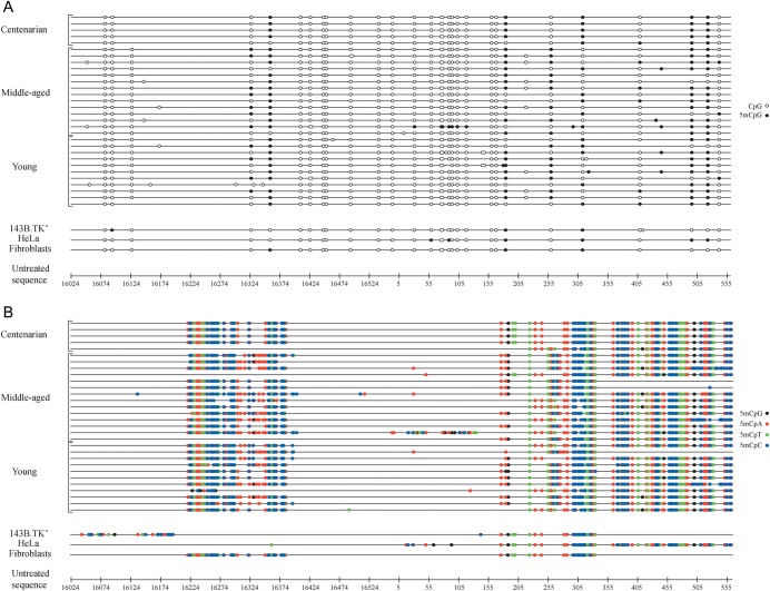
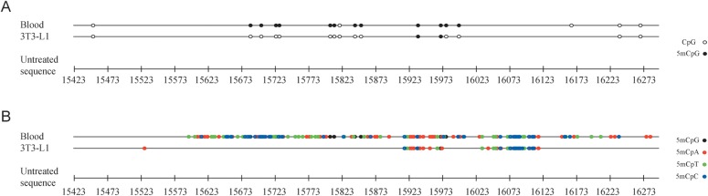
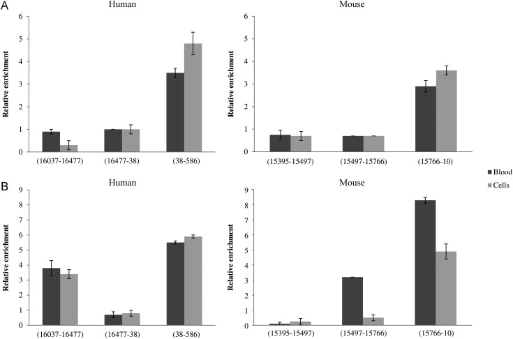
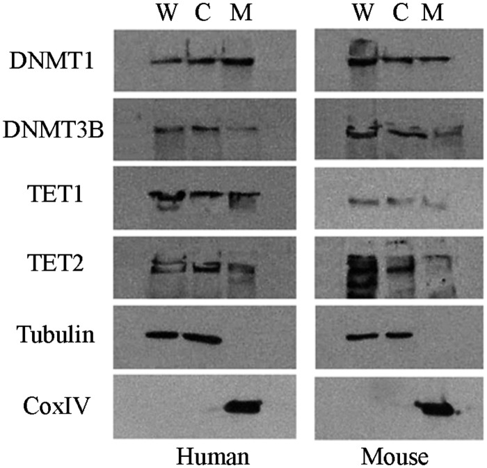
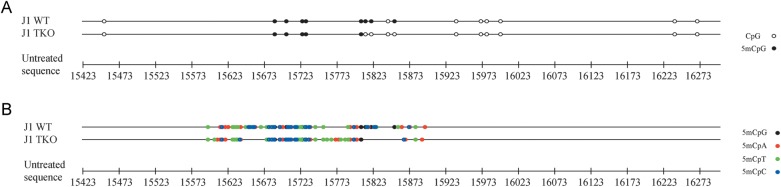
DNA methylation is a common epigenetic modification of the mammalian genome. Conflicting data regarding the possible presence of methylated cytosines within mitochondrial DNA (mtDNA) have been reported. To clarify this point, we analysed the methylation status of mtDNA control region (D-loop) on human and murine DNA samples from blood and cultured cells by bisulphite sequencing and methylated/hydroxymethylated DNA immunoprecipitation assays. We found methylated and hydroxymethylated cytosines in the L-strand of all samples analysed. MtDNA methylation particularly occurs within non-C-phosphate-G (non-CpG) nucleotides, mainly in the promoter region of the heavy strand and in conserved sequence blocks, suggesting its involvement in regulating mtDNA replication and/or transcription. We observed DNA methyltransferases within the mitochondria, but the inactivation of Dnmt1, Dnmt3a, and Dnmt3b in mouse embryonic stem (ES) cells results in a reduction of the CpG methylation, while the non-CpG methylation shows to be not affected. This suggests that D-loop epigenetic modification is only partially established by these enzymes. Our data show that DNA methylation occurs in the mtDNA control region of mammals, not only at symmetrical CpG dinucleotides, typical of nuclear genome, but in a peculiar non-CpG pattern previously reported for plants and fungi. The molecular mechanisms responsible for this pattern remain an open question.

Keywords: 5-hydromethylcytosine; 5-methylcytosine; CpG methylation; mitochondrial D-loop region; non-CpG methylation.


Figures

Figure 1.
Figure 1.
Methylation patterns of the human mitochondrial D-loop in DNA samples from blood and cultured cells. The graphical representation of bisulphite sequencing results was generated by the MethTools software (version 1.2) (http://genome.imb-jena.de/methtools). Bisulphite-generated sequence of each sample (black line) was compared with the untreated sequence of the mitochondrial D-loop, reported here in a base-pair scale. In (A), methylation of cytosine residues located within CpG nucleotides (CpG methylation) is shown. The variability among samples is due to reported polymorphisms (http://www.mitomap.org), which insert or delete cytosines or guanines based on the revised Cambridge reference sequence (rCRS), thus creating/suppressing CpGs. In (B), methylation of cytosine residues located outside of CpG nucleotides (non-CpG methylation) is shown.
Figure 2.
Figure 2.
Methylation patterns of the mouse mitochondrial D-loop in DNA samples from blood and cultured cells. In (A), methylation of cytosine residues located within CpG nucleotides (CpG methylation) is shown. In (B), methylation of cytosine residues located outside of CpG nucleotides (non-CpG methylation) is shown.
Figure 3.
Figure 3.
Methylated and hydroxymethylated mtDNA immunoprecipitation. AluI-digested DNA extracted from blood and cultured cells of human and mouse were immunoprecipitated with anti-5mC (A) and anti-5hmC (B). Samples were amplified with primers specific for DNA fragments detected as unmethylated and methylated by bisulphite sequencing. The nucleotide position of these fragments is indicated. Fold enrichment was calculated as the ratio of amplification efficiency of the immunoprecipitated sample over that of non-immune IgG and normalized to the nanograms of immunoprecipitated DNA amount. Data represent the means of six triplicate experiments with standard errors of the mean.
Figure 4.
Figure 4.
Representative western blot electrophoresis patterns of DNMTs and TETs. Whole cell lysate (W), cytosolic (C), and mitochondrial (M) protein fractions of human HeLa and murine 3T3-L1 cells. CoxIV and tubulin were analysed as mitochondrial- and cytosolic-specific markers, respectively.
Figure 5.
Figure 5.
Methylation patterns of the mitochondrial D-loop in DNA samples from wt and Dnmt1−/−, Dnmt3a−/−, and Dnmt3b−/− mouse ES cells. In (A), methylation of cytosine residues located within CpG nucleotides (CpG methylation) is shown. In (B), methylation of cytosine residues located outside of CpG nucleotides (non-CpG methylation) is shown.

References

    1. Lee T., Zhai J., Meyers C. Conservation and divergence in eukaryotic DNA methylation. Proc. Natl Acad. Sci. USA. 2010;107:9027–8. - PMC - PubMed
    1. Vanyushin B.F., Ashapkin V.V. DNA methylation in higher plants: past, present and future. Biochim. Biophys. Acta. 2011;1809:360–8. - PubMed
    1. Ramsahoye B.H., Biniszkiewicz D., Lyko F., Clark V., Bird A.P., Jaenisch R. Non-CpG methylation is prevalent in embryonic stem cells and may be mediated by DNA methyltransferase 3a. Proc. Natl Acad. Sci. USA. 2000;97:5237–42. - PMC - PubMed
    1. Lister R., Pelizzola M., Dowen R.H., et al. Human DNA methylomes at base resolution show widespread epigenomic differences. Nature. 2009;462:315–22. - PMC - PubMed
    1. Xie W., Barr C.L., Kim A., et al. Base-resolution analyses of sequence and parent-of-origin dependent DNA methylation in the mouse genome. Cell. 2012;148:816–31. - PMC - PubMed

Publication types