. 2013 Sep;87(17):9643-8.
doi: 10.1128/JVI.01506-13. Epub 2013 Jun 26.

Aberrant virion assembly and limited glycoprotein C production in varicella-zoster virus-infected neurons

Affiliations

Aberrant virion assembly and limited glycoprotein C production in varicella-zoster virus-infected neurons

Charles Grose et al. J Virol. 2013 Sep.

Abstract

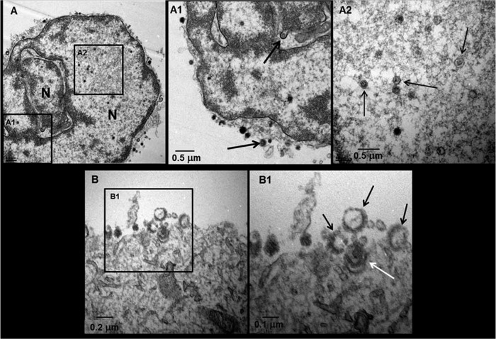
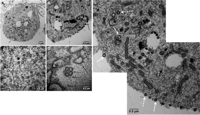
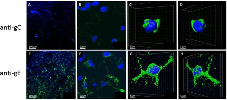
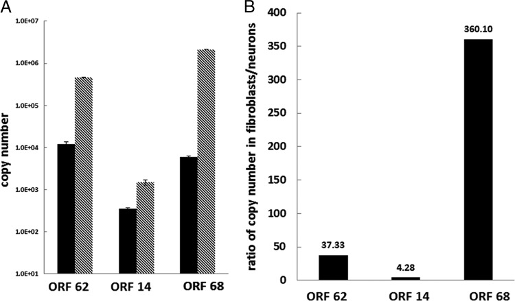
Highly pure (>95%) terminally differentiated neurons derived from pluripotent stem cells appear healthy at 2 weeks after infection with varicella-zoster virus (VZV), and the cell culture medium contains no infectious virus. Analysis of the healthy-appearing neurons revealed VZV DNA, transcripts, and proteins corresponding to the VZV immediate early, early, and late kinetic phases of replication. Herein, we further characterized virus in these neuronal cells, focusing on (i) transcription and expression of late VZV glycoprotein C (gC) open reading frame 14 (ORF14) and (ii) ultrastructural features of virus particles in neurons. The analysis showed that gC was not expressed in most infected neurons and gC expression was markedly reduced in a minority of VZV-infected neurons. In contrast, expression of the early-late VZV gE glycoprotein (ORF68) was abundant. Transcript analysis also showed decreased gC transcription compared with gE. Examination of viral structure by high-resolution transmission electron microscopy revealed fewer viral particles than typically observed in cells productively infected with VZV. Furthermore, viral particles were more aberrant, in that most capsids in the nuclei lacked a dense core and most enveloped particles in the cytoplasm were light particles (envelopes without capsids). Together, these results suggest a considerable deficiency in late-phase replication and viral assembly during VZV infection of neurons in culture.


Figures

Fig 1
Fig 1
Ultrastructure of VZV particles in infected neurons. A VZV-infected neuron with a multilobed nucleus exhibited viral particles and capsids (A). A viral capsid was seen egressing from the nucleus (A1, top arrow), and viral particles were seen on the cell surface (A1, bottom arrow). Multiple capsids were seen in the nucleus (A2, arrows). Among virus particles seen on the cell surface (B), four were near the cell surface (B1), three of which were light particles lacking a DNA core (black arrows); the fourth particle was a complete virion (white arrow).
Fig 2
Fig 2
Transmission electron microscopy of VZV-infected neurons. A cytoplasmic section of one VZV-infected neural cell body exhibited a large number of cytoplasmic viral particles with a smaller number on the cell surface (A). A section of a different infected neuron exhibited indistinct viral particles egressing onto the surface of the cell (B; arrow). At high magnification, 23 viral capsids were seen in a section of an infected cell nucleus (C), of which 7 or 8 contained a DNA core (dashed arrow), while another viral capsid lacked DNA (solid arrow). A vesicle of enveloped viral particles (D) was seen in the nucleus (dashed arrow) with several viral capsids (solid arrows). A montage of the cytoplasm and cell surface of a VZV-infected neuron showed viral particles without capsids and viral DNA (solid white arrows), viral particles with capsids but not viral DNA (dashed white arrows), and complete viral particles with capsid and DNA (black arrows) (E).
Fig 3
Fig 3
Expression of VZV glycoproteins C and E in VZV-infected neurons. VZV-infected neurons were fixed at 14 d.p.i. and stained with anti-VZV gC antibody (A to D), anti-VZV gE antibody (E to H), and Hoechst H33342 as described in the text. Images were collected on a laser-scanning confocal microscope at 100× (A and E) and 900× (B and F). The 3D images of single infected cells were generated from a z-stack using Imaris software (C, D, G, and H). Note the markedly reduced gC staining (A and B) compared to the abundant gE staining (E and F) in infected neurons. gC staining was restricted to the perinuclear endoplasmic reticulum (C and D), in contrast to extensive cytoplasmic gE staining (G and H).
Fig 4
Fig 4
VZV mRNA transcripts in VZV-infected neurons and fibroblasts. The abundance of all VZV transcripts was decreased in neurons (black bars) compared to fibroblasts (gray) (A). The ratio of VZV transcripts expressed in fibroblasts and neurons is shown in panel B.

References

    1. Weller TH, Witton HM, Bell EJ. 1958. The etiologic agents of varicella and herpes zoster; isolation, propagation, and cultural characteristics in vitro. J. Exp. Med. 108:843–868 - PMC - PubMed
    1. Gilden DH, Wroblewska Z, Kindt V, Warren KG, Wolinsky JS. 1978. Varicella-zoster virus infection of human brain cells and ganglion cells in tissue culture. Arch. Virol. 56:105–117 - PubMed
    1. Wigdahl B, Rong BL, Kinney-Thomas E. 1986. Varicella-zoster virus infection of human sensory neurons. Virology 152:384–399 - PubMed
    1. Hood C, Cunningham AL, Slobedman B, Boadle RA, Abendroth A. 2003. Varicella-zoster virus-infected human sensory neurons are resistant to apoptosis, yet human foreskin fibroblasts are susceptible: evidence for a cell-type-specific apoptotic response. J. Virol. 77:12852–12864 - PMC - PubMed
    1. Markus A, Grigoryan S, Sloutskin A, Yee MB, Zhu H, Yang IH, Thakor NV, Sarid R, Kinchington PR, Goldstein RS. 2011. Varicella-zoster virus (VZV) infection of neurons derived from human embryonic stem cells: direct demonstration of axonal infection, transport of VZV, and productive neuronal infection. J. Virol. 85:6220–6233 - PMC - PubMed

Publication types

MeSH terms

Substances

LinkOut - more resources