. 2013 Aug 1;191(3):1240-9.
doi: 10.4049/jimmunol.1300062. Epub 2013 Jun 26.

T cell-dependent IgM memory B cells generated during bacterial infection are required for IgG responses to antigen challenge

Affiliations

T cell-dependent IgM memory B cells generated during bacterial infection are required for IgG responses to antigen challenge

Jennifer L Yates et al. J Immunol. .

Abstract

Immunological memory has long considered to be harbored in B cells that express high-affinity class-switched IgG. IgM-positive memory B cells can also be generated following immunization, although their physiological role has been unclear. In this study, we show that bacterial infection elicited a relatively large population of IgM memory B cells that were uniquely identified by their surface expression of CD11c, CD73, and programmed death-ligand 2. The cells lacked expression of cell surface markers typically expressed by germinal center B cells, were CD138 negative, and did not secrete Ab ex vivo. The population was also largely quiescent and accumulated somatic mutations. The IgM memory B cells were located in the region of the splenic marginal zone and were not detected in blood or other secondary lymphoid organs. Generation of the memory cells was CD4 T cell dependent and required IL-21R signaling. In vivo depletion of the IgM memory B cells abrogated the IgG recall responses to specific Ag challenge, demonstrating that the cell population was required for humoral memory, and underwent class-switch recombination following Ag encounter. Our findings demonstrate that T cell-dependent IgM memory B cells can be elicited at high frequency and can play an important role in maintaining long-term immunity during bacterial infection.


Figures
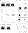
Figure 1
Figure 1. Identification of a long-term splenic B cell population elicited by bacterial infection
(A) Representative flow cytometry analysis of CD11c expression on CD19-positive B cells in the spleens of E. muris-infected, and uninfected mice. The data in the plots were generated after gating on viable cells, as determined by forward and side scatter parameters. (B) Kinetic analysis of CD11c-positive B cells in E. muris infected mice. The frequency (top panel) and number (bottom panel) of CD11c-positive B cells among total splenocytes are shown. Each datum represents an individual mouse, and horizontal lines represent the mean. The asterisks indicate statistical significance, where * p< 0.01 and ** p < 0.001, relative to the values obtained on day 0. (C) Representative flow cytometry analysis of CD11c-expressing B cells from E. muris infected mice, and from mice infected with a recently identified human isolate (Ehrlichia sp. Wisconsin HM543745), analyzed 49 days post-infection. (D) Representative flow cytometry analyses of CD11c-positive and –negative B cells obtained from E. muris infected mice. (E) Frequency and number of splenic CD11c-positive B cells in untreated, or antibiotic-treated E. muris-infected mice. The mice were treated with doxycycline for two weeks, starting on day 30 post-infection. The splenocytes were analyzed approximately one month following antibiotic treatment. The dot plots are representative of two experiments of similar design.
Figure 2
Figure 2. The CD11c-positive B cells exhibit characteristics of memory cells
(A) Representative flow cytometry analysis of CD11c-positive and -negative CD19+ B cells during E. muris infection. The histograms are representative of 5–9 mice that were analyzed at several time-points on or after day 30 post-infection. Basal cell surface staining and voltage settings were established using unstained cells; the data were omitted from the histograms for clarity. Cells obtained from uninfected mice exhibited similar cell surface marker expression as the CD11c-negative B cells obtained from infected mice (data not shown). (B) BrdU incorporation studies of splenic CD11c-positive and -negative B cell from E. muris infected mice. The mice were administered BrdU over a 4 day period, beginning on day 63 post-infection. The splenocytes were analyzed for BrdU incorporation at day 67 post-infection; the cells were also analyzed for CD73 expression, which improved population discrimination. The data are representative of 2 experiments. (C) Mutation analysis of the V region heavy chain J558 family genes from flow cytometrically-purified CD11c-negative, and –positive B cells obtained from infected mice. The segment sizes in the pie charts are proportional to the number of sequences carrying the number of mutations indicated in the periphery of the chart. The number of independent sequences analyzed is indicated in the center of the chart. The mutation frequencies, per base pair, are indicated below the pie charts. The data were compiled from two experiments. Statistical significance was determined using a two-tailed student’s T test, assuming unequal variance, by comparing the CD11c-negative and -positive B cells; the p value is indicated. A single mutation was identified in 9,396 bp from 32 clones obtained from the CD11c-negative B cells, and 87 mutations were identified in 20,247 bp from 69 analyzed clones from the CD11c-positive B cells.
Figure 3
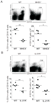
Figure 3. CD4+ T cells and IL-21 signaling were required for the generation of the CD11c-positive B cell population
(A) Flow cytometry analysis of splenic CD11c-positive B cells from wild-type and MHCII-deficient mice analyzed on day 30 post-infection is shown. The frequencies of CD11c-positive B cells in the flow cytometry dot plots represent their percentage among total spleen cells. The data in the panels below indicate the frequency of CD11c-positive cells among total splenic B cells. The average B cell frequency was 67% and 37% in the WT and MHCII-deficient strains, and the average B cell number was 1.6 × 10⁸ and 1.5 × 10⁸ total B cells in the WT and MHCII-deficient strains, respectively. (B) Flow cytometric analysis of CD11c-positive B cells from wild-type and IL-21R-deficient mice on day 70 post-infection. The frequencies of CD11c-positive B cells shown in the dot plots represent their percentage among total splenic B cells. In the experiment shown, the average B cell frequency was 35% and 18% in the WT and IL-21R-deficient strains, and the average B cell number was 5.9 × 10⁷ and 5.8 × 10⁷ B cells, respectively. The data in A and B are representative of two experiments; * p<0.05, **p< 0.005.
Figure 4
Figure 4. CD19-positive B cells were detected in the splenic marginal zone and at the edge of B cell follicles
Cryosections from uninfected (A, C), and infected mice (B, D; analyzed on day 63 post-infection) were stained with Alexa 488-conjugated CD11c (green), and CF594-conjugated CD19 (red), and Alexa 647-conjugated CD169 (blue; panels A and B) or PNA-Alexa 647 (blue; panels C and D). The images show representative images of B cells identified in several randomly selected cryosections. The scale bars represent 40 μm, and yellow arrows indicate several dual CD11c- CD19 -positive B cells.
Figure 5
Figure 5. Antigen challenge of E. muris-infected mice
Uninfected and infected mice were administered alum, or were challenged with 100 μg recombinant OMP-19 in alum; challenge of the infected mice was performed on day 30 post-infection. (A) Reciprocal OMP-19-specific serum IgG titers obtained on days 0, 3, 6, and 12 post-antigen challenge, determined by ELISA, are shown. (B) OMP-19-specific IgG subclass titers measured in the serum of infected mice that remained unchallenged, or had been challenged with OMP-19; OMP-19 was delivered in either alum or PBS, as indicated. (C) Serum OMP-19 specific antibody titers were determined on days 0, 6, and 12 post-challenge in MHCII-deficient, IL-21-deficient, and wild-type mice. The data in each of the panels are representative of two experiments. * p<0.05, ** p< 0.005
Figure 6
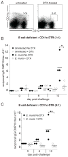
Figure 6. In vivo depletion of CD11c-positive B cells abrogated the IgG responses following antigen challenge
(A) CD11c-DTR transgenic mice were infected, and administered diphtheria toxin (DTX; 9ng/g of body weight) on day 30 post-infection. Twenty-four hours later, splenocytes were analyzed by flow cytometry. The panels shown are representative of 3 mice per group. (B) Chimeric mice were generated using a 1:1 ratio of B cell-deficient and CD11c-DTR bone marrow. The chimeric mice were infected, administered DTX on day 30 post-infection, and challenged with OMP-19, as described in Figure 5. (C) Chimeric mice were generated as in B, using a 9:1 ratio of B cell deficient to CD11c-DTR bone marrow. Serum OMP-19 titers were determined on day 12 post-challenge. The data in each of the studies shown are representative of at least two experiments; * p<0.05

Similar articles

Cited by

References

    1. McHeyzer-Williams LJ, McHeyzer-Williams MG. Antigen-specific memory B cell development. Annu Rev Immunol. 2005;23:487–513. - PubMed
    1. McHeyzer-Williams M, Okitsu S, Wang N, McHeyzer-Williams L. Molecular programming of B cell memory. Nat Rev Immunol. 2011;12:24–34. - PMC - PubMed
    1. Kaisho T, Schwenk F, Rajewsky K. The roles of gamma 1 heavy chain membrane expression and cytoplasmic tail in IgG1 responses. Science. 1997;276:412–415. - PubMed
    1. Martin SW, Goodnow CC. Burst-enhancing role of the IgG membrane tail as a molecular determinant of memory. Nat Immunol. 2002;3:182–188. - PubMed
    1. Tangye SG, Avery DT, Deenick EK, Hodgkin PD. Intrinsic differences in the proliferation of naive and memory human B cells as a mechanism for enhanced secondary immune responses. J Immunol. 2003;170:686–694. - PubMed

Publication types

MeSH terms