Review
. 2013 Sep;41(16):7589-605.
doi: 10.1093/nar/gkt556. Epub 2013 Jun 26.

DNA double-strand-break complexity levels and their possible contributions to the probability for error-prone processing and repair pathway choice

Affiliations
Review

DNA double-strand-break complexity levels and their possible contributions to the probability for error-prone processing and repair pathway choice

Agnes Schipler et al. Nucleic Acids Res. 2013 Sep.

Abstract

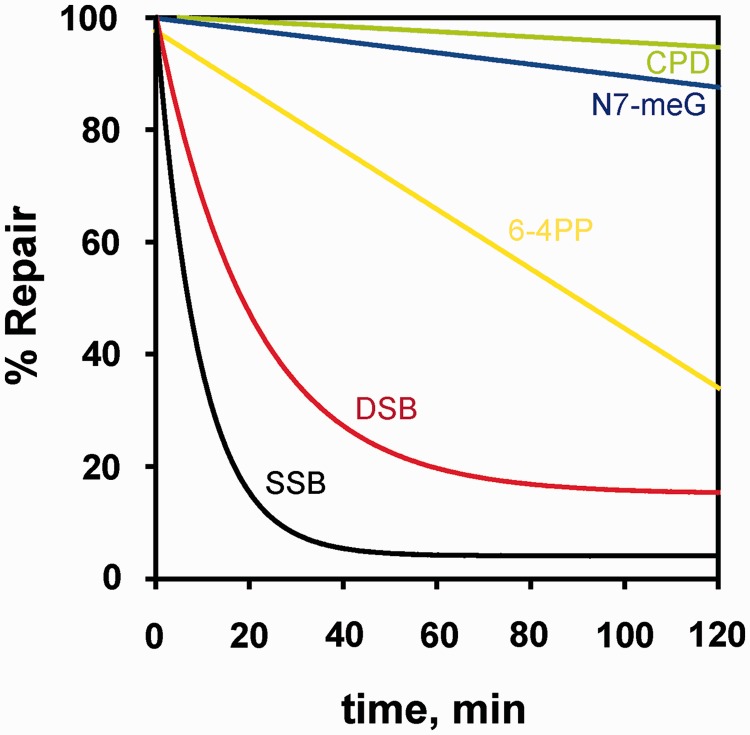
Although the DNA double-strand break (DSB) is defined as a rupture in the double-stranded DNA molecule that can occur without chemical modification in any of the constituent building blocks, it is recognized that this form is restricted to enzyme-induced DSBs. DSBs generated by physical or chemical agents can include at the break site a spectrum of base alterations (lesions). The nature and number of such chemical alterations define the complexity of the DSB and are considered putative determinants for repair pathway choice and the probability that errors will occur during this processing. As the pathways engaged in DSB processing show distinct and frequently inherent propensities for errors, pathway choice also defines the error-levels cells opt to accept. Here, we present a classification of DSBs on the basis of increasing complexity and discuss how complexity may affect processing, as well as how it may cause lethal or carcinogenic processing errors. By critically analyzing the characteristics of DSB repair pathways, we suggest that all repair pathways can in principle remove lesions clustering at the DSB but are likely to fail when they encounter clusters of DSBs that cause a local form of chromothripsis. In the same framework, we also analyze the rational of DSB repair pathway choice.


Figures

Figure 1.
Figure 1.
Kinetics of repair of different types of DNA lesions. Shown is the kinetics of removal from CHO-AA8 cells of SSBs, DSBs, 6–4 photoproducts (6–4PP), cyclobutane pyrimidine dimers (CPD) and, for human lymphocytes, of N7-meG. SSB and DSB repair was measured after exposure to 7.5 Gy and 100 Gy of γ-rays, respectively. SSBs were assayed by alkaline filter elution at pH 12.1 and DSBs by non-denaturing filter elution at pH 9.6 (2). Repair of UV-induced CPD and 6–4PP was measured in CHO cells by radioimmunoassay using damage-specific antibodies. Removal of antibody-binding sites after various repair times was determined after 10 J/m2 UV-irradiation (3). Repair of N7-meG was measured in human lymphocytes after treatment with alkylating agents (7).
Figure 2.
Figure 2.
Three scenarios of DSB misrepair. (A) DSB ends drift apart resulting in a chromosomal aberration in the form of an acentric fragment (Del). (B) Rejoining of the DSB occurs but the junction is altered. Examples for large deletions in the Hprt locus are shown. The nine exons of Hprt are indicated at the top of the right panel. Genomic regions amplified by polymerase chain reaction are shown by solid lines. Spaces between the lines represent DNA sections that are deleted [drawn from results published by (24)]. (C) Joining of incongruent ends can cause chromosomal translocations. Fluorescence in situ hybridization analysis shows a c-myc/Ig locus translocation between chromosomes 8 and 14 in a multiple myeloma cell line [image from (25)]. An exchange-type aberration in the form of a ring chromosome is also shown in panel A (Exch).
Figure 3.
Figure 3.
Illustration of the different types of DSBs as defined in the text. (A) T1-DSBs are direct DSBs induced by RE. An example for EcoRI DSB is shown that produces staggered ends with a 5′-phosphate and a 3′-OH group. (B) T2-DSBs are induced by IR and frequently comprise a 3′-phosphoglycolate and a 5′-OH at the DNA ends as shown in this example. (C) IR also induces clustered lesions from ionization clusters, defined as T3-DSBs. In this case, the direct DSB is accompanied by other types of lesions, like base damage or base loss proximal to the DSB. (D) T4-DSBs represent a non-DSB damage cluster that can convert to DSBs (indirect DSB) by enzymatic processing of the constituent base lesions. (E) T5-DSBs are also induced indirectly, up to 1 h after IR, by temperature-sensitive chemical processing of damaged sugar moieties opposing SSBs. (F) T6-DSBs are composed of clustered DSBs that can destabilize chromatin. Two possible scenarios are illustrated: in the first scenario (upper left) radiation induces two DSBs in the linker regions between a nucleosome risking nucleosome loss. The second scenario (lower right) shows higher-order packaging of nucleosomes forming a chromatin loop that is broken as shown by a radiation track. Here, loss of a larger segment of chromatin is possible. In the lower right corner of the drawing the 10-nm chromatin fiber is shown, compacted as a fractal globule (36,37); the opening of a loop from this fractal globule is indicated.
Figure 4.
Figure 4.
Distribution of DNA damage inducing events after exposure to H2O2 and IR of low and high LET·OH radicals from H2O2 are evenly distributed in space and induce, therefore, also evenly distributed DNA damage. In the case of IR, ionization events localize along the particle tracks [middle panel 0.5 and 10 keV electrons (e), right panel 4 MeV α particle] and can, therefore, induce clustered damage as indicated. Note that with increasing LET (from 10 to 0.5 keV e up to the 4 MeV α particle) the damage clustering increases. Large dots represent ionizations and small dots represent excitations along the radiation track. Monte Carlo simulated tracks are drawn for the 0.5 keV e and the α particle on the same scale as the DNA [redrawn from (58)]. The track for the 10 keV e, as well as the events shown after treatment with H2O2 are by free drawing and shown only for illustration purposes.
Figure 5.
Figure 5.
Fragment loss through 2xDSB cluster. An example of clustered DSB: two DSBs in the cluster induced in the linker region between nucleosomes. It can lead to chromatin destabilization through the loss of the DNA segment between the two DSBs. Two possible processing scenarios are illustrated. If the DSB ends stay close, the DNA molecule is restored by simple rejoining. In a second scenario (shown on the right), a small DNA fragment comprising four nucleosomes is lost from the chromatin context causing a deletion and possibly also jeopardizing, or somehow impairing, all forms of processing.
Figure 6.
Figure 6.
Key steps of DSB repair pathways (HRR, D-NHEJ and B-NHEJ) with examples of end-processing options for T3-DSBs. (A) During HRR, extensive processing of the 5′-ends takes place that can remove lesions in the vicinity of DSB ends. Although base damage remains at the 3′-end after HRR, the DSB is repaired and the remaining single base lesion can be removed by BER at a later time. (B) For D-NHEJ, limited end processing takes place at both DNA strands—5′ and 3′. As a result, lesions that span up to 10 bp from the DSB ends could be removed as well, although their presence is likely to delay this processing. (C) During B-NHEJ, even more extensive end processing takes place, and as for D-NHEJ, lesions adjacent to the DSB may be removed. B-NHEJ often results in large deletions—and translocations. (D) Illustration of Ku bound to DNA. This protein–DNA interaction was visualized using the program PyMOL (PyMOL Molecular Graphics System, Version 1.2r3pre, Schrödinger, LLC). The results are from (129). The structure illustrates that each Ku molecule binds roughly two helical turns of DNA.
Figure 7.
Figure 7.
Propensity for errors by HRR, D-NHEJ and B-NHEJ. For each repair pathway, the probability for sequence alterations at the junction is indicated with orange shading, whereas the probability for translocations is indicated with blue shading. The scale is arbitrary and serves only illustration purposes—also when comparing the two sources of errors. HRR has very low probability for both sequence alterations at the junction, as well as for translocations. D-NHEJ has low probability for translocations, but relatively high probability for sequence alterations at the junction. B-NHEJ is, on the other hand, highly error prone on all counts.

Similar articles

Cited by

References

    1. Friedberg EC, Walker GC, Siede W, Wood RD, Schultz RA, Ellenberger T. DNA Repair and Mutagenesis. 2nd edn. Washington, D.C: ASM Press; 2006.
    1. van Ankeren SC, Murray D, Meyn RE. Induction and rejoining of gamma-ray-induced DNA single- and double-strand breaks in Chinese hamster AA8 cells and in two radiosensitive clones. Radiat. Res. 1988;116:511–525. - PubMed
    1. Nairn RS, Mitchell DL, Adair GM, Thompson LH, Siciliano MJ, Humphrey RM. UV mutagenesis, cytotoxicity and split-dose recovery in a human—CHO cell hybrid having intermediate (6−4) photoproduct repair. Mutat. Res. 1989;217:193–201. - PubMed
    1. Berens TJ, Toczyski DP. Keeping it together in times of stress: checkpoint function at stalled replication forks. Mol. Cell. 2012;45:585–586. - PMC - PubMed
    1. Burrell RA, McClelland SE, Endesfelder D, Groth P, Weller MC, Shaikh N, Domingo E, Kanu N, Dewhurst SM, Gronroos E, et al. Replication stress links structural and numerical cancer chromosomal instability. Nature. 2013;494:492–496. - PMC - PubMed

Publication types