. 2013 Sep;41(16):7815-27.
doi: 10.1093/nar/gkt560. Epub 2013 Jun 26.

Structural insight into negative DNA supercoiling by DNA gyrase, a bacterial type 2A DNA topoisomerase

Affiliations

Structural insight into negative DNA supercoiling by DNA gyrase, a bacterial type 2A DNA topoisomerase

Julie Papillon et al. Nucleic Acids Res. 2013 Sep.

Abstract

Type 2A DNA topoisomerases (Topo2A) remodel DNA topology during replication, transcription and chromosome segregation. These multisubunit enzymes catalyze the transport of a double-stranded DNA through a transient break formed in another duplex. The bacterial DNA gyrase, a target for broad-spectrum antibiotics, is the sole Topo2A enzyme able to introduce negative supercoils. We reveal here for the first time the architecture of the full-length Thermus thermophilus DNA gyrase alone and in a cleavage complex with a 155 bp DNA duplex in the presence of the antibiotic ciprofloxacin, using cryo-electron microscopy. The structural organization of the subunits of the full-length DNA gyrase points to a central role of the ATPase domain acting like a 'crossover trap' that may help to sequester the DNA positive crossover before strand passage. Our structural data unveil how DNA is asymmetrically wrapped around the gyrase-specific C-terminal β-pinwheel domains and guided to introduce negative supercoils through cooperativity between the ATPase and β-pinwheel domains. The overall conformation of the drug-induced DNA binding-cleavage complex also suggests that ciprofloxacin traps a DNA pre-transport conformation.


Figures

Figure 1.
Figure 1.
Domain organization and characterization of DNA gyrase complexes. (A) Domain organization of the eukaryotic Topo2A and prokaryotic DNA gyrase. Functional regions are colored and labeled. The CTD is divergent between prokaryotes and eukaryotes (green and purple). The DNA gyrase CTD contains a conserved GyrA box motif (lilas box). The catalytic tyrosine in the WHD domain is shown as a red Y letter and is conserved through evolution. The linker connecting the DNA gyrase A and B subunits used for cryo-EM studies is represented as a gray dash line. (B) E. coli and T. thermophilus negative supercoiling enzyme assays. Increasing concentration of reconstituted E. coli or T. thermophilus DNA gyrase have been incubated at 37 or 65°C, respectively, with relaxed pUC19 plasmid in presence of 1 mM ATP and run on an agarose gel colored with ethidium bromide. DNA topoisomers are labeled on the left of each gel. SC: supercoiled DNA, R: relaxed DNA. The T. thermophilus DNA gyrase displays an optimal supercoiling activity at 65°C and supercoils DNA to a lower extent than the mesophile E. coli enzyme in the same concentration range. (C) Native mass spectrometry on the holoenzyme and DNA-bound complex showing the presence of the dimeric A2B2 form of the fusion DNA gyrase in presence of ADPNP and formation of the complex with DNA (green) as shown by the total shift of the mass spectra. The measured masses (in Da) are indicated under the name of each complex.
Figure 2.
Figure 2.
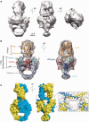
Orthogonal orientation and intertwined organization of the holoenzyme. (A) Cryo-EM map of the holoenzyme shown in three orientations. The cavities of the core GyrA and the ATPase domains appear clearly in the first two orientations in agreement with the crystal structures of these domains. (B) Model of the holoenzyme complex. The crystal structures of the ATPase domain (PDB:1EI1) and DNA binding–cleavage domain (PDB: 3NUH) deleted from the E. coli GyrB specific insertion domain [560–735] were fitted in the cryo-EM map (gray surface). The ATPase domain sits above DNA binding–cleavage domain in an orthogonal orientation. (C) Surface representation of the DNA gyrase full-length architecture and close-up of the DNA cavity interface fitted in the cryo-EM map of the holoenzyme (gray mesh). The orthogonal orientation of the two subunits leads to an intertwined dimer where one ATPase transducer helix connects to the TOPRIM domain on the opposite side through a 10 amino-acids linker (pointed by the arrows).
Figure 3.
Figure 3.
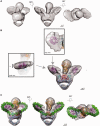
DNA stabilizes and wraps around DNA gyrase β-pinwheel CTDs. (A) Cryo-EM map of the DNA-bound complex shown in three orientations. Outside the core enzyme, the DNA-bound complex shows two additional disk-shape electron densities. (B) Fitting of the crystal structures of DNA gyrase domains in the cryo-EM map. The crystal structures of the ATPase (PDB:1EI1) and DNA binding–cleavage domain in presence of ciprofloxacin (PDB:2XCT) were fitted in the core enzyme map. The two additional densities on both side of the core enzyme can accommodate a β-pinwheel structure (PDB:3L6V), typical of DNA gyrase CTDs. The close-up of one CTD ß-pinwheel fitted in the cryo-EM map (gray surface) shows some empty extra densities large enough to position a dsDNA helix as visible on the top and side views. (C) Modeled duplex DNA wrapping around DNA gyrase. Extra densities around the β-pinwheels are large enough to accommodate DNA (green) that wraps around the two asymmetric β-pinwheels and binds in the DNA-gate cavity.
Figure 4.
Figure 4.
DNA path and domain geometry in DNA-bound complex. (A) Surface representation of the DNA binding–cleavage domain (top view). The ATPase domain has been omitted for clarity. The modeled 130 bp dsDNA is chirally wrapped around the CTD orienting the T-segment (black) in the DNA-gate groove formed by the TOPRIM-WHD and TOWER domains to form a 60° angle positive crossover when we extrapolate the T-segment path. The GyrA-box motif, colored in magenta, is facing the central cavity of the DNA binding–cleavage domain. (B) The conserved orthogonal orientation of the two monomers in the DNA-bound gyrase complex leads to an intertwined dimer with the ATPase domain sitting ∼10 Å higher on the DNA binding–cleavage domain (surface representation in yellow and blue). The linker (red) connecting the DNA binding–cleavage domain and the β-pinwheels domain is running along the modeled DNA (green), while the first blade bearing the GyrA-box motif (magenta) is facing the central DNA cavity. (C) Schematic representation of the domains orientation in the DNA-bound complex model. The DNA binding–cleavage domain (DNA- and C-gate) is depicted as a triangle, the GyrB ATPase domain (N-gate) as an ellipse, and the CTD β-pinwheel domains as disks. Magenta and orange solid and dash lines indicate the CTD and ATPase domain planes, respectively. (Left) The black horizontal line represents the DNA binding–cleavage plane in a top view. The CTDs β-pinwheels are located in an upper position and are distributed asymmetrically on each side of the DNA gate (33° versus 28°). (Right) The ATPase domain bends toward one CTD by an angle of ∼15°with respect to the vertical axis.
Figure 5.
Figure 5.
DNA gyrase supercoiling catalytic cycle in the light of DNA gyrase full architecture. (1) Before DNA binding, the ATPase domain is in a wide-open conformation and the CTDs are located in a lower position. (2, 3) Upon G-segment binding (in green), the DNA gyrase CTDs are rising up asymmetrically as one contiguous T-segment (black) wraps around the CTDs, narrowing and rotating the ATPase subunits in an upper-close position. The tilted position of the ATPase domain toward a CTD on one side may favor T-segment capture. (3, 4) ATP binding closes the ATPase domain dimeric interface and traps the T-segment to form a positive crossover with the G-segment. In this scheme, the swiveling ‘crossover-trap’ mechanism for T-segment capture could be common for all Topo2A. In this context, our structural data suggest that the ciprofloxacin antibiotic (red stars) could trap a T-segment pre-transport intermediate conformation according to the respective positions of the CTD and ATPase domain. (4, 5) The T-segment is guided from the N- to the C-gate by a lowering movement of the CTD domains through the DNA gate orthogonal opening. The G-segment is resealed after DNA gate closure promoting an efficient passage through the enzyme down to the C-gate. The second ATP is hydrolyzed to reset the enzyme for another strand passage. Extrapolation of the domains positions during the catalytic cycle is reminiscent of a ‘crawl-like’ coordinated swimming movement with sequential DNA wrapping guided by oscillations of the ATPase domain (head), T-segment transport supported by the β-pinwheel movements (arms) and orthogonal opening of the C-gate (legs).

Similar articles

Cited by

References

    1. Wang JC. Cellular roles of DNA topoisomerases: a molecular perspective. Nat. Rev. Mol. Cell Biol. 2002;3:430–440. - PubMed
    1. Nitiss JL. DNA topoisomerase II and its growing repertoire of biological functions. Nat. Rev. Cancer. 2009;9:327–337. - PMC - PubMed
    1. Pommier Y, Leo E, Zhang H, Marchand C. DNA topoisomerases and their poisoning by anticancer and antibacterial drugs. Chem. Biol. 2010;17:421–433. - PMC - PubMed
    1. Nitiss JL. Targeting DNA topoisomerase II in cancer chemotherapy. Nat. Rev. Cancer. 2009;9:338–350. - PMC - PubMed
    1. Schoeffler AJ, Berger JM. DNA topoisomerases: harnessing and constraining energy to govern chromosome topology. Q. Rev. Biophys. 2008;41:41–101. - PubMed

Publication types