. 2013 Sep;41(16):7947-59.
doi: 10.1093/nar/gkt547. Epub 2013 Jun 26.

Structural basis for S-adenosylmethionine binding and methyltransferase activity by mitochondrial transcription factor B1

Affiliations

Structural basis for S-adenosylmethionine binding and methyltransferase activity by mitochondrial transcription factor B1

Kip E Guja et al. Nucleic Acids Res. 2013 Sep.

Abstract

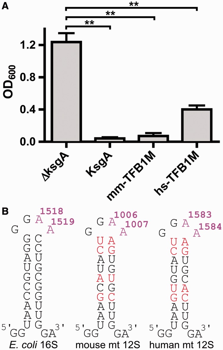
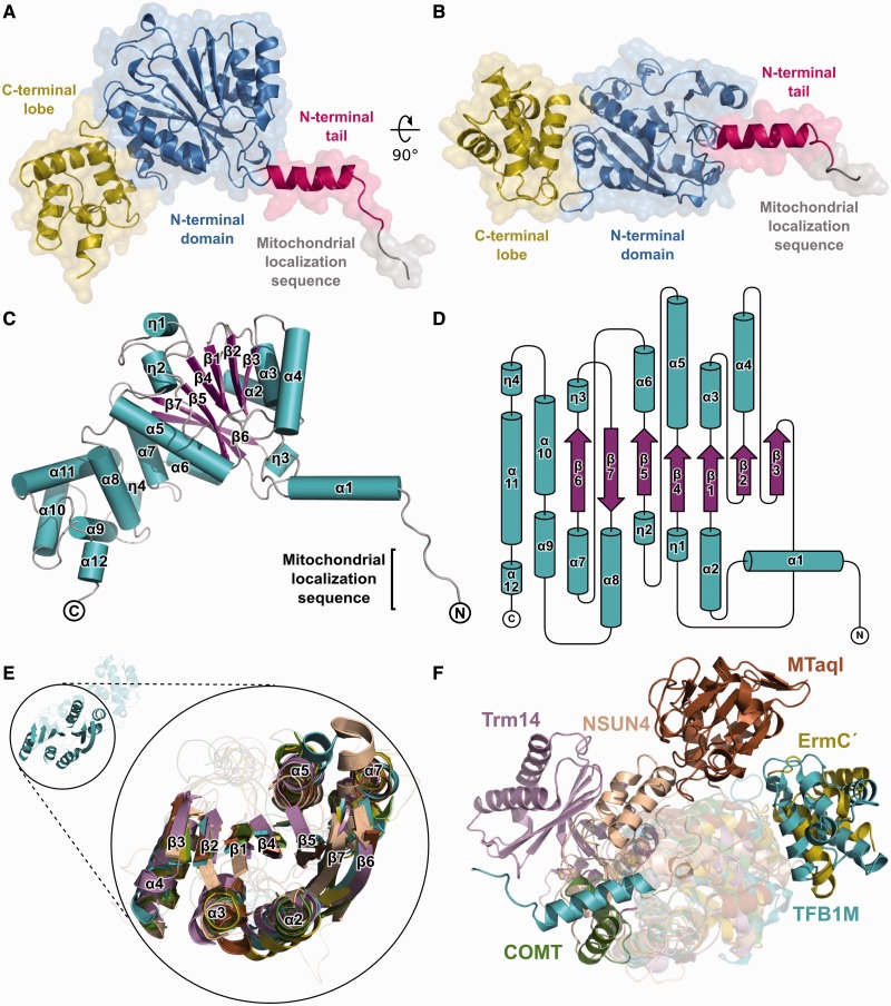
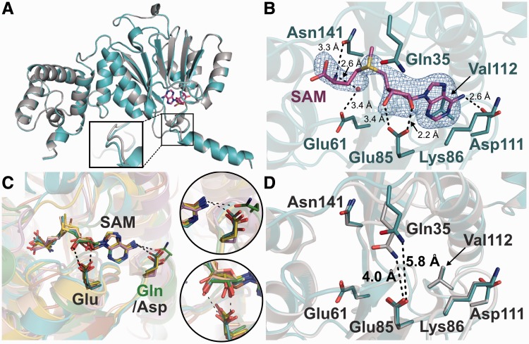
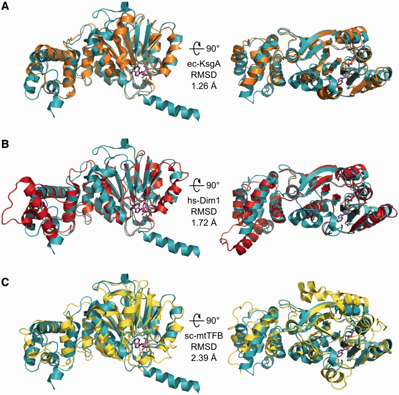
Eukaryotic transcription factor B (TFB) proteins are homologous to KsgA/Dim1 ribosomal RNA (rRNA) methyltransferases. The mammalian TFB1, mitochondrial (TFB1M) factor is an essential protein necessary for mitochondrial gene expression. TFB1M mediates an rRNA modification in the small ribosomal subunit and thus plays a role analogous to KsgA/Dim1 proteins. This modification has been linked to mitochondrial dysfunctions leading to maternally inherited deafness, aminoglycoside sensitivity and diabetes. Here, we present the first structural characterization of the mammalian TFB1 factor. We have solved two X-ray crystallographic structures of TFB1M with (2.1 Å) and without (2.0 Å) its cofactor S-adenosyl-L-methionine. These structures reveal that TFB1M shares a conserved methyltransferase core with other KsgA/Dim1 methyltransferases and shed light on the structural basis of S-adenosyl-L-methionine binding and methyltransferase activity. Together with mutagenesis studies, these data suggest a model for substrate binding and provide insight into the mechanism of methyl transfer, clarifying the role of this factor in an essential process for mitochondrial function.


Figures

Figure 1.
Figure 1.
Functional complementation of E. coli KsgA rRNA methlytransferase activity by human and mouse TFB1M. (A) The results of a kasugamycin-sensitivity assay are shown. Plotted is the optical density (OD600) after 5 h of culture growth in the presence of kasugamycin. Error bars represent the standard deviation (s.d.) of three replicates. Double asterisk indicates a P-value of ≤0.005 (see ‘Materials and Methods’ section). (B) Schematic representation of the conserved stem-loops in the 16S E. coli rRNA as well as the human and mouse 12S mitochondrial rRNA. The two dimethylated adenines are highlighted in magenta. Non-conserved residues are shown in red.
Figure 2.
Figure 2.
Overall architecture of TFB1M. (A) TFB1M adopts a methyltransferase fold with a two-domain architecture. A conserved N-terminal domain (blue) constitutes a Rossman-like methyltransferase fold and contains the active site. A C-terminal lobe (yellow) is believed to mediate RNA substrate specificity. A short N-terminal extension (magenta) is resolved in the crystal structure. The structure contains a portion of the mitochondrial localization sequence that is predicted to be cleaved on import (gray). The molecular surface of the protein is rendered transparent. A 90° rotation is shown in (B). (C) Schematic representation of mmTFB1M. β-strands are shown as magenta arrows, α-helices are shown as cyan ribbons and coiled regions are shown as gray lines. The N- and C-termini are marked. (D) Topology diagram of the protein fold using the same color coding as in (C). Secondary structure elements were identified using DSSP (43). Coiled regions are indicated by solid black lines. (E) Structural conservation of the methyltransferase domain. The inset shows the conservation of the central seven-stranded beta-sheet and the three flanking α-helices on each side in several classes of methyltransferases. Shown are another adenine N6-specific rRNA methyltransferase [ErmC′; yellow; PDB 1QAO; (44)], a large rRNA subunit methyltransferase [NSUN4, beige; PDB 4FZV; (45)], a tRNA methyltransferase [Trm14; pink; PDB 3TM4; (46)], a DNA methyltransferase [MTaqI; brown; PBD 2ADM; (47)] and a small-molecule methyltransferase [COMT, green; PDB 1VID (48)]. The secondary structure elements corresponding to TFB1M are labeled. (F) Divergence of the C-terminal lobe in different methyltransferases. The figure shows an overlay of the enzymes rendered in (E) maintaining the same color scheme. The C-terminal domain in the ErmC′ rRNA methyltransferase is similar to that in TFB1M. Other methyltransferases exhibit structurally distinct domains that are oriented differently with respect to the methyltransferase catalytic domain (see text).
Figure 3.
Figure 3.
SAM-binding pocket in TFB1M. (A) Overlay between the ligand-free (gray) and SAM-bound TFB1M (cyan) structures. Both structures are essentially identical, with an rmsd of 0.26 Å for 326 C-α atoms. The inset highlights a subtle shift in the loop connecting α-helices 1 and 2 (see text). (B) The SAM-binding pocket in TFB1M. SAM is bound in a negatively charged binding pocket in the N-terminal domain. Several residues establish contacts with the cofactor (see text). A simulated-annealing Fo-Fc omit electron density map is shown (blue), contoured at 3σ. A water molecule (red sphere) bridges the interaction between SAM and Glu61. (C) Overlay of the SAM-binding pocket in the TFB1M (cyan), ErmC’ (yellow), NSUN4 (beige), MTaqI (brown), Trm14 (pink) and COMT (green) methyltransferases (see Figure 2). The insets highlight two conserved interactions with the SAM cofactor. A conserved hydrogen bond with the N6 atom of the adenine base is observed in all methyltransferases (upper inset), although the protein residue involved in the interaction is not always the same and is not strictly conserved (Gln in COMT and Asp in all the others). An absolutely conserved glutamate residue forms at least one hydrogen bond to the ribose of the SAM cofactor in every structure examined (lower inset). (D) Overlay between the ligand-free and SAM-bound TFB1M highlighting differences in the SAM-binding pocket. The loop between α-helices 1 and 2 undergoes a subtle repositioning on SAM binding, resulting in a substantial shift of the Gln35 side chain.
Figure 4.
Figure 4.
TFB1M is structurally similar to KsgA and Dim1 methyltransferases. (A) Overlay between TFB1M (cyan) and E. coli KsgA (orange; PDB ID 1QYR) (52). (B) Overlay between TFB1M and human Dim1 (red; PDB ID 1ZQ9; A. Dong, H. Wu, H. Zeng, P. Loppnau, M. Sundstrom, C. Arrowsmith, A. Edwards, A. Bochkarev and A. Plotnikov, unpublished data). (C) Overlay between TFB1M and sc-mtTFB (yellow; PDB ID 1I4W) (53). A 90 degree rotation is shown on the right. The SAM molecule from the TB1M structure is shown in all three panels for reference.
Figure 5.
Figure 5.
Electrostatic surface potential maps of TFB1M, KsgA and mtTFB. Electrostatic surface potential maps of (A) TFB1M in complex with SAM, shown as magenta sticks, (B) E. coli KsgA [PDB ID 1QYR] and (C) sc-mtTFB [PDB ID 1I4W]. The TFB1M:SAM structure was overlaid with both KsgA and mtTFB, and the SAM molecule bound in the TFB1M structure is shown in (B) and (C) for reference (magenta). The insets on the right of the figure highlight the observed SAM-bidning pocket in TFB1M and the putative SAM-binding pockets in KsgA and mtTFB. The position of the N-terminal and C-terminal domains (and the N-terminal extension for TFB1M) is indicated. The electrostatic surface potential maps were generated with Delphi (50) and are colored from −7 kTe−1 (blue) to +7 kTe−1 (red).
Figure 6.
Figure 6.
A model for RNA binding by TFB1M. (A) Structure of the conserved stem-loop in the E. coli 16S rRNA that is methylated by KsgA. The two substrate adenine bases are shown in magenta. (B) Overview of the TFB1M:SAM structure superposed with the structure of MTaqI in complex with substrate DNA and a cofactor analog [PDB entry 1G38]. TFB1M is shown in teal, with the bound SAM molecule and substrate adenine shown as magenta and pink sticks, respectively. MTaqI is shown in yellow, with the bound DNA shown in orange. The conserved methyltransferase domain of each protein is shown as a solid cartoon, whereas the remainder of each protein and the DNA is transparent. (C) The putative catalytic center of TFB1M. The overlay of TFB1M and MTaqI suggests a putative substrate binding mechanism that involves base flipping, with the substrate adenine residue (pink) forming a π-stacking interaction with residue Phe144 (blue) of TFB1M. The methyl leaving group of SAM and N6 of the substrate adenine are separated by a distance of 2.4 Å (dashed line). (D) Model for RNA binding by TFB1M. The electrostatic surface potential of TFB1M is rendered transparent, and a single DNA strand containing the substrate adenine from the MTaqI structure is shown in orange. The black arrow indicates where the methyl group of SAM and the N6 of the adenine come into close proximity. The black asterisk denotes the location of Arg291 near the backbone of the nucleic acid. (E) Phe144 and Arg291 are important for catalysis, suggesting that they play a role in substrate binding. The results of a kasugamycin-sensitivity assay, with optical density (OD600) after 5 h of culture growth in the presence of kasugamycin is plotted in gray. The experiments were carried out using mouse TFB1M. Error bars represent the s.d. of three replicates. Double asterisk indicates a P-value of ≤0.005.

Similar articles

Cited by

References

    1. Ryan MT, Hoogenraad NJ. Mitochondrial-nuclear communications. Annu. Rev. Biochem. 2007;76:701–722. - PubMed
    1. Shutt TE, Shadel GS. A compendium of human mitochondrial gene expression machinery with links to disease. Environ. Mol. Mutagen. 2010;51:360–379. - PMC - PubMed
    1. Zhu X, Peng X, Guan MX, Yan Q. Pathogenic mutations of nuclear genes associated with mitochondrial disorders. Acta Biochim. Biophys. Sin. 2009;41:179–187. - PubMed
    1. Ylikallio E, Suomalainen A. Mechanisms of mitochondrial diseases. Ann. Med. 2012;44:41–59. - PubMed
    1. Tuppen HA, Blakely EL, Turnbull DM, Taylor RW. Mitochondrial DNA mutations and human disease. Biochim. Biophys. Acta. 2010;1797:113–128. - PubMed

Publication types