. 2013 Sep;41(16):7771-82.
doi: 10.1093/nar/gkt571. Epub 2013 Jun 26.

Zinc-finger-nucleases mediate specific and efficient excision of HIV-1 proviral DNA from infected and latently infected human T cells

Affiliations

Zinc-finger-nucleases mediate specific and efficient excision of HIV-1 proviral DNA from infected and latently infected human T cells

Xiying Qu et al. Nucleic Acids Res. 2013 Sep.

Abstract

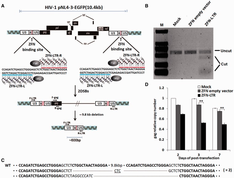
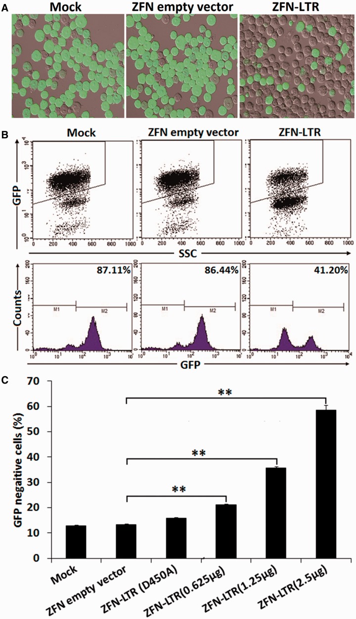
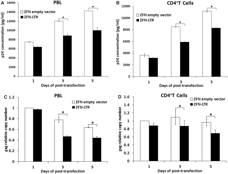
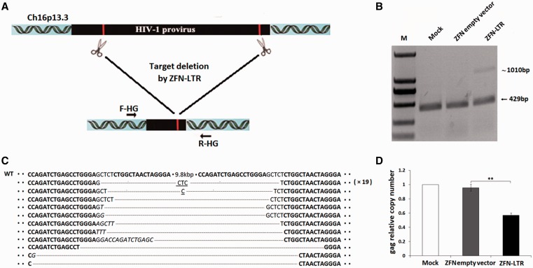
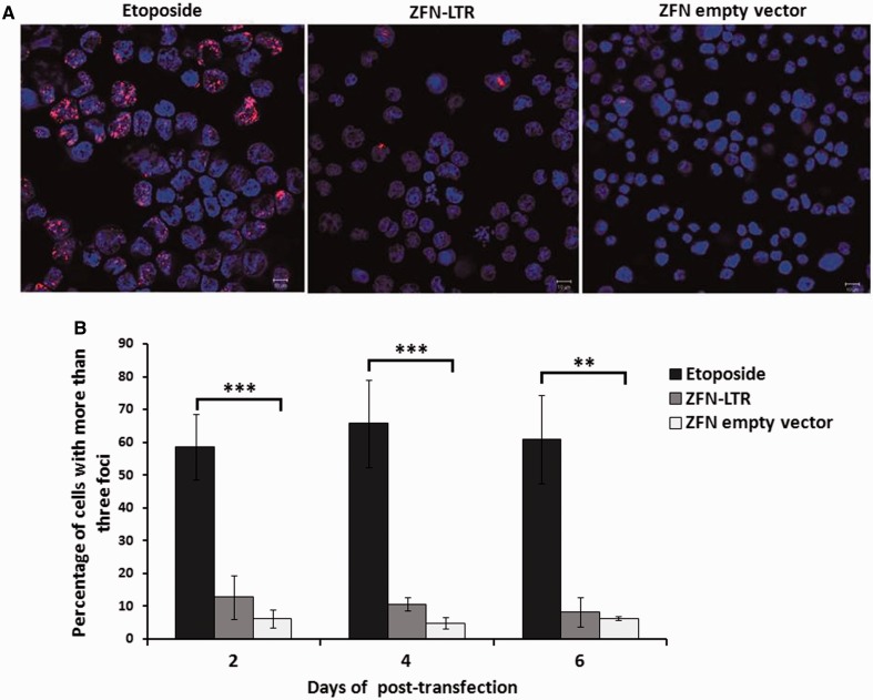
HIV-infected individuals currently cannot be completely cured because existing antiviral therapy regimens do not address HIV provirus DNA, flanked by long terminal repeats (LTRs), already integrated into host genome. Here, we present a possible alternative therapeutic approach to specifically and directly mediate deletion of the integrated full-length HIV provirus from infected and latently infected human T cell genomes by using specially designed zinc-finger nucleases (ZFNs) to target a sequence within the LTR that is well conserved across all clades. We designed and screened one pair of ZFN to target the highly conserved HIV-1 5'-LTR and 3'-LTR DNA sequences, named ZFN-LTR. We found that ZFN-LTR can specifically target and cleave the full-length HIV-1 proviral DNA in several infected and latently infected cell types and also HIV-1 infected human primary cells in vitro. We observed that the frequency of excision was 45.9% in infected human cell lines after treatment with ZFN-LTR, without significant host-cell genotoxicity. Taken together, our data demonstrate that a single ZFN-LTR pair can specifically and effectively cleave integrated full-length HIV-1 proviral DNA and mediate antiretroviral activity in infected and latently infected cells, suggesting that this strategy could offer a novel approach to eradicate the HIV-1 virus from the infected host in the future.


Figures

Figure 1.
Figure 1.
ZFN-LTR-induced excision of integrated HIV-1 proviral DNA in HIV-1-infected cells. (A) Schematic representation of ZFN-LTR-mediated genome deletions. Red lines in the 5′- and 3′-LTR of HIV-1 genome indicate ZFN-LTR target sites. ZFN-LTR-L and ZFN-LTR-R are pairs of zinc-finger proteins that have been customized to specifically recognize the left and right half-sites (indicated by blue and red lines) that are separated by a 5 bp spacer. F-LTR and R-LTR are PCR primers (arrows) located in the 5′- and 3′-LTRs, respectively, which were used for the detection of genome deletion events. (B) T7 endonuclease 1 assay of ZFN cleavage in HIV-1 infected cells. Jurkat T cells were infected with the HIV-1 pNL4-3-EGFP virus, and EGFP-positive cells were sorted by flow cytometry as HIV-1-infected cells. Then, the HIV-1-infected cells were untransfected (mock) or nucleofected with 2.5 μg of ZFN empty vector (negative control) or ZFN-LTR plasmid. At 3 days post-transfection, cells were collected for extraction of genomic DNA, and PCR was performed with the F-LTR and R-LTR primers to detect ZFN-LTR-mediated HIV-1 genomic DNA deletions. Amplicons encompassing the target sites were digested with the T7E1. The expected positions of the resulting DNA bands are indicated by an horizontal line (uncut ∼583 bp) and a bracket (cut ∼450 and ∼130 bp) at the left of the gel panels. (C) DNA sequences of PCR products. The PCR products were cloned and sequenced. ZFN-LTR target sites are shown in boldface letters. Dashes indicate deleted bases relative to the wild-type sequence. Inserted bases are shown in italics. In cases where a deletion sequence was detected more than once, the number of instances is given on the right in parentheses. (D) Analysis of HIV-1 proviral DNA deletions by quantitative real-time PCR. HIV-1-infected cells were untransfected (mock) or nucleofected with 2.5 μg of ZFN empty vector or ZFN-LTR plasmid. Genomic DNA was isolated at the indicated times and subjected to quantitative real-time PCR using gene-specific primers for HIV-1 gag and human β-globin. The relative copy numbers of gag were calculated based on the standard curve obtained by serial dilution (10–160 ng) of an infected cell DNA on the same plate. Normalization was carried out by division of gag gene amplicons in mock group of 2 days post-transfection. Data are representative of three independent experiments, and error bars represent standard errors (SD). *P < 0.05, **P < 0.01, ***P < 0.001; paired t-test.
Figure 2.
Figure 2.
ZFN-LTR-mediated excision efficiency of HIV-1 proviral DNA in infected cells. (A) Detection of EGFP expression in infected cells treated with 2.5 μg of ZFN empty vector (negative control) or ZFN-LTR plasmid or mock-treated by fluorescence microscopy. (B and C) The efficiency of HIV-1 proviral DNA excision in infected cells untransfected or transfected with 2.5 μg of ZFN empty vector or 2.5 μg of ZFN-LTR with inactive FokI (D450A) or ZFN-LTR plasmid dose titration (0.625–2.5 μg). At 3 days after transfection, the percentage of EGFP-negative cells was measured by flow cytometry. Results are presented as fluorescence histograms. Data are representative of three independent experiments, and error bars represent SD. *P < 0.05, **P < 0.01, ***P < 0.001; paired t-test.
Figure 3.
Figure 3.
ZFN-LTR-induced excision of integrated HIV-1 proviral DNA in HIV-1-infected PBL or CD4+ T cells. HIV-1/NL4-3 infected-PBLs and -CD4+ T Cells were nucleofected with 5 μg of ZFN-LTR plasmid or ZFN empty vector, and at the indicated post-transfection time points, (A and B) culture supernatants were harvested for detecting HIV-1 p24 production; (C and D) the relative copy numbers of gag were calculated based on the standard curve obtained by serial dilution (10–160 ng) of an infected cell DNA on the same plate. Normalization was carried out by division of gag gene amplicons in ZFN empty vector group of 1 day post-transfection. Data are representative of three independent experiments, and error bars represent SD. *P < 0.05, **P < 0.01, ***P < 0.001; paired t-test.
Figure 4.
Figure 4.
ZFN-LTR mediated deletions of HIV-1 proviral DNA in latently infected cell clone C11. (A) Schematic representation of ZFN-LTR-mediated genome deletions. The red lines in the 5′- and 3′-LTR of HIV-1 genome indicate ZFN-LTR target sites. F-HG and R-HG are PCR primers (arrows) located at the two ends of the integrated HIV genome, which were used for the detection of genome deletion events. (B) PCR products validating ZFN-LTR-induced HIV-1 proviral deletions. Jurkat T cells were infected with the HIV-1 pNL4-3-EGFP virus; originally, EGFP-negative cells, which can be reactivated by TSA stimulation, were then sorted by FACS and identified as HIV-1 latently infected cells. The latently infected clone C11 cells were untransfected (mock) or nucleofected with 2.5 μg of ZFN empty vector or ZFN-LTR plasmid. Genomic DNA was extracted 3 days post-transfection. PCR was performed with the F-HG and R-HG primers. (C) DNA sequences of PCR products. The PCR products were cloned and sequenced. ZFN-LTR target sites are shown in boldface letters. Dashes indicate deleted bases relative to the wild-type sequence (WT). Microhomologies are underlined, and inserted bases are shown in italics. In cases where a deletion sequence was detected more than once, the number of instances is given on the right in parentheses. (D) Quantification of integrated provirus. At 3 days post-transfection, genomic DNA from infected cells untransfected (mock) or nucleofected with 2.5 μg of ZFN empty vector or ZFN-LTR plasmid was isolated and subjected to quantitative real-time PCR using gene-specific primers for HIV-1 gag and human β-globin. The relative copy numbers of gag were calculated based on the standard curve obtained by serial dilution (10–160 ng) of an infected cell DNA on the same plate. Normalization was carried out by division of gag gene amplicons in mock group. Data are representative of three independent experiments, and error bars represent SD. *P < 0.05, **P < 0.01, ***P < 0.001; paired t-test.
Figure 5.
Figure 5.
Genotoxicity assay of ZFN-LTR based on the formation of nuclear repair foci. (A) Intranuclear 53BP1 immunostaining and confocal microscopy 2 days after Jurkat T cells were treated with 1 μM DNA-damaging agent etoposide (positive control) or nucleofected with 2.5 μg of ZFN-LTR plasmid or ZFN empty vector (negative control). Representative images of cells stained with DAPI (blue) and antibodies to 53BP1 (red) are shown. (B) The relative number of cells containing more than three foci was assessed at the indicated times after Jurkat T cells were treated with 1 μM etoposide or nucleofected with 2.5 μg of ZFN-LTR plasmid or ZFN empty vector. Error bars represent SD, *P < 0.05, **P < 0.01, ***P < 0.001; paired t-test.

Similar articles

Cited by

References

    1. Richman DD, Margolis DM, Delaney M, Greene WC, Hazuda D, Pomerantz RJ. The challenge of finding a cure for HIV infection. Science. 2009;323:1304–1307. - PubMed
    1. Nisole S, Saïb A. Early steps of retrovirus replicative cycle. Retrovirology. 2004;1:9. - PMC - PubMed
    1. Chun TW, Stuyver L, Mizell SB, Ehler LA, Mican JA, Baseler M, Lloyd AL, Nowak MA, Fauci AS. Presence of an inducible HIV-1 latent reservoir during highly active antiretroviral therapy. Proc. Natl Acad. Sci. USA. 1997;94:13193–13197. - PMC - PubMed
    1. Finzi D, Hermankova M, Pierson T, Carruth LM, Buck C, Chaisson RE, Quinn TC, Chadwick K, Margolick J, Brookmeyer R. Identification of a reservoir for HIV-1 in patients on highly active antiretroviral therapy. Science. 1997;278:1295–1300. - PubMed
    1. Trono D, Van Lint C, Rouzioux C, Verdin E, Barré-Sinoussi F, Chun TW, Chomont N. HIV persistence and the prospect of long-term drug-free remissions for HIV-infected individuals. Science. 2010;329:174–180. - PubMed

Publication types