. 2013 Aug;41(15):e150.
doi: 10.1093/nar/gkt576. Epub 2013 Jun 26.

Co-expression of RNA-protein complexes in Escherichia coli and applications to RNA biology

Affiliations

Co-expression of RNA-protein complexes in Escherichia coli and applications to RNA biology

Luc Ponchon et al. Nucleic Acids Res. 2013 Aug.

Abstract

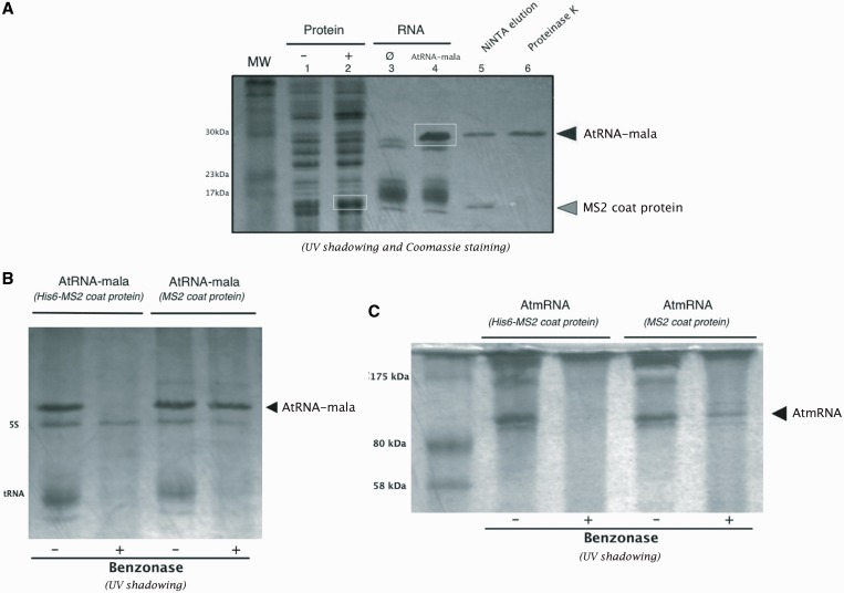
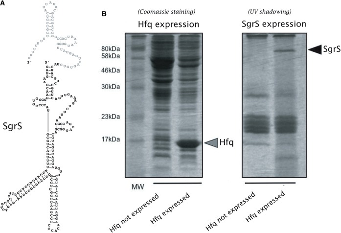
RNA has emerged as a major player in many cellular processes. Understanding these processes at the molecular level requires homogeneous RNA samples for structural, biochemical and pharmacological studies. We previously devised a generic approach that allows efficient in vivo expression of recombinant RNA in Escherichia coli. In this work, we have extended this method to RNA/protein co-expression. We have engineered several plasmids that allow overexpression of RNA-protein complexes in E. coli. We have investigated the potential of these tools in many applications, including the production of nuclease-sensitive RNAs encapsulated in viral protein pseudo-particles, the co-production of non-coding RNAs with chaperone proteins, the incorporation of a post-transcriptional RNA modification by co-production with the appropriate modifying enzyme and finally the production and purification of an RNA-His-tagged protein complex by nickel affinity chromatography. We show that this last application easily provides pure material for crystallographic studies. The new tools we report will pave the way to large-scale structural and molecular investigations of RNA function and interactions with proteins.


Figures

Figure 1.
Figure 1.
(A) Design of the pACYCT2, pBSTNAV, p44K and pProRNA plasmids described in this study. The pBSTNAV and pACYCT2 plasmids are used to co-express RNA–protein pairs using the two-plasmid strategy. (B) Co-expression of RNA–protein partners was tested for the A. aeolicus AtmRNA/MS2 coat protein, AtRNA/MS2 coat protein, E. coli tmRNA/SmpB and phi29 pRNA/gp10 in 5 ml of culture. The ‘A’ before AtmRNA and AtRNA refers to armored RNA. RNA–protein partners were expressed into the two plasmids pBSTNAV and pACYT2 system, the pProRNA system or the p44K plasmid system. The expression levels of RNA (upper gel) following phenol extraction and of total protein in crude extracts (lower gel) were analysed by SDS–PAGE. The RNA bands are visualized by SYBR Safe staining and the protein bands by Coomassie Blue staining. The white triangles indicate the overexpressed RNA and protein for each pair.
Figure 2.
Figure 2.
Armored tRNA–RNA fusion. (A) Sequences of the DNA inserted into the pBSTNAV-AtRNA plasmid: tRNALys3 scaffold in black, the MS2 operator hairpin boxed in grey, the RNA cloning site boxed in light grey. (B) Secondary structure of the corresponding processed transcript using the same color codes. The restriction sites within the RNA cloning site were selected to maintain the hairpin structure. (C) Overexpression of AtRNA (123 nt) and AtRNA-mala (155 nt) in E. coli JM101 strain. Stroke (control lane): bacteria transformed by the vector with no insert. Crude RNA minipreps were analysed by electrophoresis on a 10% polyacrylamide-urea gel and RNA visualized by UV shadowing. The white triangles indicate the overexpressed RNA.
Figure 3.
Figure 3.
The armored tRNA–RNA fusion is packaged into MS2 coat protein pseudo-particles. (A) Size exclusion chromatograms of different supernatants of crude cell extracts of E. coli JM101 expression cultures eluted on a Superose 6 column (10/300 GL, GE Healthcare). In red, overexpression of the His6 MS2 coat protein that bears a polyhistidine tag to prevent it from forming pseudo-particles; and in green, overexpression levels of the AtRNA and MS2 coat protein after benzonase treatment of the supernatant. RNA and protein were cloned into the pBSTNAV and the pACYCT2 plasmids, respectively. Arrows indicate the fractions where the MS2 coat protein was found. A chromatogram of reference proteins (thyroglobulin: 669 kDa, ferritin: 400 kDa, carbonic anhydrase: 29 kDa) is indicated in grey. V0 is the void volume of the column. (B) SDS–PAGE analysis of the fraction denoted by the green arrow in the green chromatogram of Figure 3A. The AtRNA is visualized by SYBR Safe staining (white band, lane 2) and the MS2 coat protein by Coomassie Brilliant Blue staining (black band, lane 2). A molecular weight ladder for protein is given in lane 1. The size of protein standards in kilodalton is given on the left. The black triangles indicate the AtRNA band and the MS2 coat protein band. (C) Transmission electron microscopy analysis of the fraction denoted by a green arrow in the green chromatogram of Figure 3A. Images were taken with a screen magnification of 100 000×.
Figure 4.
Figure 4.
The A. aeolicus AtmRNA encapsulated in MS2 coat protein pseudo-particles is protected against nucleases. (A) Secondary scheme of A. aeolicus AtmRNA showing the MS2 translational operator, the tRNA-like domain and the MLR in grey. (B) Overexpression of the AtmRNA alone in E. coli JM101 (left part of the upper gel) and co-expressed with the MS2 coat protein (right part of the upper gel): crude RNA minipreps were analysed by electrophoresis on a 10% polyacrylamide-urea gel and RNA visualized with SYBR Safe (upper part). Proteins in the supernatant after bacteria lysis were visualized by SDS–PAGE and Coomassie Brilliant Blue staining (lower gel) before and after IPTG induction (−/+). The black triangle indicates the AtmRNA band and the grey triangle the MS2 coat protein band. The AtmRNA/MS2 coat protein pair was cloned into pBSTNAV and pACYT2 plasmids, respectively.
Figure 5.
Figure 5.
The use of His6-MS2 coat protein simplifies the RNA purification protocol. (A) Co-expression of AtRNA-mala/His6-MS2 coat protein in E. coli JM101 strain. Crude bacteria extracts before (lane 1) and after (lane 2) IPTG induction, and crude RNA minipreps (lane 3 and 4) were separated on a 16% SDS–PAGE gel and visualized by Coomassie Brilliant Blue staining and UV shadowing. Stroke indicates the control experiment: bacteria transformed by the vector with no insert. White boxes indicate the overexpressed AtRNA-mala and the MS2 coat protein. NiNTA elution (lane 5): the AtRNA-mala/His6-MS2 coat protein complex was eluted in the same fractions upon binding to NiNTA agarose column. The MS2 coat protein can then be digested using proteinase K (lane 6). The black triangle indicates the AtRNA-mala band and the grey triangle indicates the MS2 coat protein band. The molecular weight of protein standards is given in kilodalton on the left. (B) Comparison of benzonase resistance of the AtRNA-mala because of co-expression with the MS2 protein (wild-type or His-tagged). RNA extracts in absence (−) or in presence (+) of benzonase in the lysis supernatant (sonication) were analysed by electrophoresis on a 16% SDS–PAGE gel and visualized by UV shadowing. The black triangle indicates the AtRNA-mala. (C) Comparison of benzonase resistance of the AtmRNA because of co-expression with the MS2 protein (wild-type or His-tagged). RNA extracts in absence (−) or in presence (+) of benzonase in the lysis supernatant (sonication) were analysed by electrophoresis on a 16% SDS–PAGE gel and visualized by UV shadowing. The black triangle indicates the AtmRNA.
Figure 6.
Figure 6.
Co-expression of SgrS/Hfq in E. coli. (A) Predicted secondary structure of E. coli K12 SgrS calculated with Mfold (66) showing, in grey, the 3′ portion of SgrS that corresponds to the polyU tail of rho-independent terminator. (B) Crude extracts of protein (gel on the left) in absence or presence of Hfq expression (−/+) were analysed by electrophoresis on a 12% SDS–PAGE gel. Crude extract of RNA (gel on the right) in absence or presence of Hfq expression was analysed by electrophoresis on a 12% SDS–PAGE gel. Protein and RNA were visualized by either Coomassie Brilliant Blue staining (left) or UV shadowing (right). The black and grey triangles indicate SgrS band and Hfq, respectively. The SgrS/Hfq pair was cloned in plasmid p44K. Lane 1 (gel on the left) shows protein molecular weight markers and associated molecular masses.

Similar articles

Cited by

References

    1. Breaker RR. Natural and engineered nucleic acids as tools to explore biology. Nature. 2004;432:838–845. - PubMed
    1. Roth A, Breaker RR. The structural and functional diversity of metabolite-binding riboswitches. Annu. Rev. Biochem. 2009;78:305–334. - PMC - PubMed
    1. Guo P. RNA nanotechnology: engineering, assembly and applications in detection, gene delivery and therapy. J. Nanosci. Nanotechnol. 2005;5:1964–1982. - PMC - PubMed
    1. Guo P. The emerging field of RNA nanotechnology. Nat. Nanotechnol. 2010;5:833–842. - PMC - PubMed
    1. Gumireddy K, Young DD, Xiong X, Hogenesch JB, Huang Q, Deiters A. Small-molecule inhibitors of microrna miR-21 function. Angew. Chem. Int. Ed Engl. 2008;47:7482–7484. - PMC - PubMed

Publication types

MeSH terms