. 2013 Jun 12:19:1332-40.
Print 2013.

Oxidative stress response signaling pathways in trabecular meshwork cells and their effects on cell viability

Affiliations

Oxidative stress response signaling pathways in trabecular meshwork cells and their effects on cell viability

Nanako Awai-Kasaoka et al. Mol Vis. .

Abstract

Purpose: To clarify the primary oxidative stress response signaling pathways in trabecular meshwork (TM) cells and their effects on cell viability.

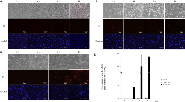
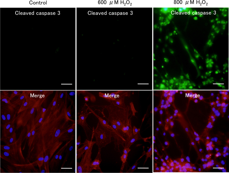
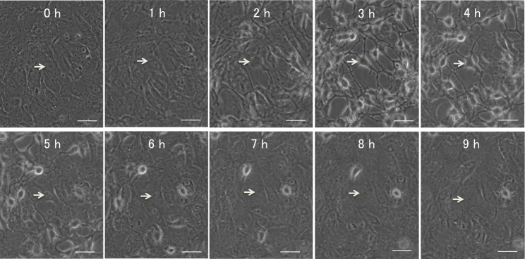
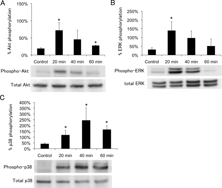
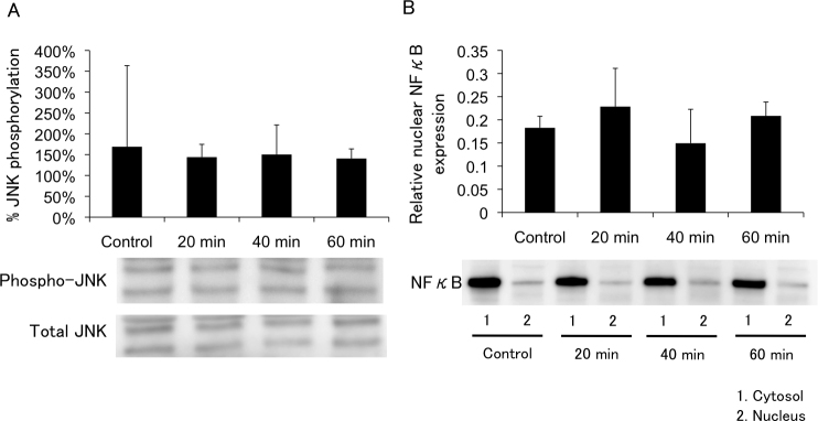
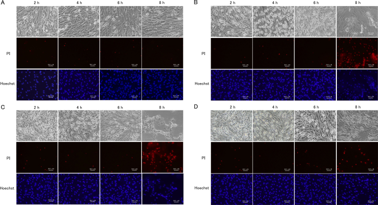
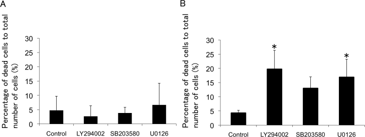
Methods: Porcine TM cells were treated with 600 μM or 800 μM H₂O₂, and their time-dependent morphologic changes were observed. Phosphorylation of protein kinase B (Akt), extracellular regulated kinase (ERK)1/2, p38, and c-Jun NH2-terminal kinase (JNK) was evaluated by western blot analysis. The intracellular localization of NFκB was evaluated by western blot analysis. One-hour pretreatments with LY294002, U0126, and SB203580, with the inhibitors of PI3K, ERK1/2, and p38, respectively, were conducted to evaluate the roles of these molecules in the cellular reaction against H₂O₂. Cell viability was assessed using propidium iodide and anticleaved caspase-3 antibody.

Results: TM cells treated with 600 μM H₂O₂ showed morphologic changes at 2 h that were partially recovered at 8 h after treatment. TM cells treated with 800 μM H₂O₂ did not recover, and the viability was significantly decreased. Both doses of H₂O₂ activated Akt, ERK1/2, and p38 in TM cells at 20 min after treatment, but not JNK or NFкB until 1 h after treatment. Inhibitors of PI3K, ERK1/2, and p38 suppressed recovery from the morphologic changes induced by 600 μM H₂O₂. Of these three inhibitors, the PI3K and ERK1/2 inhibitors decreased TM cell viability under oxidative stress.

Conclusions: In TM cells, the PI3K-Akt, ERK, and p38 signaling pathways are primary oxidative stress response pathways involved in the mechanism of recovery from cellular morphologic changes induced by H₂O₂ treatment accompanied by actin cytoskeletal changes.


Figures

Figure 1
Figure 1
Cellular morphological changes in porcine trabecular meshwork (PTM) cells treated with medium alone, or with 600 μM or 800 μM H2O2. PTM (1x10⁵) cells were seeded into 12-well plates. Three days later, the medium was changed to serum-free medium overnight, and then PTM cells were treated as followed for various amounts of time: A: medium alone, B: 600 μM H2O2 C: 800 μM H2O2. The unfixed cells were stained with propidium iodide to detect cell death (red; middle panel). Nuclei of total cells were stained with Hoechst 33342 (blue; lower panel). The time course for mean percentages of dead cells (propidium iodide-positive cells) to total number of cells (Hoechst 33342-positive cells) is shown (n=3; D). Thus, 800 μM H2O2 increased the number of dead PTM cells for 8 h after treatment, but PTM cells treated with 600 μM H2O2 recovered from the morphologic changes without significant cell death. Scale bar represents 50 μm.
Figure 2
Figure 2
Immunocytochemical staining of H2O2-treated porcine trabecular meshwork (PTM) cells. PTM cells treated with 600 or 800 μM H2O2 for 6 h were fixed, permeabilized, and stained with anticleaved caspase-3 antibody (green; upper panels). F-actin was stained with phalloidin-TRITC (red; lower panels). Cell nuclei were counterstained with DAPI (blue). Scale bar represents 50 μm.
Figure 3
Figure 3
Time-lapse images of porcine trabecular meshwork (PTM) cells treated with 600 μM H2O2. Time-lapse sequence of PTM cells after treatment with 600 μM H2O2 were recorded every 15 min. The figure with “0 hour” (left upper) shows PTM cells immediately after H2O2 treatment. Arrow indicates an affected cell in the sequence. Scale bar represents 50 μm.
Figure 4
Figure 4
Time course of H2O2-induced Akt (Ser473), ERK1/2, p38 phosphorylation. Porcine trabecular meshwork (PTM) cells (1x10⁵) in 12-well plates were treated with medium alone (control) or 600 μM H2O2 for various amounts of time. Time course H2O2-induced Akt (Ser473), ERK1/2, p38 phosphorylation were indicated panels A, B, and C, respectively. Levels of protein bands were quantified, and the ratios of phosphorylated protein to total protein were calculated (n=3). *p<0.05 compared with medium alone. Bars indicate the standard deviations.
Figure 5
Figure 5
Time course of H2O2-induced JNK phosphorylation and NFκB. Porcine trabecular meshwork (PTM) cells were treated with medium alone (control) or with 600 μM H2O2. Cytosol or nucleus fraction was extracted from TM cells. Cytoplasmic proteins were separated by sodium dodecyl sulfate PAGE, transferred onto a polyvinylidene fluoride transfer membrane, and probed with antibody to p-JNK, JNK (A), and NFκB (B). Levels protein bands were quantified, and the expression ratios of phosphorylated protein to total protein for JNK and nucleus to cytosol for NFκB were calculated (n=3). *p<0.05 compared with medium alone. Bars indicate the standard deviations.
Figure 6
Figure 6
Effects of PI3K-Akt, ERK1/2, p38 blockade in porcine trabecular meshwork (PTM) cells treated with medium alone or 600 μM H2O2 for various amounts of time. PTM (1x10⁵) cells were seeded into 12-well plates. Three days later, the medium was changed to serum-free medium overnight. PTM cells were pretreated with vehicle (A), LY294002 (10 μM; B), U0126 (10 μM; C), SB203580 (10 μM; D), inhibitors of PI3K, ERK1/2, or p38, respectively, for 1 h, and then treated with medium alone (A) or 600 μM H2O2 (BD) for various amounts of time. The cells were stained with propidium iodide to detect cell death, (red; middle panel). Nuclei were stained with Hoechst 33342 (blue; lower panel).
Figure 7
Figure 7
Sensitivity of porcine trabecular meshwork (PTM) cells to H2O2 for 8 h after pretreatment with LY294002, U0126, and SB203580. The mean percentages of dead cells (propidium iodide-positive cells) to total number of cells (Hoechst 33342-positive cells) were estimated with medium alone (A) or 600 μM H2O2 (B) treatment for 8 h after pretreatment with each inhibitor or vehicle (n=4). *p<0.05 compared with control. Bars indicate the standard deviations.

References

    1. Leske MC, Connell AM, Wu SY, Hyman LG, Schachat AP. Risk factors for open-angle glaucoma. The Barbados Eye Study. Arch Ophthalmol. 1995;113:918–24. - PubMed
    1. Leske MC, Heijl A, Hussein M, Bengtsson B, Hyman L, Komaroff E. Factors for glaucoma progression and the effect of treatment: the early manifest glaucoma trial. Arch Ophthalmol. 2003;121:48–56. - PubMed
    1. Gordon MO, Beiser JA, Brandt JD, Heuer DK, Higginbotham EJ, Johnson CA, Keltner JL, Miller JP, Parrish RK, 2nd, Wilson MR, Kass MA. The Ocular Hypertension Treatment Study: baseline factors that predict the onset of primary open-angle glaucoma. Arch Ophthalmol. 2002;120:714–20. - PubMed
    1. Townsend DJ, Brubaker RF. Immediate effect of epinephrine on aqueous formation in the normal human eye as measured by fluorophotometry. Invest Ophthalmol Vis Sci. 1980;19:256–66. - PubMed
    1. Jocson VL, Sears ML. Experimental aqueous perfusion in enucleated human eyes. Results after obstruction of Schlemm's canal. Arch Ophthalmol. 1971;86:65–71. - PubMed

Publication types

MeSH terms

LinkOut - more resources