. 2013 Jun 26:7:18.
doi: 10.3389/fnana.2013.00018. eCollection 2013.

Cellular and molecular basis of cerebellar development

Affiliations

Cellular and molecular basis of cerebellar development

Salvador Martinez et al. Front Neuroanat. .

Abstract

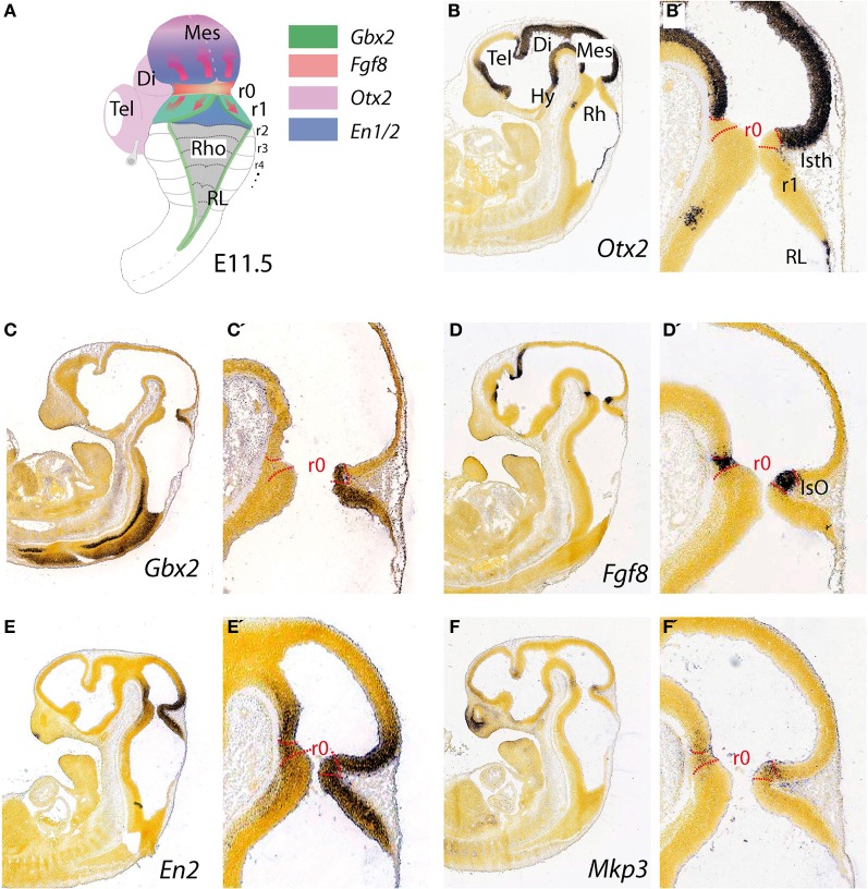
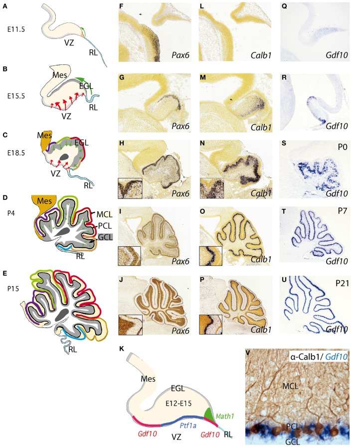
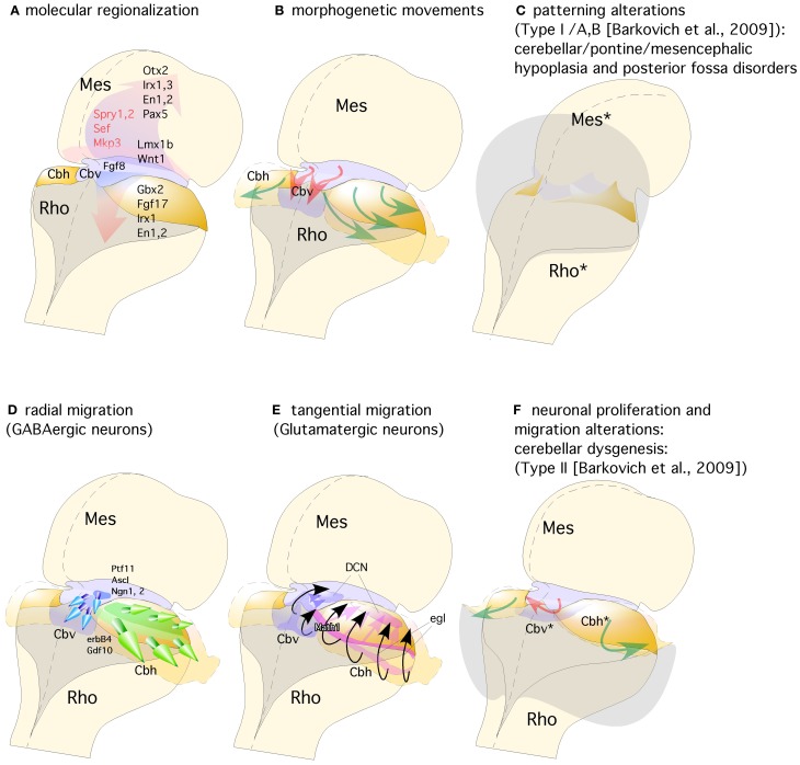
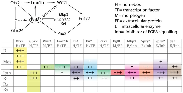
Historically, the molecular and cellular mechanisms of cerebellar development were investigated through structural descriptions and studying spontaneous mutations in animal models and humans. Advances in experimental embryology, genetic engineering, and neuroimaging techniques render today the possibility to approach the analysis of molecular mechanisms underlying histogenesis and morphogenesis of the cerebellum by experimental designs. Several genes and molecules were identified to be involved in the cerebellar plate regionalization, specification, and differentiation of cerebellar neurons, as well as the establishment of cellular migratory routes and the subsequent neuronal connectivity. Indeed, pattern formation of the cerebellum requires the adequate orchestration of both key morphogenetic signals, arising from distinct brain regions, and local expression of specific transcription factors. Thus, the present review wants to revisit and discuss these morphogenetic and molecular mechanisms taking place during cerebellar development in order to understand causal processes regulating cerebellar cytoarchitecture, its highly topographically ordered circuitry and its role in brain function.

Keywords: Fgf8; caudal mesencephalon; cerebellum; isthmic constriction; isthmic organizer; isthmus; morphogenesis; rostral hindbrain.


Figures

Figure 1
Figure 1
Topographical location and main molecular characterization of the mid-hindbrain boundary at E11.5. (A) A dorsal view of an E11.5 mouse embryo illustrating the isthmic constriction (isth) located between the mesencephalon and rhombomere 1 (r1). Moreover, rhombomeres, r0 and r1, which give rise to the cerebellum are highlighted in (A). The different color codes depict the expression pattern of the most important genes related to the morphogenetic activity and the capacity of the IsO (We only consider Fgf8-positive territory at this constriction as IsO; see also Martínez, 2001). Expression patterns of genes that are illustrated in (A) and their boundaries are also shown by in situ hybridization (ISH) at E11.5 for Otx2 (B,B′), Gbx2 (C,C′), Fgf8 (D,D′) En2 (E,E′), and Mkp3 (F,F′). Panels (B–F′) were taken from Allen Institute for Brain Science public resources (http://www.brain-map.org/).
Figure 2
Figure 2
Development of the cerebellum from the rhombencephalic alar plate from early stages to adulthood. (A–E) Cerebellar morphology/anatomy at different stages of development. The cerebellar folia development is showed from E11.5 onwards identifying the corresponding folia primordia bulges as color code lines from rostral to caudal (see also Sudarov and Joyner, 2007). Tangential migration from the rhombic lip (RL) is indicated as a green arrow corresponding to excitatory granule precursor cells that are specified by the expression of Math1 (K). These cells build up the external granule cell layer and from neonatal stages to P14, they descend radially forming the internal granule cell layer (GCL) located below Purkinje cell layer (PCL). The red arrows highlight the radial migration of cells originating from the ventricular zone (VZ). This is where Purkinje cells (PCs) and GABAergic cells of the cerebellum are born and specified by the early expression of Ptf1a (K); see also Dastjerdi et al. (2012). (F–U) Represent in situ hybridizations (ISH) of corresponding markers for each cerebellar cell type. Pax6 labeling granule cells (F–J). From (H) to (J) the insert shows the position of Pax6-positive cells at the EGL and later on, in the GCL (J). In the same manner (L–P) represent Calbindin (Calb1) showing the early location of PCs and their migration from the VZ to the final Purkinje cell monolayer, PCL (see also Sotelo, 2004). (Q–U) shows the expression pattern of Gdf10 (a marker for Bergmann glial cells) by means of ISH during cerebellar development. (V) Here we show the expression of Gdf10 in Bergmann glial cells in adult mice, together with Calbindin- positive Purkinje cells. (K) represents the molecular specification of the different cell types (excitatory vs. inhibitory) in the cerebellum including the two germinal centers ventricular zone (VZ) and rhombic lip (RL). Panels (F–J) and (L,M) were taken from Allen Institute for Brain Science public resources (http://www.brain-map.org/).
Figure 3
Figure 3
Representation of a dorsal view of mid-hindbrain junction, where the isthmic segment is colored in light purple (which will generate also cerebellar vermis: Cbv) and the cerebellar plates in yellow (which will generate cerebellar hemispheres). (A) The isthmic organizer expressing Fgf8 (pink arrows) induces the expression of Sprouty, Sef, and Mkp3 in this region; and is also required for other genes differentially expressed in the midbrain (Mes) or rhombencephalic (Rho) neuroepithelium. (B) The spatio-temporal expression of these genes regulates the normal morphogenesis and growth of the cerebellar vermis (Cbv; red arrows) and hemispheres (Cbh; green arrows). (C) Failure in proper isthmic organizer development (due to a lack of morphogenetic signaling or disruption of gene expression) can result in cerebellar (Rho*) and mesencephalic (Mes*) hypoplasia due to a strong increase of cell death with posterior fossa disorders and fourth ventricle dilatation (gray shadow). (D) Radial migration of GABAergic neurons in the cerebellar vermis (Cbv; blue arrows) and hemispheres (Cbh; Green arrows). (E) Rhombic lip specification is regulated by Math1. Tangential migration of glutamatergic neurons of the deep cerebellar nuclei (DCN) and granule cells (egl) are represented by pink and black arrows, respectively. (F) When normal development of cortical cerebellar cells is disrupted, the structural phenotype is classified as cerebellar dysgenesis (Cbv* and Cbh*), with enlargement of the fourth ventricle and reversion of cerebellar-choroidal junction (arrows).
Figure 4
Figure 4
The upper scheme represents the functional interaction (induction/inhibition) of genes that, together with Fgf8, are involved in the molecular maintenance of isthmic region at E9.5. The table below summarizes the expression intensity and range of genes along the AP axis of the neural tube focusing on the isthmus. The color code depicts their mRNA expression range from the isthmus toward rostral or caudal regions.

References

    1. Airey D. C., Lu L., Williams R. W. (2001). Genetic control of the mouse cerebellum: identification of quantitative trait loci modulating size and architecture. J. Neurosci. 21, 5099–5109 - PMC - PubMed
    1. Alcaraz W. A., Gold D. A., Raponi E., Gent P. M., Concepcion D., Hamilton B. A. (2006). Zfp423 controls proliferation and differentiation of neural precursors in cerebellar vermis formation. Proc. Natl. Acad. Sci. U.S.A. 103, 19424–19429 10.1073/pnas.0609184103 - DOI - PMC - PubMed
    1. Altman J., Bayer S. A. (1997). Development of the Cerebellar System in Relation to its Evolution, Structure and Functions. New York, NY: CRC Press
    1. Alvarado-Mallart R. M. (1993). Fate and potentialities of the avian mesencephalic/metencephalic neuroepithelium. J. Neurobiol. 24, 1341–1355 10.1002/neu.480241007 - DOI - PubMed
    1. Alvarez-Otero R., Sotelo C., Alvarado-Mallart R. M. (1993). Chick/quail chimeras with partial cerebellar grafts: an analysis of the origin and migration of cerebellar cells. J. Comp. Neurol. 333, 597–615 10.1002/cne.903330411 - DOI - PubMed

LinkOut - more resources