. 2013 Jun 21;8(6):e66290.
doi: 10.1371/journal.pone.0066290. Print 2013.

The GDNF System Is Altered in Diverticular Disease - Implications for Pathogenesis

Affiliations

The GDNF System Is Altered in Diverticular Disease - Implications for Pathogenesis

Martina Böttner et al. PLoS One. .

Abstract

Background & aims: Absence of glial cell line-derived neurotrophic factor (GDNF) leads to intestinal aganglionosis. We recently demonstrated that patients with diverticular disease (DD) exhibit hypoganglionosis suggesting neurotrophic factor deprivation. Thus, we screened mRNA expression pattern of the GDNF system in DD and examined the effects of GDNF on cultured enteric neurons.

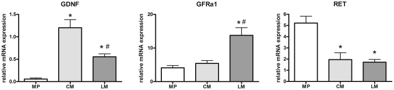
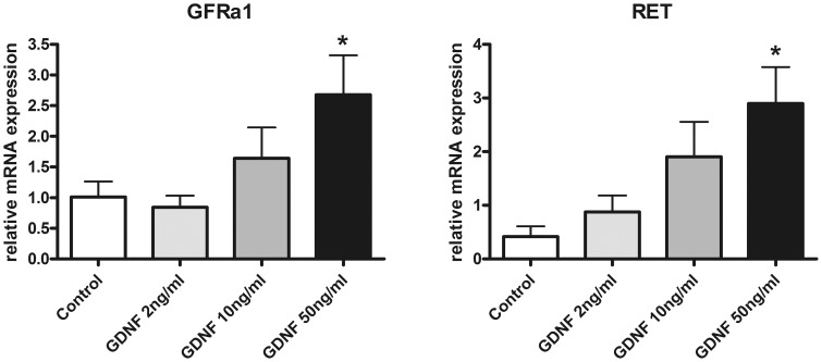
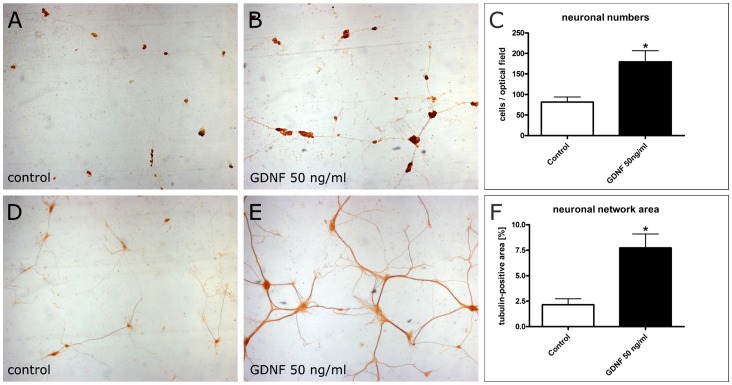
Methods: Colonic specimens obtained from patients with DD (n = 21) and controls (n = 20) were assessed for mRNA expression levels of the GDNF system (GDNF, GDNF receptors GFRα1 and RET). To identify the tissue source of GDNF and its receptors, laser-microdissected (LMD) samples of human myenteric ganglia and intestinal muscle layers were analyzed separately by qPCR. Furthermore, the effects of GDNF treatment on cultured enteric neurons (receptor expression, neuronal differentiation and plasticity) were monitored.

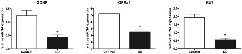
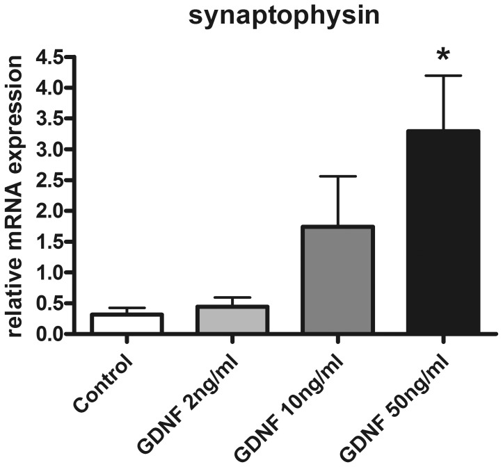
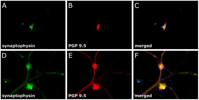
Results: mRNA expression of GDNF and its receptors was significantly down-regulated in the muscularis propria of patients with DD. LMD samples revealed high expression of GDNF in circular and longitudinal muscle layers, whereas GDNF receptors were also expressed in myenteric ganglia. GDNF treatment of cultured enteric neurons increased mRNA expression of its receptors and promoted neuronal differentiation and plasticity revealed by synaptophysin mRNA and protein expression.

Conclusions: Our results suggest that the GDNF system is compromised in DD. In vitro studies demonstrate that GDNF enhances expression of its receptors and promotes enteric neuronal differentiation and plasticity. Since patients with DD exhibit hypoganglionosis, we propose that the observed enteric neuronal loss in DD may be due to lacking neurotrophic support mediated by the GDNF system.


Conflict of interest statement

Competing Interests: The authors have declared that no competing interests exist.

Figures

Figure 1
Figure 1. mRNA expression of the GDNF system in the muscularis propria of the human colon.
mRNA expression of GDNF (A) and its receptors GFRα1 (B) and RET (C) is significantly down-regulated in patients with DD compared to controls. mRNA levels are determined by qPCR, expression of target genes is normalized to mRNA expression of the house-keeping gene HPRT. Data are shown as mean +/− SEM, n = 20 (controls) and n = 21 (DD), *p<0.05 vs. control.
Figure 2
Figure 2. Site-specific mRNA expression of the GDNF system in the human colon.
mRNA levels of GDNF (A), GFRa1 (B), and RET (C) in myenteric plexus (MP) and in the circular (CM) and longitudinal (LM) smooth muscle layer isolated by LMD. The main source for GDNF mRNA are the smooth muscle layers, whereas the GFRα1 is expressed mainly in the longitudinal muscle layer but also in myenteric plexus and RET is predominantly expressed by myenteric plexus and to a lesser extent by smooth muscle layers. mRNA expression levels are normalized to mRNA expression of the house-keeping gene HPRT. Data are shown as mean +/− SEM. GDNF: n = 10 (MP), n = 15 (CM), n = 14 (LM), GFRa1: n = 10 (MP), n = 15 (CM), n = 15 (LM), RET: n = 10 (MP), n = 15 (CM), n = 14 (LM). *p<0.05 vs. MP, #p<0.05 vs. CM.
Figure 3
Figure 3. mRNA expression of GDNF receptors in enteric nerve cell cultures following GDNF treatment.
Treatment with GDNF for 1 week increases mRNA expression of GDNF receptors GFRα1 (A) and RET (B) in enteric nerve cell cultures. mRNA expression levels are normalized to mRNA expression of the house-keeping gene HPRT. Data are shown as mean +/− SEM, n = 14–15 per experimental group, *p<0.05 vs. control.
Figure 4
Figure 4. Effects of GDNF on neuronal number and differentiation of enteric nerve cell cultures.
Rat enteric nerve cells were cultured for 1 week without (A, D) or with 50 ng/ml GDNF (B, E). Immunocytochemistry for the neuronal marker HuC/D (A, B) shows that neuronal aggregates are more numerous and larger after GDNF treatment. The morphometric analysis (C) confirms the increased neuronal number following GDNF treatment. Immunocytochemistry for tubulin ßIII (D, E) visualizes neuronal processes which are more densly distributed and ramified after GDNF treatment. The morphometric analysis (F) confirms the increased area of the neuronal network following GDNF treatment. Magnification 4×. Data are shown as mean +/− SEM, n (HuC/D immunocytochemistry) = 11–12, n (ßIII tubulin immunocytochemistry) = 7–8 per experimental group, *p<0.05 vs. control.
Figure 5
Figure 5. Synaptophysin mRNA expression of enteric nerve cell cultures after GDNF treatment.
GDNF increases mRNA expression of synaptophysin in rat enteric nerve cell cultures. Expression levels were measured after one week and are normalized to expression of the house-keeping gene HPRT. Data are shown as mean +/− SEM, n = 11–12 per experimental group, *p<0.05 vs. control.
Figure 6
Figure 6. Synaptophysin immunoreactivity of enteric nerve cell cultures after GDNF treatment.
Rat enteric nerve cells were cultured for 3 week without (A-C) or with 50 ng/ml GDNF (D-F). Dual label immunocytochemistry for synaptophysin (green, A, D) and the pan-neuronal marker PGP 9.5 (B, E) was performed. In the merged pictures (C, F) cellular nuclei are stained with DAPI (blue). Cell cultures treated with GDNF display punctuate and granular synaptophysin immunoreactivity along ramifying nerve fibers most likely resembling accumulated synaptic vesicles, whereas in untreated cell cultures immunoreactive signals were confined to neuronal somata. Magnification: 40×.

Similar articles

Cited by

References

    1. Jun S, Stollman N (2002) Epidemiology of diverticular disease. Best Pract Res Clin Gastroenterol 16: 529–542. - PubMed
    1. Simpson J, Spiller R (2002) Colonic diverticular disease. Clin Evid: 436–444. - PubMed
    1. Böttner M, Wedel T (2012) Abnormalities of neuromuscular anatomy in diverticular disease. Dig Dis 30: 19–23. - PubMed
    1. Strate LL, Liu YL, Aldoori WH, Syngal S, Giovannucci EL (2009) Obesity increases the risks of diverticulitis and diverticular bleeding. Gastroenterology 136: 115–122 e111. - PMC - PubMed
    1. Humes DJ, Simpson J, Smith J, Sutton P, Zaitoun A, et al. (2012) Visceral hypersensitivity in symptomatic diverticular disease and the role of neuropeptides and low grade inflammation. Neurogastroenterol Motil 24: 318–e163. - PubMed

Publication types

MeSH terms

Substances