. 2013 Jun 21;8(6):e66796.
doi: 10.1371/journal.pone.0066796. Print 2013.

Identification of Host Kinase Genes Required for Influenza Virus Replication and the Regulatory Role of MicroRNAs

Affiliations

Identification of Host Kinase Genes Required for Influenza Virus Replication and the Regulatory Role of MicroRNAs

Abhijeet Bakre et al. PLoS One. .

Abstract

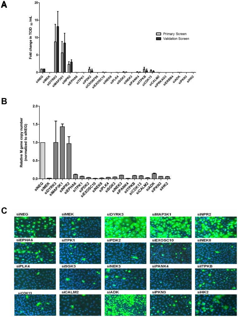
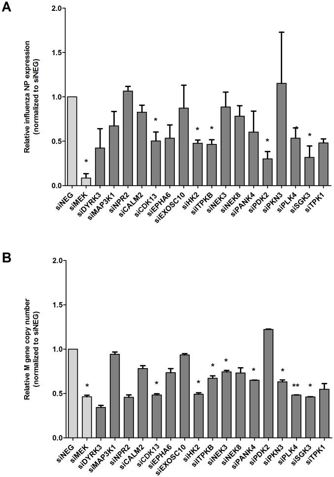
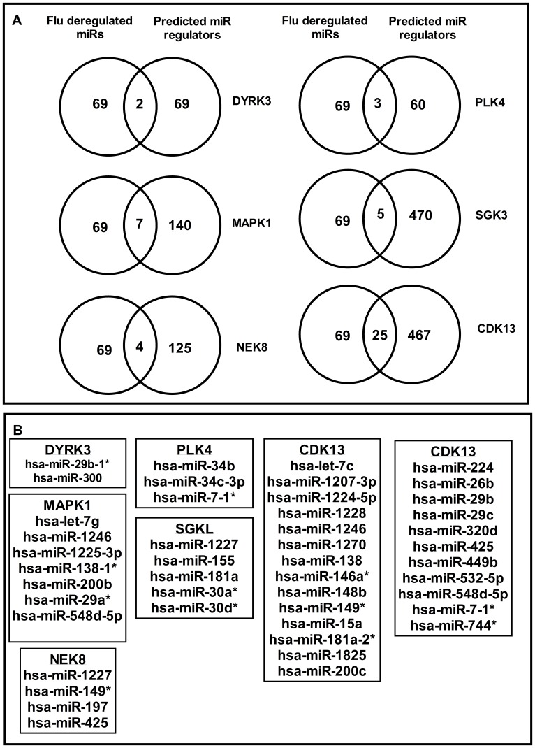
Human protein kinases (HPKs) have profound effects on cellular responses. To better understand the role of HPKs and the signaling networks that influence influenza virus replication, a small interfering RNA (siRNA) screen of 720 HPKs was performed. From the screen, 17 HPKs (NPR2, MAP3K1, DYRK3, EPHA6, TPK1, PDK2, EXOSC10, NEK8, PLK4, SGK3, NEK3, PANK4, ITPKB, CDC2L5 (CDK13), CALM2, PKN3, and HK2) were validated as essential for A/WSN/33 influenza virus replication, and 6 HPKs (CDK13, HK2, NEK8, PANK4, PLK4 and SGK3) were identified as vital for both A/WSN/33 and A/New Caledonia/20/99 influenza virus replication. These HPKs were found to affect multiple host pathways and regulated by miRNAs induced during infection. Using a panel of miRNA agonists and antagonists, miR-149* was found to regulate NEK8 expression, miR-548d-3p was found to regulate MAPK1 transcript expression, and miRs -1228 and -138 to regulate CDK13 expression. Up-regulation of miR-34c induced PLK4 transcript and protein expression and enhanced influenza virus replication, while miR-34c inhibition reduced viral replication. These findings identify HPKs important for influenza viral replication and show the miRNAs that govern their expression.


Conflict of interest statement

Competing Interests: The authors have declared that no competing interests exist.

Figures

Figure 1
Figure 1. Validation of human protein kinase genes affecting influenza virus replication.
A549 cells were reverse transfected with 50 nM of a non-target negative control siRNA (siNEG) or with siHPK and infected at a MOI = 0.01 with A/WSN/33. (A) 72 hours after infection, supernatants were harvested and the viral titers were determined by TCID50 assay on MDCK cells (B) After a 48 hour infection, the effect of siRNA silencing on influenza virus replication was measured by quantifying the levels of influenza M gene expression. The RNA from siRNA-transfected and WSN-infected A549s was isolated and used for quantification with an influenza M-specific primer/probe set. Light gray bars indicate controls. Data show mean±SEM from 3 independent experiments. (C) After a 48 hour infection, cells were fixed, permeabilized and incubated with an anti-influenza virus NP monoclonal antibody and subsequently with an Alexa-488 goat anti-mouse secondary antibody and DAPI. The intracellular distribution of the viral RNPs (NP, green) and cellular nuclei (DAPI, blue) are shown.
Figure 2
Figure 2. Human protein kinase genes affect H1N1 A/New Caledonia/20/99 virus replication.
A549 cells were reverse transfected with 50 nM of siRNA specific for validated HPK genes and after 48 h the cells were infected with A/New Caledonia/20/99 at an MOI of 0.01 in the presence of 1 µg/ml TPCK-trypsin. After 48 h of infection, RNA was extracted and the effect of siRNA silencing of HPK genes on viral genome replication was measured by quantifying (A) influenza NP expression and (B) the level of influenza M gene. Data show mean±SEM of 3 independent experiments. *p<0.05 and **p<0.001 compared to control.
Figure 3
Figure 3. Identifying miRNA regulators of HPKs important for influenza replication.
(A) Venn diagrams showing miRNAs common to computationally predicted miRNA regulators and influenza deregulated miRNAs. (B) miRNAs that are shared between computationally predicted HPK regulators and miRNAs deregulated during influenza infection.
Figure 4
Figure 4. miRNA regulators of NEK8.
A549 cells were transfected with 25 nM of miR-1227, -149* and -197 inhibitor/mimic for 48 hrs followed by RNA extraction and RT-qPCR with NEK8 specific primers. Expression data was normalized to 18S rRNA expression and shown as mean±SEM of independent experiments. *p<0.05 compared to control.
Figure 5
Figure 5. miRNA regulators of MAP3K1.
(A) A549 cells were transfected with 25 nM of miR-548d, -29a and -138 inhibitor/mimic for 48 hrs followed by RNA extraction and RT-qPCR with MAP3K1 specific primers. Expression data was normalized to 18S rRNA expression and shown as mean±SEM of independent experiments. *p<0.05 compared to control. (B) A549 cells mock/transfected with miR-548d inhibitor/mimic for 48 hrs were fixed with 4% formaldehyde in PBS and stained for MAPK1 protein using biotinylated rabbit anti-MAP3K1 antibody (Abcam ab69533) and detected with Streptavidin-Alexa488. Cells were analyzed by Arrayscan Cellomics VTI scanner and data analyzed by GraphPad Prism. (C) A549 cells mock/transfected with miR-548d inhibitor/mimic for 48 hrs were infected with A/WSN/33 (MOI = 0.001) for 48 hrs and stained for influenza NP protein using mouse-anti-NP coupled to Alexa-488 and analyzed as above in (B). Data show mean±SEM of two independent experiments. *p<0.05.
Figure 6
Figure 6. miRNA regulators of PLK4 (A) A549 cells were transfected with 25 nM of miR-34c inhibitor/mimic for 48 hrs followed by RNA extraction and RT-qPCR with PLK4 specific primers.
Expression data was normalized to 18S rRNA expression and shown as mean±SEM of independent experiments. *p<0.05 compared to control. (B) A549 cells mock/transfected with miR-34c inhibitor/mimic for 48 hrs were fixed with 4% formaldehyde in PBS and stained for PLK4 protein using biotinylated rabbit anti-PLK4 antibody (Abcam ab71394) and detected with Streptavidin-Alexa488. Cells were analyzed by Arrayscan Cellomics VTI scanner and data analyzed by GraphPad Prism. (C) A549 cells mock/transfected with miR-34c inhibitor/mimic for 48 hrs were infected with A/WSN/33 (MOI = 0.001) for 48 hrs and stained for influenza NP protein using mouse-anti-NP coupled to Alexa-488 and analyzed as above in (B). Data show mean±SEM of two independent experiments. *p<0.05.

References

    1. Thompson WW, Shay DK, Weintraub E, Brammer L, Bridges CB, et al. (2004) Influenza-associated hospitalizations in the United States. JAMA 292: 1333–1340. - PubMed
    1. Thompson WW, Shay DK, Weintraub E, Brammer L, Cox N, et al. (2003) Mortality associated with influenza and respiratory syncytial virus in the United States. JAMA 289: 179–186. - PubMed
    1. MMWR (2010) Estimates of Deaths Associated with Seasonal Influenza - United States, 1976–2007. Atlanta, GA: Centers for Disease Control and Prevention. 1057–1062 p. - PubMed
    1. Regoes RR, Bonhoeffer S (2006) Emergence of drug-resistant influenza virus: population dynamical considerations. Science 312: 389–391. - PubMed
    1. Beigel J, Bray M (2008) Current and future antiviral therapy of severe seasonal and avian influenza. Antiviral Res 78: 91–102. - PMC - PubMed

MeSH terms