Review
. 2013 Jul 15;453(2):155-66.
doi: 10.1042/BJ20130316.

CRISPR interference: a structural perspective

Affiliations
Review

CRISPR interference: a structural perspective

Judith Reeks et al. Biochem J. .

Abstract

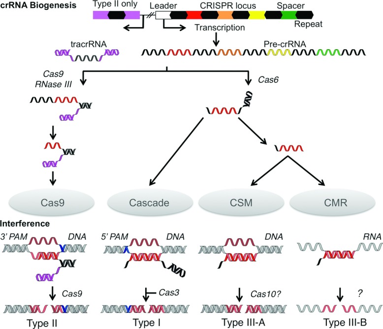
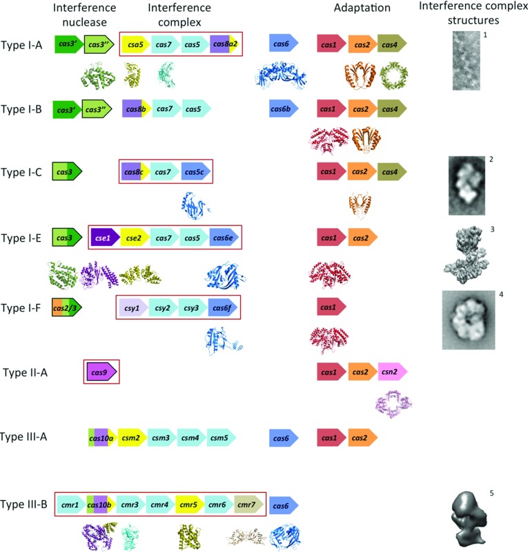
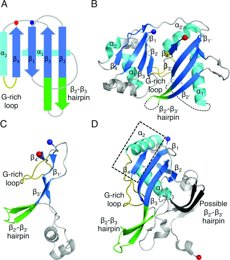
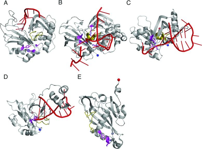
CRISPR (cluster of regularly interspaced palindromic repeats) is a prokaryotic adaptive defence system, providing immunity against mobile genetic elements such as viruses. Genomically encoded crRNA (CRISPR RNA) is used by Cas (CRISPR-associated) proteins to target and subsequently degrade nucleic acids of invading entities in a sequence-dependent manner. The process is known as 'interference'. In the present review we cover recent progress on the structural biology of the CRISPR/Cas system, focusing on the Cas proteins and complexes that catalyse crRNA biogenesis and interference. Structural studies have helped in the elucidation of key mechanisms, including the recognition and cleavage of crRNA by the Cas6 and Cas5 proteins, where remarkable diversity at the level of both substrate recognition and catalysis has become apparent. The RNA-binding RAMP (repeat-associated mysterious protein) domain is present in the Cas5, Cas6, Cas7 and Cmr3 protein families and RAMP-like domains are found in Cas2 and Cas10. Structural analysis has also revealed an evolutionary link between the small subunits of the type I and type III-B interference complexes. Future studies of the interference complexes and their constituent components will transform our understanding of the system.


Figures

Figure 1
Figure 1. Schematic representation of crRNA biogenesis and CRISPR interference
Processing events involving nucleic acids are coloured; repeats (black), spacers (red–green) and tracrRNA (magenta). For clarity, a single spacer (red) was used to illustrate the processes, although in actual systems all spacers are processed. Targets are shown in other red shades (lighter for the complementary strand and darker for the non-complementary). The PAMs are shown in blue. The pre-crRNA and interference nucleases are indicated along with the interference complexes.
Figure 2
Figure 2. The CRISPR/Cas systems and their respective proteins
Typical gene identities are shown for CRISPR/Cas subtypes according to the recent classification by Makarova et al. [12]. The genes are ordered by function: interference (left) and adaptation (right). The interference proteins are subdivided into the interference nuclease (left, outlined in black), proteins of the interference complex (middle, boxed in red) and pre-crRNA nucleases (right, although some are integral subunits of the interference complexes). The genes are coloured according to conserved domain and protein folds: catalytic RAMPs are shown in blue, non-catalytic RAMPs in light blue, HD nuclease domains in light green, Cas3 helicase domains in dark green, the large subunits in various shades of purple and the small subunits in yellow. Subtypes I-D and II-B are not shown as there is no directly relevant structural data. EM images and structures of the interference complexes (or subcomplex for I-A) are adapted from references 1 [13], 2 [14], 3 EMD-5314, 4 [15] and 5 [20].
Figure 3
Figure 3. The structures of catalytic RAMP proteins
(A) Topology diagram of a RAMP domain. The β-strands are shown in blue and the α-helices in cyan. The glycine-rich loop found in many RAMPs is shown in yellow and the β2–β3 hairpin observed in some RAMPs is shown in green. The N- and C-termini are shown as blue and red spheres respectively. (B) The structure of TtCas6e (PDB code 1WJ9) highlighting the two RAMP domains that may have arisen from a pseudo-duplication event. Secondary structural elements are labelled as described in the text. Conserved RAMP elements are coloured as in (A) and non-conserved elements in grey. Disordered regions are shown as broken black lines. (C) The atypical C-terminal domain of PaCas6f (PDB code 2XLK) that probably diverged from the standard RAMP fold. The recognizable features are labelled. (D) The structure of BhCas5c (PDB code 4F3M), a catalytic variant of the typically non-catalytic Cas5 family. The short β4 strand and parallel α2 helix are boxed in black. The possible β2′–β3′ hairpin in the C-terminal domain is shown in black.
Figure 4
Figure 4. RNA binding and catalysis by Cas6 and Cas5c
The structures of (A) PfuCas6 (PDB code 3PKM), (B) SsoCas6 (PDB code 4ILL), (C) TtCas6e (PDB code 2Y8W) and (D) PaCas6f (PDB code 2XLK) in complex with RNA (red). The glycine-rich loop is shown in yellow and the catalytic residues as magenta sticks. (E) The structure of BhCas5c (PDB 4F3M) highlighting the position of the active site (magenta). The four structures are shown to the same scale and same orientation. A three-dimensional representation of this Figure is available at http://www.biochemj.org/bj/453/0155/bj4530155add.htm.
Figure 5
Figure 5. The structure of Cas7, the core subunit of Cascade
(A) The structure of SsoCas7 (PDB code 3PS0) where the central RAMP domain is extended by an αβα motif (orange) and flanked by two unique domains (grey). The proposed crRNA-binding cleft located across the face of the β-sheet is indicated. (B) Topology diagram of SsoCas7 showing the connectivity of the RAMP fold relative to the other domains.
Figure 6
Figure 6. The small subunits of interference complexes
Comparison of T. thermophilus Cmr5 (PDB code 2ZOP, left), T. thermophilus Cse2 (PDB code 2ZCA, middle) and S. solfataricus Csa5 (PDB code 3ZC4, right). The N-terminal domain of Cse2 (light orange) is superimposed on Cmr5 (blue) and the C-terminal domain of Cse2 (yellow) is superimposed on Csa5 (green).
Figure 7
Figure 7. The large subunits of interference complexes
(A) The structure of PfuCas10bdHD (PDB code 3UNG) in complex with ADP (red sticks). The ferredoxin-like folds are coloured as for RAMPs and the additional adenylate cyclase elements are shown in orange. D4 is shown in yellow to highlight its homology with the small subunits. The three metal ions are shown as grey spheres. Inset: schematic diagram showing the relative positions of the four domains (D1–D4) with the cyclase-like domains in blue and the small subunit-like domain in yellow. (B) The structure of the Cas10bdHD–Cmr3 complex (PDB code 4H4K) with Cmr3 shown in navy blue and Cas10bdHD as in (A). The putative crRNA-binding cleft is indicated with a solid black line. (C) The structure of Cse1 from T. thermophilus (PDB code 4AN8) with the disordered loop L1 indicated.
Figure 8
Figure 8. The structure of Cmr3
(A) The structure of PfuCmr3 (PDB code 4H4K) showing the RAMP elements and the structural insertion in the N-terminal domain (orange). (B) Topology diagram of PfuCmr3 highlighting the conserved RAMP features and the connectivity of the insertion domain.
Figure 9
Figure 9. The structures of Cas protein HD domains
(A) The structure of TtCas3HD (PDB code 3SKD) with the conserved HD superfamily helices in green and numbered. The Ni2+ ion is shown as a dark grey sphere. Residues 222–260 are not shown as they are predicted to belong to the helicase domain. (B) A homology model of the HD domain of Cas10a from S. thermophilus created using PHYRE2 and consisting of residues 4–79. The four HD domain helices are coloured in green and labelled. (CE) Views of the active sites of (C) TtCas3HD, (D) MjaCas3″ (PDB code 3S4L) and (E) SthCas10aHD. The HD superfamily motifs are shown as sticks with motif numbers in parentheses and the metal ions as grey spheres with site numbers in white.

References

    1. Mojica F. J. M., Diez-Villasenor C., Garcia-Martinez J., Soria E. Intervening sequences of regularly spaced prokaryotic repeats derive from foreign genetic elements. J. Mol. Evol. 2005;60:174–182. - PubMed
    1. Grissa I., Vergnaud G., Pourcel C. The CRISPRdb database and tools to display CRISPRs and to generate dictionaries of spacers and repeats. BMC Bioinf. 2007;8:172. - PMC - PubMed
    1. Mojica F. J. M., Diez-Villasenor C., Soria E., Juez G. Biological significance of a family of regularly spaced repeats in the genomes of Archaea, bacteria and mitochondria. Mol. Microbiol. 2000;36:244–246. - PubMed
    1. Jansen R., van Embden J. D. A., Gaastra W., Schouls L. M. Identification of genes that are associated with DNA repeats in prokaryotes. Mol. Microbiol. 2002;43:1565–1575. - PubMed
    1. Pourcel C., Salvignol G., Vergnaud G. CRISPR elements in Yersinia pestis acquire new repeats by preferential uptake of bacteriophage DNA, and provide additional tools for evolutionary studies. Microbiology. 2005;151:653–663. - PubMed

Publication types

MeSH terms