Review
. 2013 Jul 15;453(2):167-78.
doi: 10.1042/BJ20130457.

Brown adipose tissue: development, metabolism and beyond

Affiliations
Review

Brown adipose tissue: development, metabolism and beyond

Tim J Schulz et al. Biochem J. .

Abstract

Obesity represents a major risk factor for the development of several of our most common medical conditions, including Type 2 diabetes, dyslipidaemia, non-alcoholic fatty liver, cardiovascular disease and even some cancers. Although increased fat mass is the main feature of obesity, not all fat depots are created equal. Adipocytes found in white adipose tissue contain a single large lipid droplet and play well-known roles in energy storage. By contrast, brown adipose tissue is specialized for thermogenic energy expenditure. Owing to its significant capacity to dissipate energy and regulate triacylglycerol (triglyceride) and glucose metabolism, and its demonstrated presence in adult humans, brown fat could be a potential target for the treatment of obesity and metabolic syndrome. Undoubtedly, fundamental knowledge about the formation of brown fat and regulation of its activity is imperatively needed to make such therapeutics possible. In the present review, we integrate the recent advancements on the regulation of brown fat formation and activity by developmental and hormonal signals in relation to its metabolic function.


Figures
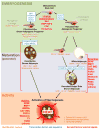
Figure 1
Figure 1. Distinct stages of brown adipocyte formation are regulated by instructive signals acting through endocrine, paracrine or autocrine mechanisms
Lineage chart summarizing the external signals that regulate the formation of constitutive and recruitable brown adipocytes during distinct stages of their formation: (1) Stem/progenitor cell lineage determination during embryonic development (green background; ‘Embryogenesis’); (2) Brown adipocyte differentiation from a committed progenitor cell into either constitutive or recruited brown adipocytes (violet background; ‘postnatal Maturation’); (3) thermogenic activity of mature, i.e. fully differentiated brown adipocytes (orange background; ‘Activity’). A common mesodermal stem cell with multi-lineage potential gives rise to fibroblast-like brown adipogenic progenitors of distinct lineages: Constitutive brown adipocytes of the common myo-adipogenic lineage, that also gives rise to skeletal muscle, and a predominantly adipogenic progenitor, that can generate mature white and recruited brown/beige/brite adipocytes residing within white adipose tissue depots. Distinct molecular markers specific to the individual cell types are shown in orange. Transcriptional regulators are shown in blue. Secreted signals that are involved in lineage determination or brown adipogenic maturation processes are shown in red, summarizing signals that may act through endocrine pathways, i.e. produced and acting in distal locations such as the CNS, and signals that may act as locally secreted paracrine or autocrine factors.
Figure 2
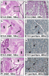
Figure 2. Brown adipose tissue emerges during late stages of embryogenesis
a, b, c, d, Hematoxylin and eosin (H&E) staining of transversal sections of the interscapular areas of mouse embryos at different embryonic stages. Arrows indicate morphologically distinguishable cBAT (a: embryonic day (E)14.5; b: E15.5; c: E16.5; d: postnatal day (P)1); original magnification: 100x; cBAT-pads indicated by arrows; § indicates spinal column). e, Same section of E14.5 (panel a) at greater magnification shows that only developing skeletal muscle, but not cBAT, can be observed at this stage (original magnification: 400x). f, g, h, Perilipin-immunohistochemistry (IHC), a marker for lipid droplets in maturing adipocytes, was performed in the same embryonic stages where cBAT was first observed (f: E15.5; g: E16.5; h: P1; original magnification: 400x). Perilipin-positive developing brown adipocytes are shown in brown, examples indicated by arrowheads. All stainings were performed as described before and using the same materials [39].
Figure 3
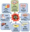
Figure 3. Brown adipocyte physiology is controlled by crosstalk mechanisms through secreted factors from multiple anatomical sites
Different organs produce and secrete instructive signals that are integrated within anatomical locations of brown adipocytes to regulate the development, differentiation, and possibly the thermogenic activity of this type of cell, thereby affecting thermoregulation and systemic energy metabolism. Figure was produced using Servier Medical Art (http://www.servier.com).

References

    1. Cypess AM, Kahn CR. Brown fat as a therapy for obesity and diabetes. Curr Opin Endocrinol Diabetes Obes. 2010;17:143–149. - PMC - PubMed
    1. Tseng YH, Cypess AM, Kahn CR. Cellular bioenergetics as a target for obesity therapy. Nat Rev Drug Discov. 2010;9:465–482. - PMC - PubMed
    1. Gesta S, Tseng YH, Kahn CR. Developmental origin of fat: tracking obesity to its source. Cell. 2007;131:242–256. - PubMed
    1. Bartelt A, Heeren J. The holy grail of metabolic disease: brown adipose tissue. Curr Opin Lipidol. 2012;23:190–195. - PubMed
    1. Jackson AS, Stanforth PR, Gagnon J, Rankinen T, Leon AS, Rao DC, Skinner JS, Bouchard C, Wilmore JH. The effect of sex, age and race on estimating percentage body fat from body mass index: The Heritage Family Study. Int J Obes Relat Metab Disord. 2002;26:789–796. - PubMed

Publication types

LinkOut - more resources