. 2013 Jun 27;11(1):45.
doi: 10.1186/1478-811X-11-45.

Insulin receptor membrane retention by a traceable chimeric mutant

Affiliations

Insulin receptor membrane retention by a traceable chimeric mutant

Jimena Giudice et al. Cell Commun Signal. .

Abstract

Background: The insulin receptor (IR) regulates glucose homeostasis, cell growth and differentiation. It has been hypothesized that the specific signaling characteristics of IR are in part determined by ligand-receptor complexes localization. Downstream signaling could be triggered from the plasma membrane or from endosomes. Regulation of activated receptor's internalization has been proposed as the mechanism responsible for the differential isoform and ligand-specific signaling.

Results: We generated a traceable IR chimera that allows the labeling of the receptor at the cell surface. This mutant binds insulin but fails to get activated and internalized. However, the mutant heterodimerizes with wild type IR inhibiting its auto-phosphorylation and blocking its internalization. IR membrane retention attenuates AP-1 transcriptional activation favoring Akt activation.

Conclusions: These results suggest that the mutant acts as a selective dominant negative blocking IR internalization-mediated signaling.


Figures
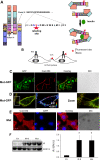
Figure 1
Figure 1
Covalent modification of IR-B at the plasma membrane. A. Tag position and insulin binding scheme according to proposed model [33,34]. L1, Large leucine rich domain 1; CR, cysteine rich domain; L2, large leucine rich domain 2; FnIII domains 1, 2 and 3; ID, inter-domain; TM, transmembrane domain; TK, tyrosine kinase; CT, C-terminus. Inserted tag shows: in grey, residues added for cloning reasons; in red, Ser recognized by ACP-S; in blue, conserved flanking amino acids for enzyme recognition [30]. B. Labeling strategy. C-E. HeLa cells expressing Mut-GFP (C-D) or Mut (E) were labeled with 0.2 μM ACP-S and 1 μM fluorescent CoA and imaged by confocal (Mut-GFP) or wide field microscopy (Mut). Scale bars: 10 μm. D. Manders map. F. Cells transiently expressing wild type IR-B or the mutant were assayed by Western blot 48 h after transfection using anti-IR-β-subunit or anti-ERK1/2. Quantification was performed by densitometry. Results are expressed as the mean ± s.e.m. (*: p≤0.05; n≥3).
Figure 2
Figure 2
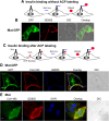
Insulin binding assays. A. Strategy for insulin binding and QD655 labeling. B. HeLa cells expressing Mut-GFP were incubated with 50 nM BAC-Ins and 1 nM QD655, fixed and imaged by confocal microscopy. C. Insulin binging after ACP labeling. D-E. Cells expressing the mutants were labeled with 0.2 μM ACP-S and 1 μM fluorescent CoA and then with 50 nM BAC-Ins and 1 nM QD655. Imaging was performed by confocal (D) or wide field microscopy (E). Scale bars: 10 μm.
Figure 3
Figure 3
Chimeras characterization. A. HeLa cells expressing Mut-GFP (A), Mut (B), IR-B-SCFP (C) or IR-B (D) were stimulated with 100 nM rhIns for 10 min and fixed. Immunofluorescence assays were performed with anti-phospho-IR and a secondary antibody conjugated with Alexa fluor 555. Cells expressing Mut were labeled with 0.2 μM ACP-S and 1 μM CoA-488 before stimulation. E. Cells expressing wt IR-B or the mutant were stimulated with 100 nM rhIns for 5 min and lysates were analyzed by Western blot using anti-pY20 and anti-IR-β subunit. Quantification was performed by densitometry. The ratio pY20 / IR was measured for each lane and fold induction was calculated with respect to basal levels. Results are expressed as the mean ± s.e.m. (*: p=0.002; n≥3). ´p´ means phospho-antibodies.
Figure 4
Figure 4
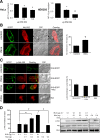
Mutant retention at the plasma membrane. A. HeLa cells expressing Mut labeled with 0.2 μM ACP-S and 1 μM CoA-488 were incubated with 50 nM BAC-Ins and 1 nM QD655. Cells were fixed or acid treated before fixation. Right panel: cells were incubated at 37°C for 30 min and fixed with or without acid treatment. B. Cells expressing IR-B were labeled with 50 nM BAC-Ins and 1 nM QD655, incubated or not at 37°C for 30 min and fixed or previously acid treated. Samples were imaged by confocal microscopy. Scale bars: 10 μm.
Figure 5
Figure 5
Dimers wild type / mutant at the plasma membrane and signaling. A. HeLa cells expressing Mut, IR-B-SCFP or both were treated with 2 μM ACP-S and 5 μM CoA-biotin for 30 min. Lysates were incubated with SA-agarose beads for 1 h at 4°C. Precipitates and total fractions were analyzed by Western blot with anti-IR-β subunit. B. HeLa cells were co-transfected with 0.1 μg pcDNA3-IR-B and different amounts of the mutant or EV, stimulated with 100 nM rhIns for 5 min and assayed by Western blot. Quantification was performed by densitometry measuring pY20 signal with respect to the first lane (basal) (*: p<0.05, n=4). C. HeLa cells co-expressing Mut and IR-B were labeled with 2 μM ACP-S and 2 μM CoA-488 and then incubated with 50 nM BAC-Ins and 1 nM QD655. Cells were directly fixed or incubated for 30 min at 37°C and then fixed or acid treated before fixation. Samples were imaged by confocal microscopy. Scale bars: 10 μm. D. Internalization analysis. Results are expressed as the mean ± s.e.m (p<0.005; n=6-32 cells).
Figure 6
Figure 6
Mutant IR blocks insulin induced AP-1 activity without affecting Akt activation. A. HeLa or HEK293 cells co-expressing AP1-Luc, IR-B and different amounts of the mutant were starved for 24 h and then stimulated with 100 nM rhIns for 16 h. Luciferase activity was measured and fold induction was calculated (*: p≤0.03, **: p=0.003; n≥3). B. Akt translocation to the plasma membrane: HeLa cells co-expressing IR-B-SCFP and Akt-HA were starved overnight and then stimulated with 100 nM rhIns for 5 min. After fixation samples were stained with anti-Akt and a secondary antibody conjugated with Alexa fluor 555. Images were acquired by confocal microscopy and quantification of Akt re-distribution was evidenced (*: p<10-6; n=34-40 cells). Scale bars: 10 μm. C. Similar experiment was performed using anti-phospho-Akt in cells co-expressing Akt-HA with: i) IR-B-SCFP, ii) Mut iii) both (n=15-23 cells). D. Cells co-transfected with 0.1 μg pcDNA3-IR-B and different amounts of the mutant or EV were stimulated with 100 nM rhIns for 5 min and assayed by Western blot. Quantification was performed by densitometry measuring phospho-Akt signal normalized to the first lane (basal) (*: p<0.05, n≥3). ´p´ means phospho-antibodies. Results are expressed as the mean ± s.e.m.

Similar articles

Cited by

References

    1. Ebina Y, Ellis L, Jarnagin K, Edery M, Graf L, Clauser E, Ou J, Masiarz F, Kan YW, Goldfine ID. et al.The human insulin receptor cDNA: the structural basis for hormone-activated transmembrane signaling. Cell. 1985;40:747–758. doi: 10.1016/0092-8674(85)90334-4. - DOI - PubMed
    1. Ullrich A, Bell JR, Chen EY, Herrera R, Petruzelli LM, Dull TJ, Gray A, Coussens L, Liao Y, Tsubokawa M. et al.Human insulin receptor and its relationship to the tyrosine kinase family of oncogenes. Nature (London) 1985;313:756–761. doi: 10.1038/313756a0. - DOI - PubMed
    1. Frasca F, Pandini G, Scalia P, Sciacca L, Mineo R, Costantino A, Goldfine ID, Belfiore A, Vigneri R. Insulin receptor isoform A, a newly recognized, high-affinity insulin-like growth factor II receptor in fetal and cancer cells. Mol Cell Biol. 1999;19:3278–3288. - PMC - PubMed
    1. Mosthaf L, Vogt B, Haring HU, Ullrich A. Altered expression of insulin receptor types A and B in the skeletal muscle of non-insulin-dependent diabetes mellitus patients. Proc Natl Acad Sci USA. 1991;88:4728–4730. doi: 10.1073/pnas.88.11.4728. - DOI - PMC - PubMed
    1. Kellerer M, Sesti G, Seffer E, Obermaier-Kusser B, Pongratz DE, Mosthaf L, Häring HU. Altered pattern of insulin receptor isotypes in skeletal muscle membranes of type 2 (noninsulin-dependent) diabetic subjects. Diabetologia. 1993;36:628–632. doi: 10.1007/BF00404072. - DOI - PubMed

LinkOut - more resources