. 2013 Jun 28;14(1):67.
doi: 10.1186/1465-9921-14-67.

The role of uPAR in epithelial-mesenchymal transition in small airway epithelium of patients with chronic obstructive pulmonary disease

Affiliations

The role of uPAR in epithelial-mesenchymal transition in small airway epithelium of patients with chronic obstructive pulmonary disease

Qin Wang et al. Respir Res. .

Abstract

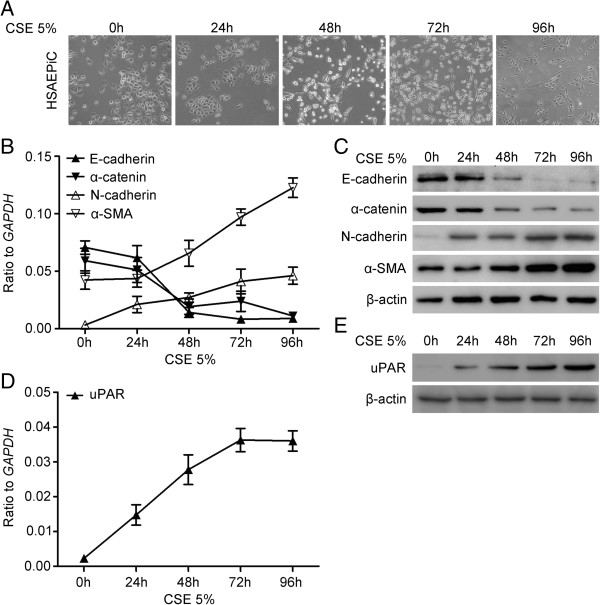
Background: Epithelial-mesenchymal transition (EMT) plays a crucial role in small airway fibrosis of patients with chronic obstructive pulmonary disease (COPD). Increasing evidence suggests that the urokinase plasminogen activator receptor (uPAR) is involved in the pathogenesis of COPD. Increased uPAR expression has been implicated in the promotion of EMT in numerous cancers; however the role of uPAR in EMT in small airway epithelial cells of patients with COPD remains unclear. In this study, we investigated the degree of EMT and uPAR expression in lung epithelium of COPD patients, and verified the effect of uPAR on cigarette smoke extract (CSE)-induced EMT in vitro.

Methods: The expression of EMT biomarkers and uPAR was assessed in lung epithelium specimens from non-smokers (n = 25), smokers (n = 25) and non-smokers with COPD (n = 10) and smokers with COPD (n = 18). The role of uPAR on CSE-induced EMT in human small airway epithelial cells (HSAEpiCs) was assessed by silencing uPAR expression in vitro.

Results: Markers of active EMT and uPAR expression were significantly increased in the small airway epithelium of patients with COPD compared with controls. We also observed a significant correlation between uPAR and vimentin expression in the small airway epithelium. In vitro, CSE-induced EMT in HSAEpiCs was associated with high expression of uPAR, and targeted silencing of uPAR using shRNA inhibited CSE-induced EMT. Finally, we demonstrate that the PI3K/Akt signaling pathway is required for uPAR-mediated EMT in HSAEpiCs.

Conclusions: A uPAR-dependent signaling pathway is required for CSE-induced EMT, which contributes to small airway fibrosis in COPD. We propose that increased uPAR expression in the small airway epithelium of patients with COPD participates in an active EMT process.


Figures

Figure 1
Figure 1
Immunohistochemistry for EMT biomarkers in small airways epithelium of patients with chronic obstructive pulmonary disease (COPD). Small airways sections from non-smokers (n = 25), smokers (n = 25), non-smokers with COPD (n = 10), and smokers with COPD (n = 18) were immunostained. (A) Epithelial marker E-cadherin (brown) and mesenchymal marker vimentin (brown) staining are observed in small airways epithelium from a non-smoker and a patient with COPD with serial sections. Black arrows show the main distribution of the positive immunostaining regions. (B) Mesenchymal marker vimentin (brown) from non-smokers (a), smokers (b), non-smokers with COPD (c) and smokers with COPD (d). (C) Quantification of mesenchymal marker vimentin positive cells in the small airways epithelium. The number of positive epithelial cells per mm reticular basement membrane (Rbm) in small airways. P values in figure were obtained by Mann–Whitney U test analyses. Images were obtained using a 100 × oil-immersion objective.
Figure 2
Figure 2
Urokinase plasminogen activator receptor (uPAR) immunostaining in small airways epithelium of patients with COPD. (A) uPAR staining in the epithelium of small airways from non-smokers (a), smokers (b), non-smokers with COPD (c) and smokers with COPD (d) (original × 200). (B) Quantification of uPAR protein levels in the small airways epithelium. The mean staining density in small airways.
Figure 3
Figure 3
Correlations between the expression of uPAR or mesenchymal marker vimentin positive cells in small airways and functional parameters. (A) The expression of uPAR in small airways of 78 subjects was correlated with FEV1% of predicted, as an index of airflow obstruction severity in COPD. (B) Mesenchymal marker vimentin positive cells per mm in small airways was correlated with FEV1% of predicted. (C) The expression of uPAR in small airways was correlated with vimentin positive cells per mm.
Figure 4
Figure 4
Cigarette smoke extract (CSE) induces epithelial-mesenchymal transition and increases uPAR levels in cultured human small airway epithelial cells (HSAEpiC). (A) Cells were cultured for variable time points with 5% CSE. Cell images were captured by phase-contrast microscopy. (B) Epithelial markers E-cadherin and α-catenin, mesenchymal markers N-cadherin and α-smooth muscle actin-SMA) mRNA levels were determined by Real-time PCR (mean ± SEM; n = 3). (C) Epithelial markers E-cadherin and α-catenin, mesenchymal markers N-cadherin and α-SMA protein levels were determined by Western blot. The original extracts were subjected to Western blot analysis for β-actin, as a loading control. (D) UPAR mRNA level was determined by Real-time PCR (mean ± SEM; n = 3). (E) uPAR protein level was determined by Western blot.
Figure 5
Figure 5
uPAR is required for CSE-induced EMT in HSAEpiC cells. (A) Cells expressing empty vector, shuPAR1 cells, and shuPAR2 cells were cultured for 24 h. UPAR mRNA was determined by Real-time PCR (mean ± SEM; n = 3). (B) uPAR protein level was determined by Western blot. (C) Empty vector and shuPAR2 cells were cultured for 72 h with 5% CSE. Cell images were captured by phase-contrast microscopy. (D) Parental cells, empty vector and shuPAR2 cells were cultured for 72 h without or with 5% CSE. Cell extracts were subjected to Western blot analysis for epithelial markers E-cadherin and α-catenin, mesenchymal markers N-cadherin and α-SMA. β-actin was used as a loading control.
Figure 6
Figure 6
PI3K/Akt signaling pathway is required for uPAR-mediated EMT in HSAEpiC Cells. (A) Cells were cultured for 72 h without or with 5% CSE. Cell extracts were subjected to Western blot to detect phosphorylated Akt and total Akt. (B) Parental cells, empty vector and shuPAR2 cells were cultured for 72 h without or with 5% CSE. Cell extracts were subjected to Western blot analysis for phosphorylated Akt (p-Akt), phosphorylated GSK-3β (p-GSK-3β), Snail, and β-actin. (C) Cells were treated with 10 μM of the PI3K inhibitor LY294002 or with vehicle (control) for 72 h without or with 5% CSE. Cell extracts were subjected to Western blot analysis to detect p-Akt, p-GSK-3β, Snail, E-cadherin, α-SMA and β-actin (representative of three studies).

Comment in

References

    1. Vestbo J, Hurd SS, Agusti AG, Jones PW, Vogelmeier C, Anzueto A, Barnes PJ, Fabbri LM, Martinez FJ, Nishimura M. et al.Global strategy for the diagnosis, management, and prevention of chronic obstructive pulmonary disease: GOLD executive summary. Am J Respir Crit Care Med. 2013;187:347–365. doi: 10.1164/rccm.201204-0596PP. - DOI - PubMed
    1. Celli BR, MacNee W. Standards for the diagnosis and treatment of patients with COPD: a summary of the ATS/ERS position paper. Eur Respir J. 2004;23:932–946. doi: 10.1183/09031936.04.00014304. - DOI - PubMed
    1. McDonough JE, Yuan R, Suzuki M, Seyednejad N, Elliott WM, Sanchez PG, Wright AC, Gefter WB, Litzky L, Coxson HO. et al.Small-airway obstruction and emphysema in chronic obstructive pulmonary disease. N Engl J Med. 2011;365:1567–1575. doi: 10.1056/NEJMoa1106955. - DOI - PMC - PubMed
    1. Hogg J. Peripheral lung remodelling in asthma and chronic obstructive pulmonary disease. Eur Respir J. 2004;24:893–894. doi: 10.1183/09031936.04.00110704. - DOI - PubMed
    1. Scotton CJ, Chambers RC. Molecular targets in pulmonary fibrosis: the myofibroblast in focus. Chest. 2007;132:1311–1321. doi: 10.1378/chest.06-2568. - DOI - PubMed

Publication types

Substances