. 2013 Jun 24;25(6):623-35.
doi: 10.1016/j.devcel.2013.05.021.

Fuz mutant mice reveal shared mechanisms between ciliopathies and FGF-related syndromes

Affiliations

Fuz mutant mice reveal shared mechanisms between ciliopathies and FGF-related syndromes

Jacqueline M Tabler et al. Dev Cell. .

Abstract

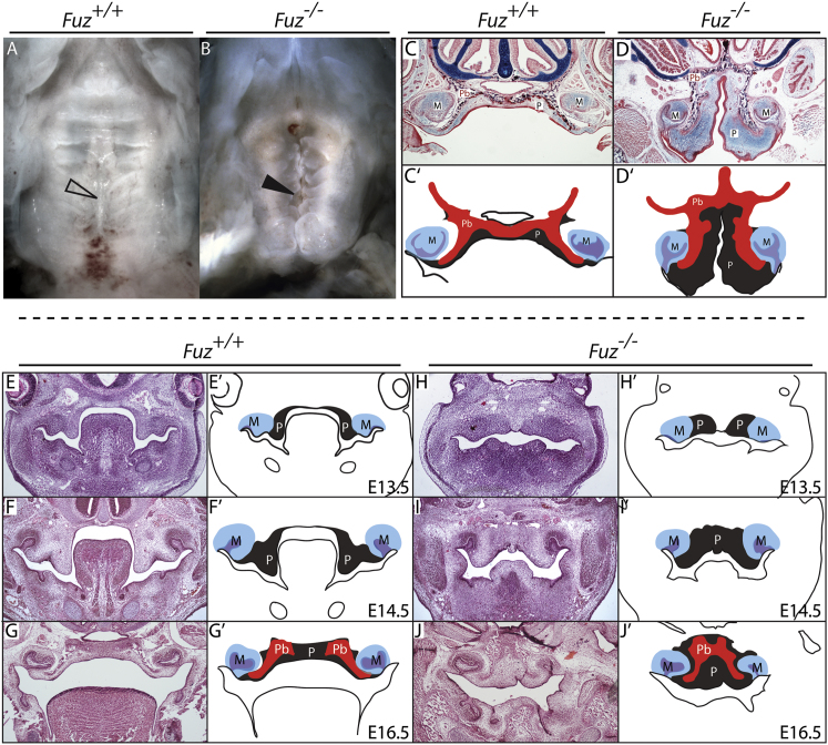
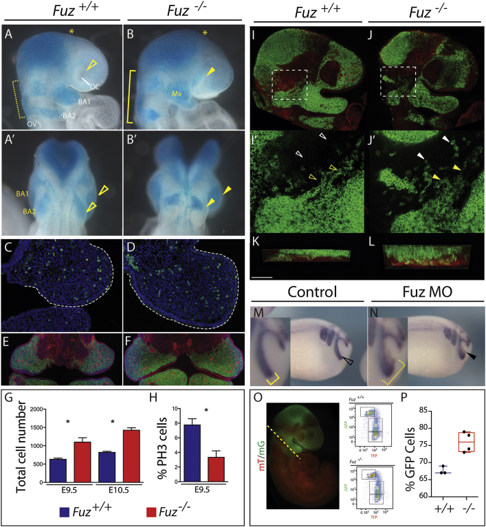
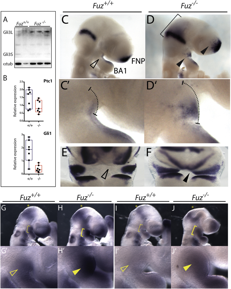
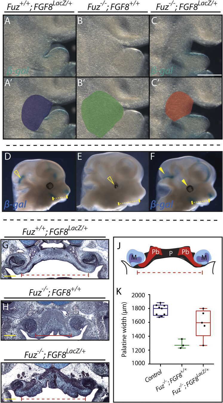
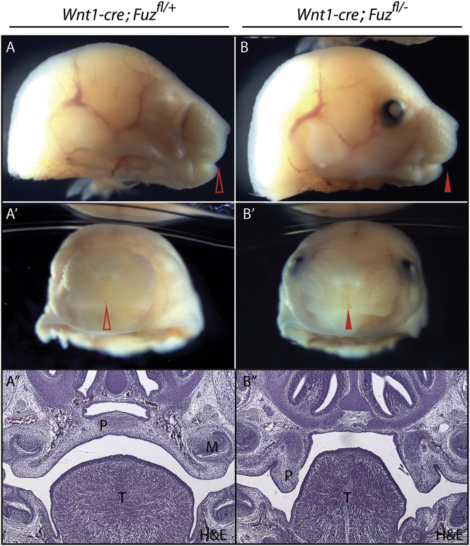
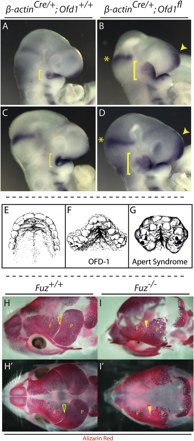
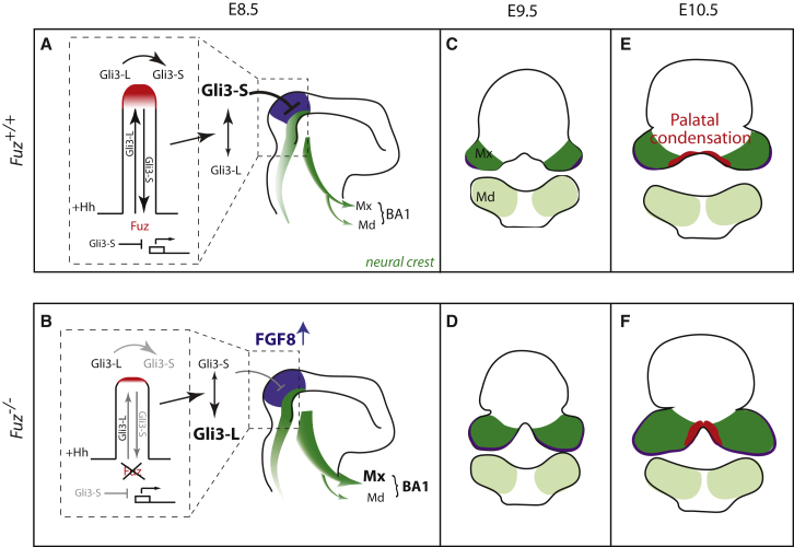
Ciliopathies are a broad class of human disorders with craniofacial dysmorphology as a common feature. Among these is high arched palate, a condition that affects speech and quality of life. Using the ciliopathic Fuz mutant mouse, we find that high arched palate does not, as commonly suggested, arise from midface hypoplasia. Rather, increased neural crest expands the maxillary primordia. In Fuz mutants, this phenotype stems from dysregulated Gli processing, which in turn results in excessive craniofacial Fgf8 gene expression. Accordingly, genetic reduction of Fgf8 ameliorates the maxillary phenotypes. Similar phenotypes result from mutation of oral-facial-digital syndrome 1 (Ofd1), suggesting that aberrant transcription of Fgf8 is a common feature of ciliopathies. High arched palate is also a prevalent feature of fibroblast growth factor (FGF) hyperactivation syndromes. Thus, our findings elucidate the etiology for a common craniofacial anomaly and identify links between two classes of human disease: FGF-hyperactivation syndromes and ciliopathies.


Figures

Figure 1
Figure 1
Fuz Mutant Mice Are a Model for High Arched Palate (A and B) Ventral views of E17.5 palates. Mutants exhibit a narrow palate (arrowheads) and disrupted rugal organization compared to littermates. (C and D) Sirius red/alcian blue staining of E17.5 coronal sections. Bilateral palatine bones are formed in both control and mutant, abutting at the midline (black). Mutant palatine bones (Pb) are mediolaterally shortened with an increase in the ventral extension. Note enlarged palatal mesenchyme (P). Molars (M) appear normal. (C′ and D′) Schematics depicting skewed palatal anatomy. (E–J) Hemotoxylin and eosin staining of coronal sections at indicated stages. (E′–J′) Schematics of sections identifying palatal mesenchyme (gray, P), molars (blue and purple, M), and palatal bone condensations (red, Pb). In (H) through (J′), mutant palatal condensations are medially constrained and do not extend into the oral cavity. Palatine bone can be observed in both mutants and controls by E16.5. See also Figure S1.
Figure 2
Figure 2
Increase in NC in Maxillary Compartment Wnt1-cre-driven LacZ (blue) or GFP (green) marks NC contributions. (A and B) Lateral views of E9.25 embryos. BA1 and BA2 NC streams are wider compared to controls (B compared to A, yellow bracket). Increased NC disrupts the optic cup (OC) in Fuz mutants compared to controls (B compared to A, arrowhead). BA2 NC stream is also increased in size and has failed to migrate as far as BA2 control NC. Mx, maxillary compartment of BA1. OV, optic vesicle. (A′ and B′) Dorsal views. Maxillary BA1 is enlarged compared to controls (B compared to A, top arrowhead). BA2 has failed to migrate sufficiently compared to controls (bottom arrowhead). (C and D) PH3 staining (green) and DAPI (blue) of E9.0, maxillary compartment. Mutant maxilla is enlarged (white dotted line). (E and F) Coronal sections of E10.5 Wnt1-cre; R26RmT/mG; Fuz+/+, or Wnt1-cre; R26RmT/mG;Fuz/ maxillae. Wnt1-cre-driven membrane-GFP (green) marks NC contributions. Epithelial membrane-Tomato (red) highlights all other tissue derivatives. Mutant maxillae are larger compared to controls. Enlarged maxillae comprise NC-derived mesenchyme. (G) Quantification of DAPI-positive cells from representative sections of E9.5 and 10.5 embryos. Note increase in cell number in mutant (red) maxilla compared to wild-type (blue) (p < 0.004). Error bars indicate SD. (H) Quantification of E9.5 maxillary PH3-positive cells compared to total cell number. A significant decrease in the percentage of PH3-positive cells is observed in mutant maxillae (red) compared to controls (blue) (p < 0.01). Error bars indicate SD. (I and J) Single confocal z-sections of Wnt1-cre; R26RmT/mG;Fuz+/+ or Wnt1-cre; R26RmT/mG;Fuz/ E9.5 embryos. Wnt1-cre-driven membrane-GFP (green) marks NC contributions. (I′–J′) Magnified maximum projections of (I) and (J), indicated by a white dotted box. In controls, chains of Wnt1-cre-driven membrane-GFP-positive NC are observed (yellow arrowheads), with few isolated cells between brain and maxillary compartment (white arrowheads). Isolated NC cells are observed in mutant embryos, indicated by a white arrowhead in (J′), and NC chains are disorganized. (K and L) Z-projections of (I′) and (J′). The thickness of NC streams anterior to the prospective trigeminal ganglion is increased in mutants (WT = 41 ± 20 μm thick; mutant = 79 ± 16 μm deep). The immediately underlying membrane-Tomato-positive mesenchymal cells are shown in red, at the bottom of the image. The overlying epithelium is not included. Scale bar, 100 μm. This doubling in thickness is consistent with increased NC invasion into BA1. (M and N) Twist in situ hybridization of stage 22 embryos injected unilaterally with Fuz MO. In controls, Twist is expressed in three streams, where the anterior NC stream surrounds the optic placode, indicated in (M) by open arrowhead and inset (bracket). Ectopic Twist expression is observed in the optic placode in Fuz morphants, indicated in (N) by arrowhead and inset (bracket). (O) Representative epifluorescence image of an E9 Wnt1-cre; R26mT/mG embryo. Lineage-traced NC cells are labeled GFP (green), and nonrecombined cells are Tomato positive (red). Dashed line illustrates where embryos were bisected caudal to BA1. Wnt1-cre; R26RmT/mG; Fuz+/+, or Wnt1-cre; R26RmT/mG;Fuz/ embryo heads were dissociated and GFP positive, Tomato, or double-labeled cells were analyzed by flow cytometry. Representative flow cytometry plots from single E9 Wnt1-cre; R26mT/mG; Fuz+/+, or Wnt1-cre; R26RmT/mG;Fuz/ dissociated heads. mG, Wnt1-cre-driven membrane-GFP; mT, membrane-Tomato. (P) The percentage of Wnt1-Cre-driven GFP-positive cells was significantly increased in mutants (79% ± 2%) compared to controls (68% ± 1%) (p = 0.0103). Whiskers represent maximum/minimum data values with median and quartiles represented in the box.
Figure 3
Figure 3
Gli3 Processing and Hh and FGF Signaling Are Altered in Fuz Mutants (A) Western blot analysis of activator and repressor forms of Gli3 (Gli3-L and Gli3-S, respectively) in E9.0 embryos. Increased Gli3-L and reduced Gli3-S was detected in Fuz/ embryos in (A), lanes 3–5, compared to controls in (A), lanes 1 and 2. Alpha-tubulin was analyzed as a loading control. (B) qPCR of Hh target genes Patched 1 (Ptc1) and Gli1 from E9.0 Fuz+/+ and Fuz/ heads. Relative mRNA levels are normalized to β-actin. Ptc1 and Gli1 are decreased in mutants (red) compared to controls (blue). Whiskers represent maximum/minimum data values with median and quartiles represented in the box. (C and D) Lateral views of Fgf8 expression in E9.0 embryos. Fgf8 is expressed in the midhindbrain boundary (MHB), frontonasal prominence (FNP), and the BA1 epithelium. In mutants, Fgf8 expression domains are expanded, indicated by bracket and arrowheads. (C′ and D′) Magnified view of BA1 in control and mutant embryos. Dotted line indicates extent of maxillary compartment. Note enlarged maxillary prominence. (E and F) Frontal view of Fgf8 expression in E10.5 embryos. Note mediolateral expansion of maxillary Fgf8 expression in the mutant, indicated by black arrowhead. (G and H) Lateral views of Erm1 mRNA expression in E9.5 embryos. Erm1 is expressed in the MHB, FNP, and BA1. In mutants, expression of Erm1 is expanded ventrally from the MHB (yellow asterisk) and in BA1 (yellow bracket). (G′ and H′) Magnified view of BA1. Erm1 expression is increased in mutant maxillae (yellow arrowheads). (I and J) Lateral views of Pea3 expression in E9.5 embryos. Pea3 is expressed in the MHB, FNP, and BA1. In mutants, expression of Pea3 is expanded in BA1 (yellow bracket). (I′ and J′) Magnified view of BA1 in control and mutant embryos. Pea3 expression is increased in mutant maxillae (yellow arrowhead). See also Figure S2.
Figure 4
Figure 4
Fgf8 Reduction Rescues Maxillary Hyperplasia and Palatal Width in Compound Mutants (A–C) Lateral views of BA1. Maxillary size is expanded in Fuz/ mutants in (B) compared to Fgf8LacZ/+ in (A). Maxillary expansion is rescued in Fuz/; Fgf8LacZ/+ embryos in (C). Fgf8 expression is also rescued in compound mutants as β-gal staining reveals similar BA1 epithelial expression in compound mutants and Fgf8 heterozygotes. (A′–C′) Maxillary size is schematized for each genotype. (D–F) Lateral view, E12.5. Maxillary size is expanded in mutants in (E) compared to Fgf8LacZ/+ in (D). Maxillary expansion is rescued in Fuz/; Fgf8LacZ/+ embryos in (F). Normal Fgf8 expression (β-gal/blue) is also restored in compound mutants. Brain overgrowth and eye defects are rescued in compound mutants (arrowheads). (G–I) Trichrome staining of coronal sections of E16.5 embryos. Palatal width and bone angle are decreased in mutant embryos in (H) compared to controls in (G). Palatal width in Fuz/; Fgf8LacZ/+ in (I) appears normal compared to controls in (H). Angle of palatine bone is partially rescued when compared to controls, as shown in (I) compared to (G) and (H). (J) Schematic of palatal anatomy. (K) Quantification of palatal width in E16.5 Fuz+/+; Fgf8LacZ/+(blue), Fuz/; Fgf8+/+ (green), and Fuz/; Fgf8LacZ/+(red) embryos. Palatal width is significantly decreased in Fuz/; Fgf8+/+ compared to controls, while Fgf8 heterozygosity rescues palatal width in Fuz mutants (p < 0.0005, one-way analysis of variance). Whiskers represent maximum/minimum data values with median and quartiles represented in the box. See also Figure S3.
Figure 5
Figure 5
Maxillary Hyperplasia Is Not Due to Fuz Function in the NC (A and B) Lateral views of E17.5 Wnt1-cre; Fuzfl/+ and Wnt1-cre; Fuzfl/ embryos. The control embryo in (A) is an albino and lacks pigment in the eye. Arrowheads indicate rostral midline. (A′ and B′) Frontal views of E17.5 Wnt1-cre; Fuzfl/+ and Wnt1-cre; Fuzfl/ embryos. A cleft lip is observed in conditional null embryos [(B′) compared to (A′), indicated by arrowheads]. (A″ and B″) Hemotoxylin and eosin (H&E) staining of coronal sections of E17.5 Wnt1-cre; Fuzfl/+ and Wnt1-cre; Fuzfl/ embryos. Sections show the anterior secondary palate (P), tongue (T), and molars (M). Palatal shelves are elevated and fused across the midline in control embryos; however, a failure in shelf elevation is observed in Wnt1-cre; Fuzfl/ embryos [(B″) compared to (A″), indicated by P]. The tongue also appears smaller and irregularly shaped in mutants [(B″) compared to (A″) indicated by T].
Figure 6
Figure 6
Ofd-1 Mutants Also Show Expanded Maxillary Compartments and Cranial FGF8 Expression Domains (A–D) Lateral views of E9.5 embryos showing cranial Fgf8 mRNA expression domains. The 21 and 23 somite ofd-1 mutants have enlarged maxillae compared to stage-matched control embryos, indicated by yellow brackets in (B) and (D) compared to (A) and (C), respectively. Maxillary Fgf8 expression is expanded in mutants compared to controls [brackets in (B) and (D) compared to (A) and (C), as well as expression in the frontonasal process, indicated by arrowheads]. (E–G) OFD-1 and Apert syndrome patients exhibit high arched palate. In (E), a ventral view of a normal palate shows a hard palate with shallow, anterior bilateral rugae and smooth posterior palate (after photo by Millicent Odunze: http://plasticsurgery.about.com/od/Cleft-Lip-And-Palate/ss/What-Is-A-Cleft-Palate_3.htm). In (F), a ventral view is shown of the palate from OFD 1 (OFD-1) patient (after Tagliani et al., 2010; Figure 2F). (G) shows a ventral view of palate from the Apert syndrome patient (after Rynearson, 2000). Both (F) ciliopathic and (G) FGF-related high arched palates are narrow with a deep medial cleft extending from the anterior hard palate. Rugal-like swellings are more numerous and are extant with the medial cleft. Soft tissue swellings and dental crowding are observed in both groups. (H–I′) Loss of Fuz causes coronal craniosynostosis. E17.5 heads stained with Alizarin red. (H) and (I) show dorsolateral views. (H′) and (I′) show dorsal views. Alizarin red negative coronal suture (open arrowheads) separates frontal (F) and parietal (P) bones in wild-type embryos. Coronal suture is absent in mutant embryos (closed arrowheads).
Figure 7
Figure 7
Proposed Model for Palatal Defects in Fuz Mutants (A and B) Schematics depict E8.5 embryos. In Hedgehog signaling, anterograde transport delivers Gli3L (Gli3-activator) to the distal CLAMP-positive ciliary tip (red). Gli3-L is processed into Gli3-S (Gli3 repressor) which undergoes retrograde transport, repressing transcription of targets such as FGF8. Anterior NC (dark green) arises from the posterior mesencephalon and anterior hindbrain (purple), and is normally limited by a balance of Hh and FGF signals. NC then migrates into BA1, comprising maxillary and mandibular compartments (Mx and Md, respectively). When Fuz function is lost, ciliary transport and the distal compartment are severely disrupted leading to disruption of Gli processing and an expansion in cranial NC numbers, specifically maxillary NC. (C–F) In (C) and (D), schematics depict coronal sections of E9.5 embryos. In coronal sections, the maxilla forms as two bilateral prominences and Fgf8 is expressed in lateral maxillary epithelia, indicated by purple in (C) through (F). In mutants, the maxillary compartment is enlarged and epithelial Fgf8 is expanded. In (E) and (F), schematics depict coronal sections of E10.5 embryos. Palatal condensations (red) are medial to FGF8 expression domains. In wild-type animals, these flank midline mesenchyme but remain separated. In Fuz mutants, expansion of FGF8 causes a medial shift of the palatal condensations. Subsequently, the normal bilateral palatal primordia join at the midline.

Similar articles

Cited by

References

    1. Agochukwu N.B., Solomon B.D., Doherty E.S., Muenke M. Palatal and oral manifestations of Muenke syndrome (FGFR3-related craniosynostosis) J. Craniofac. Surg. 2012;23:664–668. - PMC - PubMed
    1. Anderson P.J., Hall C., Evans R.D., Harkness W.J., Hayward R.D., Jones B.M. The cervical spine in Crouzon syndrome. Spine (Phila Pa 1976) 1997;22:402–405. - PubMed
    1. Aoto K., Nishimura T., Eto K., Motoyama J. Mouse GLI3 regulates Fgf8 expression and apoptosis in the developing neural tube, face, and limb bud. Dev. Biol. 2002;251:320–332. - PubMed
    1. Arts H.H., Bongers E.M., Mans D.A., van Beersum S.E., Oud M.M., Bolat E., Spruijt L., Cornelissen E.A., Schuurs-Hoeijmakers J.H., de Leeuw N. C14ORF179 encoding IFT43 is mutated in Sensenbrenner syndrome. J. Med. Genet. 2011;48:390–395. - PubMed
    1. Beales P.L., Elcioglu N., Woolf A.S., Parker D., Flinter F.A. New criteria for improved diagnosis of Bardet-Biedl syndrome: results of a population survey. J. Med. Genet. 1999;36:437–446. - PMC - PubMed

Publication types

MeSH terms

Substances