. 2013 Jun 24;25(6):645-54.
doi: 10.1016/j.devcel.2013.05.023.

The Drosophila nuclear lamina protein otefin is required for germline stem cell survival

Affiliations

The Drosophila nuclear lamina protein otefin is required for germline stem cell survival

Lacy J Barton et al. Dev Cell. .

Abstract

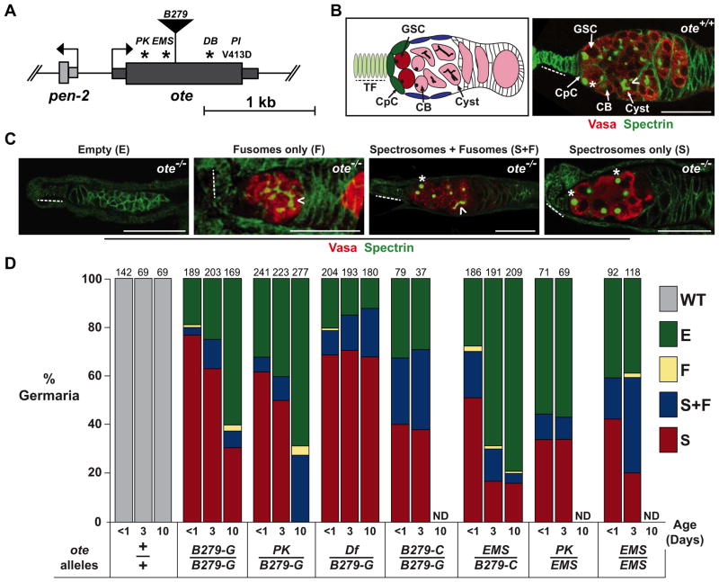
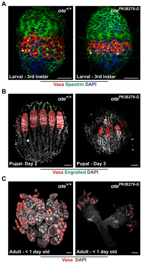
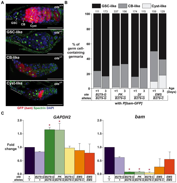
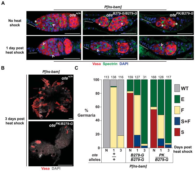
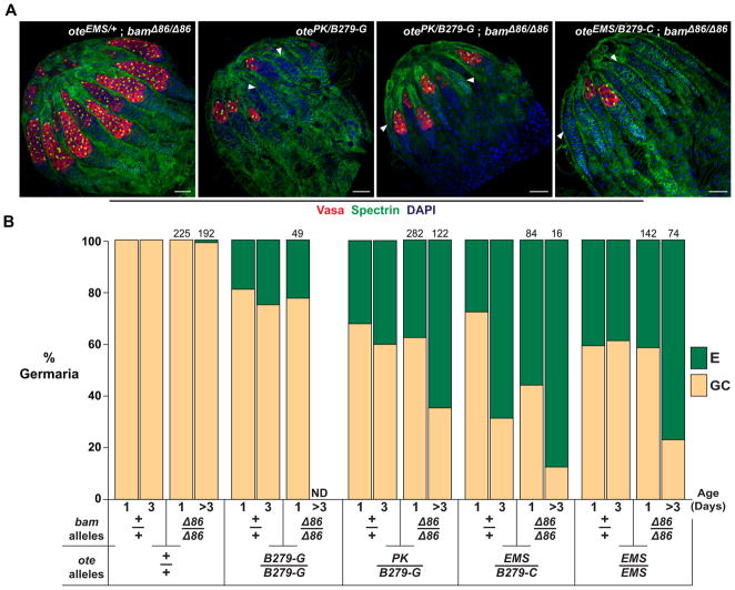
LEM domain (LEM-D) proteins are components of an extensive protein network that assembles beneath the inner nuclear envelope. Defects in LEM-D proteins cause tissue-restricted human diseases associated with altered stem cell homeostasis. Otefin (Ote) is a Drosophila LEM-D protein that is intrinsically required for female germline stem cell (GSC) maintenance. Previous studies linked Ote loss with transcriptional activation of the key differentiation gene bag-of-marbles (bam), leading to the model in which Ote tethers the bam gene to the nuclear periphery for gene silencing. Using genetic and phenotypic analyses of multiple ote(-/-) backgrounds, we obtained evidence that is inconsistent with this model. We show that bam repression is maintained in ote(-/-) GSCs and that germ cell loss persists in ote(-/-), bam(-/-) mutants, together demonstrating that GSC loss is independent of bam transcription. We show that the primary defect in ote(-/-) GSCs is a block of differentiation, which ultimately leads to germ cell death.


Figures

Figure 1
Figure 1. Loss of Otefin causes a complex GSC phenotype
A. Schematic of the ote gene structure. The thick rectangle represents the ote coding region with the positions of the missense (PK, EMS, DB, PI) mutations and transposon insertion (B279) indicated. Asterisks indicate premature stop codons. B. Left: Shown is a schematic of a germarium. Terminal filament (TF) cells (light green, dashed line) and cap cells (CpC) (dark green) comprise the germline stem cell (GSC) niche. The GSCs (dark red) are positioned adjacent to the niche. Cystoblasts (CB) and cysts (pink) are positioned distal to the niche. Right: Shown is a wild type (ote+/+) germaria stained for Vasa (red) and Spectrin (green). A GSC is indicated by an asterisk and identified by the position near the niche and the presence of a spectrosome. A developing cyst is indicated by a chevron and identified by the presence of a fusome. C. Shown are representative images of each ote−/− germaria class in one-day-old ovaries stained for Vasa (red) and Spectrin (green), with class names noted above the image. All images show germaria oriented with anterior to the left. Scale bars represent 25 μm. D. Quantification of germarial class prevalence in several ote−/− backgrounds. WT: germaria with germ cells with spectrosomes, followed directly by germ cells with fusomes and then egg chambers. E: Empty germaria lacking germ cells, F: germaria with only fusome-containing germ cells, S+F: germaria with germ cells containing both spectrosomes and fusomes, and S: germaria with only spectrosome-containing germ cells. The total number of germaria counted is shown above each bar. In each case, ten or more ovaries were studied, obtained in two independent experiments. ND (Not Determined). See also Figure S1.
Figure 2
Figure 2. Germ cell differentiation is blocked in ote−/− ovaries
A. Shown are developing gonads obtained from female wandering 3rd instar larvae stained for Vasa (red), Spectrin (green) and DAPI (blue). Scale bars represent 25 μm. B. Shown are developing gonads obtained from females aged two and three days after pupation. Gonads were stained for Vasa (red), Engrailed (green) and DAPI (blue). Scale bars represent 25 μm. C. Shown are ovaries obtained from newly eclosed females stained for Vasa (red) and DAPI (grey). Scale bars represent 100 μm. All gonads are oriented with anterior towards the top. Asterisks: spectrosome.
Figure 3
Figure 3. Germ cells lacking Ote exhibit repression of bam transcription
A. Shown are germaria from less than one-day-old ote+/+ and ote−/− females carrying the P[bam-GFP] reporter construct that were stained for GFP (red), Spectrin (green) and DAPI (blue). Top Panel: The level of GFP signal found in ote+/+ GSCs, CBs and Cysts is indicated. Bottom Panels: Three GFP signal intensities observed in ote−/− germaria are listed in the top left corner of each image and are categorized as GSC-like, CB-like and Cyst-like. The GSC-like category of ote−/− germaria lacked detectable GFP signal in all germ cells. Scale bars represent 25 μm. Dashed line: TF. B: Quantification of ote−/− germaria categorized by GFP signal. Genotypes are listed below each bar. The total number of germaria counted from ten or more ovaries in two independent experiments is shown above each bar. C. Graphs showing quantitative real time PCR analyses of RNAs obtained from two hour old wild type (ote+/+and oteB279-G/+) and ote−/− ovaries. Fold change is set relative to value obtained in ote+/+ ovary RNA. Error bars indicate standard deviation from three biological replicates. (*, P<0.05, Student’s t-test). See also Figure S2.
Figure 4
Figure 4. Ectopic bam expression accelerates ote−/− germ cell loss
A. Shown are confocal images of germaria obtained from ote+/+ and ote−/− females carrying the P[hs-bam] transgene that were stained for Vasa (red), Spectrin (green) and DAPI (blue). The top panels show germaria from non-heat shocked females and the bottom panels show germaria from females that were heat shocked one day prior to ovary staining. Genotypes are listed in the top right corner of each image. Scale bars represent 25 μm. Dashed line: TF. Asterisk: spectrosome. Chevron: fusome. B. Shown are ovaries obtained from ote+/+ (top) and ote−/− (bottom) females carrying P[hs-bam] stained for Vasa (red) and DAPI (grey) three days after heat shock. Scale bars represent 250 μm. C. Shown is quantification of germarial class prevalence in females carrying the P[hs-bam] transgene. The germarial class designations are the same as described in Figure 1. N: non-heat shocked females aged for two days. The number of germaria scored from ten or more ovaries in two independent experiments is shown above each bar.
Figure 5
Figure 5. Loss of Bam does not rescue GSC loss in ote−/− ovaries
A. Shown are confocal images of ovaries obtained from one-day-old females stained for Vasa (red), Spectrin (green) and DAPI (blue). The anterior of each ovary is oriented toward the top left corner. Arrowheads indicate empty germaria. Genotypes are listed in the top right corner. Scale bars represent 25 μm. B. Shown is a graph of the quantification of the prevalence of germaria without (empty, E) or with germ cells (germ cell containing, GC) in ovaries of wild type and mutant females. Ovary genotypes are listed below each bar. Data for the ote+/+, bam+/+ and ote−/−, bam+/+ genotypes represent a re-categorization of data presented in Figure 1D, wherein the WT, F, S+F and S classes of germaria are encompassed in the GC category. The total number of bam−/− germaria scored is indicated above each bar. ND (Not Determined).

References

    1. Ashery-Padan R, Ulitzur N, Arbel A, Goldberg M, Weiss AM, Maus N, Fisher PA, Gruenbaum Y. Localization and posttranslational modifications of otefin, a protein required for vesicle attachment to chromatin, during Drosophila melanogaster development. Mol Cell Biol. 1997;17:4114–4123. - PMC - PubMed
    1. Bakay M, Wang Z, Melcon G, Schiltz L, Xuan J, Zhao P, Sartorelli V, Seo J, Pegoraro E, Angelini C, et al. Nuclear envelope dystrophies show a transcriptional fingerprint suggesting disruption of Rb-MyoD pathways in muscle regeneration. Brain. 2006;129:996–1013. - PubMed
    1. Bakhrat A, Pritchett T, Peretz G, McCall K, Abdu U. Drosophila Chk2 and p53 proteins induce stage-specific cell death independently during oogenesis. Apoptosis. 2010;15:1425–1434. - PMC - PubMed
    1. Baxley RM, Soshnev AA, Koryakov DE, Zhimulev IF, Geyer PK. The role of the Suppressor of Hairy-wing insulator protein in Drosophila oogenesis. Dev Biol. 2011;356:398–410. - PMC - PubMed
    1. Cai M, Huang Y, Ghirlando R, Wilson KL, Craigie R, Clore GM. Solution structure of the constant region of nuclear envelope protein LAP2 reveals two LEM-domain structures: one binds BAF and the other binds DNA. Embo J. 2001;20:4399–4407. - PMC - PubMed

Publication types

MeSH terms