

Preparation of developing Xenopus muscle for sarcomeric protein localization by high-resolution imaging
- PMID: 23806641
- PMCID: PMC3871942
- DOI: 10.1016/j.ymeth.2013.06.015
Preparation of developing Xenopus muscle for sarcomeric protein localization by high-resolution imaging
Abstract
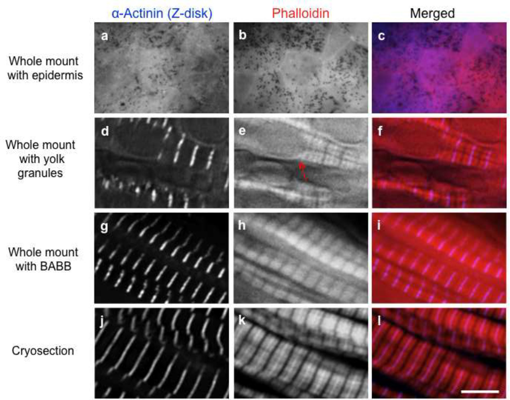
Mutations in several sarcomeric proteins have been linked to various human myopathies. Therefore, having an in vivo developmental model available that develops quickly and efficiently is key for investigators to elucidate the critical steps, components and signaling pathways involved in building a myofibril; this is the pivotal foundation for deciphering disease mechanisms as well as the development of myopathy-related therapeutics. Although striated muscle cell culture studies have been extremely informative in providing clues to both the distribution and functions of sarcomeric proteins, myocytes in vivo develop in an irreproducible 3D environment. Xenopus laevis (frog) embryos are cost effective, compliant to protein level manipulations and develop relatively quickly (⩽ a week) in a petri dish, thus providing a powerful system for de novo myofibrillogenesis studies. Although fluorophore-conjugated phalloidin labeling is the gold standard approach for investigating actin-thin filament architecture, it is well documented that phalloidin-labeling can be challenging and inconsistent within Xenopus embryos. Therefore we highlight several techniques that can be utilized to preserve both antibody and fluorophore-conjugated phalloidin labeling within Xenopus embryos for high-resolution fluorescence microscopy.
Keywords: Immunofluorescence microscopy; Myofibrillogenesis; Phalloidin; Sarcomere; Xenopus laevis.
Copyright © 2013. Published by Elsevier Inc.
Figures
References
-
- Muntz L. Myogenesis in the trunk and leg during development of the tadpole of Xenopus laevis (Daudin 1802) J. Embryol. Exp. Morph. 1975;33:757–774. - PubMed
-
- Boucaut J, Thierry D. Fibronectin in early amphibian embryos. Cell Tissue Res. 1983;234:135–145. - PubMed
-
- Fagotto F, Gumbiner BM. Beta-catenin localization during Xenopus embryogenesis: accumulation at tissue and somite boundaries. Development. 1994;120:3667–3679. - PubMed
-
- Schohl A, Fagotto F. Beta-catenin, MAPK and Smad signaling during early Xenopus development. Development. 2000;129:37–52. - PubMed
-
- Kieserman EK, Lee C, Gray RS, Park TJ, Wallingford JB. High-magnification in vivo imaging of Xenopus embryos for cell and developmental biology. Cold Spring Harbor Protocols. 2010;5 pdb-prot5426. - PubMed
Publication types
MeSH terms
Substances
Grants and funding
LinkOut - more resources
Full Text Sources
Other Literature Sources
