. 2013:3:2093.
doi: 10.1038/srep02093.

Diacylglycerol lipase α manipulation reveals developmental roles for intercellular endocannabinoid signaling

Affiliations

Diacylglycerol lipase α manipulation reveals developmental roles for intercellular endocannabinoid signaling

Erik Keimpema et al. Sci Rep. 2013.

Abstract

Endocannabinoids are small signaling lipids, with 2-arachidonoylglycerol (2-AG) implicated in modulating axonal growth and synaptic plasticity. The concept of short-range extracellular signaling by endocannabinoids is supported by the lack of trans-synaptic 2-AG signaling in mice lacking sn-1-diacylglycerol lipases (DAGLs), synthesizing 2-AG. Nevertheless, how far endocannabinoids can spread extracellularly to evoke physiological responses at CB₁ cannabinoid receptors (CB₁Rs) remains poorly understood. Here, we first show that cholinergic innervation of CA1 pyramidal cells of the hippocampus is sensitive to the genetic disruption of 2-AG signaling in DAGLα null mice. Next, we exploit a hybrid COS-7-cholinergic neuron co-culture system to demonstrate that heterologous DAGLα overexpression spherically excludes cholinergic growth cones from 2-AG-rich extracellular environments, and minimizes cell-cell contact in vitro. CB₁R-mediated exclusion responses lasted 3 days, indicating sustained spherical 2-AG availability. Overall, these data suggest that extracellular 2-AG concentrations can be sufficient to activate CB₁Rs along discrete spherical boundaries to modulate neuronal responsiveness.


Figures

Figure 1
Figure 1. DAGLα localization in the fetal cholinergic basal forebrain.
(a) Schema of 2-AG signaling during fetal development and in adulthood. Note the lack of both MGL in growth cones and glial 2-AG inactivation during development, allowing 2-AG spread. (b,b1) CB1R mRNA in the medial septum (ms) at embryonic day (E)18.5 and postnatal day 1 (P1). (b2,b3) The lack of hybridization signal in sense control experiments (neocortex, P1) confirmed detection specificity. (c,c1) A subpopulation of ChAT+ septal neurons was immunoreactive for CB1Rs in neonates. (c2–c4) In the neonatal hippocampus, cholinergic afferents likely harbored CB1Rs given the lack of physical signal separation for ChAT and CB1R immunoreactivities. Solid and open arrowheads pinpoint the presence and lack of co-localization for select marker combinations, respectively. (c5) Colocalization coefficients for cholinergic (ChAT+) and CB1R+ boutons in the CA1 subfield of the neonatal hippocampus. Data on dual-labeled terminal subsets were expressed as the percentage of all cholinergic or CB1R-containing synapses per field of view. (d–d1) A subpopulation of p75NTR+ neurons expressed DAGLα. p75NTR+ neurons, assumed to acquire cholinergic phenotype, were generally situated proximal to DAGLα+ cells (arrowheads). (d2–d4) Representative images of cholinergic neurites coursing along DAGLα+ neurons and of adjacent p75NTR+-DAGLα cell pairs in the medial septum. (d5) Quantitative analysis of the portion of p75NTR+ processes that apposed DAGLα+ perikarya (representative configuration: d3,d4), and of p75NTR+ perikarya in the vicinity (<10 μm) of a DAGLα+ cell. (e,e1) p75NTR+ fibers (dashed lines) were found interspersed with DAGLα+ processes in the E18.5/P1 corpus callosum. Arrowheads indicate physical separation between fibers. (e2) Distances between p75NTR+ and DAGLα+ parallel fibers. Abbreviations: cc, corpus callosum; ml, midline; ls, lateral septum; pyr, pyramidal cells. Data were expressed as means ± s.e.m.; Scale bars = 100 μm (b1), 20 μm (c), 10 μm (c2,d2–d4), 5 μm (e1), 2 μm (c4).
Figure 2
Figure 2. Genetic deletion of DAGLα alters cholinergic innervation of the adult mouse hippocampus.
(a,a1) DAGLα knock-out mice (DAGLα-KO) presented altered cholinergic afferentation of the hippocampus, manifesting as the loss of perisomatic “baskets” around CA1 pyramidal cells (*). Note the complete lack of DAGLα staining in the DAGLα-KO mouse, confirming staining specificity and genotype. (a2) Paired high-resolution images of complete (in wild-type) or fragmented (in DAGLα-KO and DAGLβ-KO) perisomatic “baskets” (arrowheads) in the CA1 pyramidal layer. (b,b1) The number of cholinergic perisomatic “baskets” decreased significantly in the CA1 pyramidal layer of DAGLα-KO but not DAGLβ-KO mice (b). In contrast, the cumulative density of ChAT+ presynaptic profiles did not change (b1), emphasizing disrupted synapse targeting rather than impaired synaptogenesis. Grey and white circles differentiate knock-out animals on C57Bl6 background (n = 2–3/group) obtained from Tanimura et al. and Gao et al., respectively. (c). Particle profiling tools revealed no appreciable difference amongst the size of ChAT+ synaptic profiles from DAGL knock-outs. Abbreviations: n, nucleus; n.s., non-significant; Pyr, pyramidal layer; **p < 0.01, *p < 0.05. Scale bars = 20 μm (a,a1), 5 μm (a2).
Figure 3
Figure 3. DAGLα overexpression does not affect COS-7 physiology and induces 2-AG accumulation.
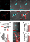
(a–b2) Endogenously produced DAGLα in non-transfected COS-7 (“parent”) cells is undetectable by indirect immunohistochemistry (open arrowheads; a–a2). In contrast, DAGLα, when highly expressed in COS-7 cells transfected with a DAGLα-V5 vector, accumulates in the plasma membrane (solid arrowheads; b–b2). (c) COS-7-DAGLα cells expressed DAGLα protein and (c1) synthesized 2-AG in a tetrahydrolipstatin (THL)-sensitive fashion. (d–d3) DAGLα overexpression did not influence COS-7 cell morphology (d,d1), including their surface area (d2) and density in culture (d3). Data were normalized to mean values from COS-7 “parent” cells. Abbreviations: n.s. = non significant. Scale bars = 20 μm (b,d1).
Figure 4
Figure 4. Extracellular 2-AG promotes spherical exclusion of cholinergic growth cones (GCs).
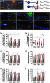
(a) Cholinergic neurons from C56Bl6 mice co-cultured with COS-7-DAGLα cells allowed measurement of GC exclusion triggered by extracellular 2-AG. (a1) Schema of the in vitro method and the parameters determined and plotted in (b2–f). (b,b1) Representative images of cholinergic GCs apposing control (“parent”) or DAGLα-overexpressing COS-7 cells (COS-7-DAGLα). Arrows point to filopodia. Dotted lines indicate the membrane surface of COS-7(-DAGLα) cells with arrowheads pinpointing their proximal segment facing the GCs. (b2) Percentage of GCs that were unable to make contact upon DAGLα overexpression. (b3) The distance (in μm) of cholinergic GCs from COS-7(-DAGLα) cells. GCs gradually approached COS-7 cells in control. DAGLα overexpression occluded this response, suggesting GC repulsion by extracellular 2-AG. DAGLα overexpression did not affect the angle at which the GC faced the proximal surface of the COS-7 cell (c), neurite outgrowth per se (d) or the distance of cholinergic somata from COS-7 cells (e). Upon making contact, cholinergic neurites were no longer affected by DAGLα overexpression (f). Data were averaged from 3 (1,2DIV) or 4 (3DIV) separate experiments; n = 25–77 cells/group. Abbreviations: DIV, days in vitro; N, neuron; n, nucleus. Data were expressed as means ± s.e.m. except for (b3) showing a combination of individual data points and mean values (horizontal lines); **p < 0.01, *p < 0.05, +p < 0.1. Scale bars = 10 μm (a), 3 μm (b).
Figure 5
Figure 5. Pharmacological inhibition of DAGLα or CB1Rs prevents chemorepulsion of cholinergic growth cones (GC).
(a) Representative photomicrographs of cholinergic neurite-COS-7(-DAGLα) cell contacts upon inhibiting DAGLs (THL, O-3841) or CB1R antagonism (AM 251, O-2050) at 3DIV. Arrows point to cholinergic neurites. Dotted lines indicate the membrane surface of COS-7(-DAGLα) cells. (b,c) Disrupting excess 2-AG signaling prevented chemorepulsion of cholinergic GCs. None of the ligands affected the angle of approach (d), neurite outgrowth (e), or cholinergic-to-COS-7 cell distance (f). Data were expressed as means ± s.e.m. except for (c) where individual data points and mean values (horizontal lines) were plotted. Data were averaged from duplicate samples from 2 independent experiments; n = 12–30 cells/group. **p < 0.01, *p < 0.05, +p < 0.1. Scale bar = 20 μm (a).

Similar articles

Cited by

References

    1. Kano M., Ohno-Shosaku T., Hashimotodani Y., Uchigashima M. & Watanabe M. Endocannabinoid-mediated control of synaptic transmission. Physiol Rev. 89, 309–380 (2009). - PubMed
    1. Gao Y. et al. Loss of retrograde endocannabinoid signaling and reduced adult neurogenesis in diacylglycerol lipase knock-out mice. J. Neurosci. 30, 2017–2024 (2010). - PMC - PubMed
    1. Peterfi Z. et al. Endocannabinoid-mediated long-term depression of afferent excitatory synapses in hippocampal pyramidal cells and GABAergic interneurons. J. Neurosci. 32, 14448–14463 (2012). - PMC - PubMed
    1. Kreitzer A. C. & Regehr W. G. Retrograde signaling by endocannabinoids. Curr. Opin. Neurobiol. 12, 324–330 (2002). - PubMed
    1. Grueter B. A., Brasnjo G. & Malenka R. C. Postsynaptic TRPV1 triggers cell type-specific long-term depression in the nucleus accumbens. Nat. Neurosci. 13, 1519–1525 (2010). - PMC - PubMed

Publication types

LinkOut - more resources