Review
. 2014 Apr;71(7):1149-70.
doi: 10.1007/s00018-013-1388-z. Epub 2013 Jun 27.

α-Amylase: an enzyme specificity found in various families of glycoside hydrolases

Affiliations
Review

α-Amylase: an enzyme specificity found in various families of glycoside hydrolases

Štefan Janeček et al. Cell Mol Life Sci. 2014 Apr.

Abstract

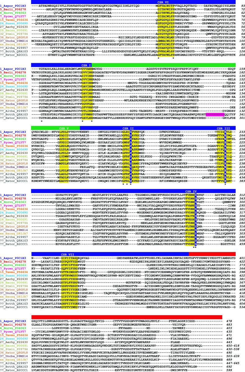
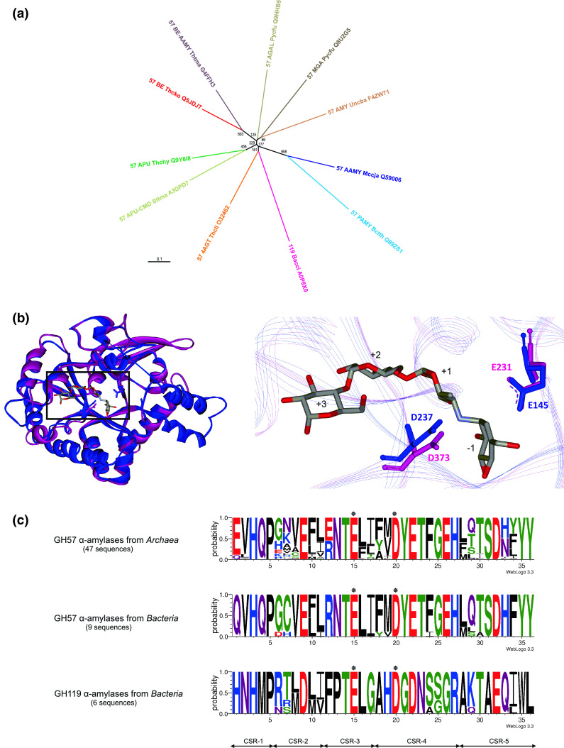
α-Amylase (EC 3.2.1.1) represents the best known amylolytic enzyme. It catalyzes the hydrolysis of α-1,4-glucosidic bonds in starch and related α-glucans. In general, the α-amylase is an enzyme with a broad substrate preference and product specificity. In the sequence-based classification system of all carbohydrate-active enzymes, it is one of the most frequently occurring glycoside hydrolases (GH). α-Amylase is the main representative of family GH13, but it is probably also present in the families GH57 and GH119, and possibly even in GH126. Family GH13, known generally as the main α-amylase family, forms clan GH-H together with families GH70 and GH77 that, however, contain no α-amylase. Within the family GH13, the α-amylase specificity is currently present in several subfamilies, such as GH13_1, 5, 6, 7, 15, 24, 27, 28, 36, 37, and, possibly in a few more that are not yet defined. The α-amylases classified in family GH13 employ a reaction mechanism giving retention of configuration, share 4-7 conserved sequence regions (CSRs) and catalytic machinery, and adopt the (β/α)8-barrel catalytic domain. Although the family GH57 α-amylases also employ the retaining reaction mechanism, they possess their own five CSRs and catalytic machinery, and adopt a (β/α)7-barrel fold. These family GH57 attributes are likely to be characteristic of α-amylases from the family GH119, too. With regard to family GH126, confirmation of the unambiguous presence of the α-amylase specificity may need more biochemical investigation because of an obvious, but unexpected, homology with inverting β-glucan-active hydrolases.


Figures

Fig. 1
Fig. 1
Tertiary structures of representative family GH13 α-amylases. The structures from following subfamilies and origins are shown: a GH13_1, Aspergillus oryzae (PDB code: 2TAA; [31]); b GH13_5, Bacillus licheniformis (PDB code: 1BLI; [249]); c GH13_6, Hordeum vulgare—barley isozyme AMY-1 (PDB code: 1P6W; [129]); d GH13_37, uncultured bacterium AmyP (modeled structure; residues Leu6-Thr491) obtained from the Phyre server [245] based on the Flavobacterium sp. 92 GH13 cyclomaltodextrinase (PDB code: 3EDE; [205]) as template; e unclassified, A. haloplanktis (PDB code: 1G94; [12]); f unclassified, Halothermothrix orenii AmyB (PDB code: 3BC9; [70]); g unclassified, Bacteroides thetaiotaomicron (PDB code: 3K8L; [38]). The individual domains are colored as follows: catalytic (β/α)8-barrel blue, domain B green, domain C red, N-terminal domain cyan; starch-binding domain of CBM58 family magenta. The GH13 catalytic triad, i.e., catalytic nucleophile—Asp (top), proton donor—Glu (left) and transition-state stabilizer—Asp (right)—is highlighted in the (β/α)8-barrel domain in each structure in a similar position. Saccharide molecules are colored yellow to emphasize: c an additional surface binding site as “a pair of sugar tongs” with the side-chain of tyrosine (in black) involved in binding; e a heptasaccharide occupying the subsites from −4 to +3 as a transglycosylation product from acarbose (a pseudotetrasaccharide); and g a maltopentaose bound by the SBD of the family CBM58 inserted within the domain B. The structures were retrieved from the Protein Data Bank (PDB; [250]) and visualized with the program WebLabViewerLite (Molecular Simulations, Inc.)
Fig. 2
Fig. 2
Amino acid sequence alignment of GH13 α-amylases representing the individual α-amylase subfamilies. The aligned sequences span the typical GH13 α-amylase’s domain arrangement consisting of catalytic (β/α)8-barrel with domain B (inserted between the strand β3 and helix α3) and domain C (succeeding the TIM-barrel). The name of an enzyme is composed of the GH13 subfamily number (“xx” for the recently described α-amylase from Bacillus aquimaris and “??” for α-amylases from A. haloplanktis, Bacteroides thetaiotaomicron, and Halothermothrix orenii AmyB that currently are still not assigned any GH13 subfamily in CAZy) followed by the abbreviation of the source (organism) and the UniProt accession number. Note there are two α-amylases from Halothermothrix orenii, the AmyA assigned to subfamily GH13_36 (UniProt Q8GPL8) and the AmyB not assigned as yet to any GH13 subfamily (UniProt B2CCC1). The organisms are abbreviated as follows: Aspor, Aspergillus oryzae; Bacli, Bacillus licheniformis; Horvu, Hordeum vulgare; Pycwo, Pyrococcus woesei; Tenmo, Tenebrio molitor; Sussc, Sus scrofa (pancreas); Aerhy, Aeromonas hydrophila; Bacsu, Bacillus subtilis; Stmli, Streptomyces limosus; Hator, Halothermothrix orenii; Uncba, uncultured bacterium; Bacaq, Bacillus aquimaris; Altha, A. haloplanktis; and Bctth, Bacteroides thetaiotaomicron. The seven CSRs typical for the family GH13 [46] and the catalytic triad are highlighted in yellow and blue, respectively. The residues conserved invariantly are marked by an asterisk below the alignment. The individual α-amylase family GH13 domains are indicated as a colored lane above the alignment: blue catalytic domain A, green domain B, red C-terminal domain C. The positions corresponding to the deleted SBD of the family CBM58 in ??_Bctth_Q8A1G3 (SusG α-amylase) are signified by a magenta lane. All sequences were retrieved from the UniProt knowledge database [251]. The sequence alignments were done using the program Clustal-W2 [252] and then manually tuned in order to maximize sequence similarities
Fig. 3
Fig. 3
Evolutionary trees of GH13 α-amylases representing the individual α-amylase subfamilies. The trees are based on the alignment of: a catalytic (β/α)8-barrel including domain B with succeeding domain C (653 positions; aligned in Fig. 2); and b seven GH13 characteristic CSRs (52 residues; highlighted in Fig. 2). The names of the α-amylases are explained in the legend to Fig. 2. The evolutionary trees were calculated as a Phylip-tree type using the neighbor-joining clustering [253] and the bootstrapping procedure [254] (the number of bootstrap trials used was 1,000) implemented in the ClustalX package [252], and then displayed with the program TreeView [255]
Fig. 4
Fig. 4
a Evolutionary tree showing the α-amylase representative of the family GH57 among the other GH57 enzyme specificities together with the α-amylase representative of the family GH119. The tree is based on the alignment of the five GH57 characteristic CSRs (36 residues). The name of an enzyme is composed of the GH57 or GH119 family number followed by the abbreviation of specificity (in capitals), abbreviation of source (organism) and the UniProt accession number. All sequences were retrieved from the UniProt knowledge database [251]. The specificities are abbreviated as follows: AAMY α-amylase, PAMY putative α-amylase-like protein, MGA maltogenic amylase, AMY unspecified amylase, APU amylopullulanase, APU-CMD amylopullulanase-cyclomaltodextrinase, BE branching enzyme, BE-AAMY α-amylase-branching enzyme, 4AGT 4-α-glucanotransferase, AGAL α-galactosidase. The organisms are abbreviated as follows: Bacci, Bacillus circulans; Bccth, Bacteroides thetaiotaomicron; Mccja, Methanocaldococcus jannaschii; Pycfu, Pyrococcus furiosus; Sttma, Staphylothermus marinus; Thchy, Thermococcus hydrothermalis; Thcko, Thermococcus kodakaraensis; Thcli, Thermococcus litoralis; Thtma, Thermotoga maritima; Uncba, uncultured bacterium. The evolutionary tree was calculated as a Phylip-tree type using the neighbor-joining clustering [253] and the bootstrapping procedure [254] (the number of bootstrap trials used was 1,000) implemented in the ClustalX package [252], and then displayed with the program TreeView [255]. b Structural models of α-amylases from families GH57 and GH119. Left superimposed modeled structures of the GH57 Methanocaldococcus jannaschii α-amylase (blue) and GH119 Bacillus circulans α-amylase (magenta). The α-helical bundle succeeding the catalytic (β/α)7-barrel was modeled only in the GH57 α-amylase. The rectangle indicates a detailed view on the right. Right a close-up focused on predicted catalytic residues of the α-amylases from GH57 (Glu145 and Asp237) and GH119 (Glu231 and Asp373). Both models were superimposed with the real structure of GH57 Thermococcus litoralis 4-α-glucanotransferase (PDB code: 1K1Y; [212]; not shown). The structural models of α-amylases were obtained from the Phyre server [245] based on the GH57 4-α-glucanotransferase template as follows: residues Met1-Tyr356 of the GH57 α-amylase [216] and Thr121-Asp429 of the GH119 α-amylase [242]. The superimposed part covers 218 Cα-atoms with a 0.75 Å root-mean square deviation; the superimposition was done using the MultiProt server [256]. Acarbose-occupying subsites −1 through +3 [257] from the complex with GH57 4-α-glucanotransferase structure [212] is shown. The structures were visualized with the program WebLabViewerLite (Molecular Simulations, Inc.). c Sequence logos of α-amylases from families GH57 from Archaea and Bacteria and GH119. CSR-1, residues 1–5; CSR-2, residues 6–11; CSR-3, residues 12–17; CSR-4, residues 18–27; CSR-5, residues 28–36. Asterisks signify the catalytic nucleophile (glutamic acid) in position 15 (in CSR-3) and proton donor (aspartic acid) in position 20 (in CSR-4). The logos are based on identifying the CSRs in both families [217, 242] as follows: for 59 α-amylase sequences (47 from Archaea and nine from Bacteria) from family GH57 and for six bacterial GH119 α-amylases. Sequence logos were created using the WebLogo 3.0 server [258]
Fig. 5
Fig. 5
Structure of the “α-amylase” of family GH126. a GH126 structure of the α-amylase from Clostridium perfringens (PDB code: 3REN; [17]) showing the (α/α)6-barrel with highlighted residues (colored by element) involved in catalysis: Glu84 (general acid), Asp136 (general base), and Tyr194 (contributing to catalysis). b Superimposition of Clostridium perfringens GH126 α-amylase (blue) with Clostridium thermocellum GH8 endoglucanase CelA (red PDB code: 1KWF; [247]). The superimposed part covers 193 Cα-atoms with a 1.93 Å root-mean square deviation; the superimposition was done using the MultiProt server [256]. The detailed view focuses on the catalytic residues in the structure of GH8 endoglucanase CelA (Glu95 and Asp278 with Tyr215) and the proposed residues in the GH126 α-amylase (Glu84 and Asp136 with Tyr194). Cellopentaose occupying subsites −3 through +2 [257] in complex with GH8 endoglucanase CelA is shown. A comparison with the family GH15 glucoamylase, i.e., an α-glucan-active enzyme with (α/α)6-barrel catalytic fold employing the inverting mechanism [259], reveals less similarity since the superimposed part between the GH126 α-amylase and Aspergillus niger GH15 glucoamylase [260] covered only 146 Cα-atoms with a 1.97 Å root-mean square deviation. The structures were retrieved from the PDB [250] and visualized with the program WebLabViewerLite (Molecular Simulations, Inc.)

References

    1. Henrissat B. A classification of glycosyl hydrolases based on amino acid sequence similarities. Biochem J. 1991;280:309–316. - PMC - PubMed
    1. Henrissat B, Bairoch A. New families in the classification of glycosyl hydrolases based on amino acid sequence similarities. Biochem J. 1993;293:781–788. - PMC - PubMed
    1. Henrissat B, Bairoch A. Updating the sequence-based classification of glycosyl hydrolases. Biochem J. 1996;316:695–696. - PMC - PubMed
    1. Cantarel BL, Coutinho PM, Rancurel C, Bernard T, Lombard V, Henrissat B. The Carbohydrate-Active EnZymes database (CAZy): an expert resource for glycogenomics. Nucleic Acids Res. 2009;37:D233–D238. - PMC - PubMed
    1. Henrissat B, Davies GJ. Structural and sequence-based classification of glycoside hydrolases. Curr Opin Struct Biol. 1997;7:637–644. - PubMed

Publication types

LinkOut - more resources