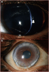
Keratoglobus
- PMID: 23807384
- PMCID: PMC3772364
- DOI: 10.1038/eye.2013.130
Keratoglobus
Abstract
Keratoglobus is a rare noninflammatory corneal thinning disorder characterised by generalised thinning and globular protrusion of the cornea. It was first described as a separate clinical entity by Verrey in 1947. Both congenital and acquired forms have been shown to occur, and may be associated with various other ocular and systemic syndromes including the connective tissue disorders. Similarities have been found with other noninflammatory thinning disorders like keratoconus that has given rise to hypotheses about the aetiopathogenesis. However, the exact genetics and pathogenesis are still unclear. Clinical presentation is characterised by progressive diminution resulting from irregular corneal topography with increased corneal fragility due to extreme thinning. Conservative and surgical management for visual rehabilitation and improved tectonic stability have been described, but remains challenging. In the absence of a definitive standard procedure for management of this disorder, various surgical procedures have been attempted in order to overcome the difficulties. This article reviews the aetiological factors, differential diagnosis, histopathology, and management options of keratoglobus.
Figures


Similar articles
-
[Keratoglobus: review of the literature].J Fr Ophtalmol. 2005 Dec;28(10):1145-9. doi: 10.1016/s0181-5512(05)81154-4. J Fr Ophtalmol. 2005. PMID: 16395211 Review. French.
-
Keratoconus and related noninflammatory corneal thinning disorders.Surv Ophthalmol. 1984 Jan-Feb;28(4):293-322. doi: 10.1016/0039-6257(84)90094-8. Surv Ophthalmol. 1984. PMID: 6230745 Review.
-
Keratoconus.Surv Ophthalmol. 1998 Jan-Feb;42(4):297-319. doi: 10.1016/s0039-6257(97)00119-7. Surv Ophthalmol. 1998. PMID: 9493273 Review.
-
Intrastromal corneal ring implants for corneal thinning disorders: an evidence-based analysis.Ont Health Technol Assess Ser. 2009;9(1):1-90. Epub 2009 Apr 1. Ont Health Technol Assess Ser. 2009. PMID: 23074513 Free PMC article.
-
Megalocornea, anterior megalophthalmos, keratoglobus and associated anterior segment disorders: A review.Clin Exp Ophthalmol. 2021 Jul;49(5):477-497. doi: 10.1111/ceo.13958. Epub 2021 Jun 27. Clin Exp Ophthalmol. 2021. PMID: 34114333 Review.
Cited by
-
Spontaneous Corneal Clearing after Descemet Membrane Rupture and Near-Total Detachment in Keratoglobus: A Case Report.Case Rep Ophthalmol. 2024 Mar 8;15(1):189-195. doi: 10.1159/000537742. eCollection 2024 Jan-Dec. Case Rep Ophthalmol. 2024. PMID: 38464399 Free PMC article.
-
[Brittle cornea syndrome type 1 caused by compound heterozygosity of two mutations in the ZNF469 gene].Ophthalmologe. 2019 Aug;116(8):780-784. doi: 10.1007/s00347-018-0796-8. Ophthalmologe. 2019. PMID: 30338343 German.
-
Replace or Regenerate? Diverse Approaches to Biomaterials for Treating Corneal Lesions.Biomimetics (Basel). 2024 Mar 28;9(4):202. doi: 10.3390/biomimetics9040202. Biomimetics (Basel). 2024. PMID: 38667213 Free PMC article. Review.
-
Limbal Stem Cell-Sparing Corneoscleroplasty with Peripheral Intralamellar Tuck: A New Surgical Technique for Keratoglobus.Case Rep Ophthalmol. 2017 Apr 28;8(1):279-287. doi: 10.1159/000471789. eCollection 2017 Jan-Apr. Case Rep Ophthalmol. 2017. PMID: 28559840 Free PMC article.
-
[Penetrating excimer laser keratoplasty after acute keratoglobus in osteogenesis imperfecta].Ophthalmologie. 2023 Jul;120(7):771-775. doi: 10.1007/s00347-023-01827-3. Epub 2023 Mar 1. Ophthalmologie. 2023. PMID: 36859561 German. No abstract available.
References
-
- Feder RS, Kshettry P.Noninflammatory ectatic disordersIn: Krachmer JH, Mannis MJ, Holland EJ (eds)Cornea: Fundamentals, diagnosis and management 3rd VolElsevier Mosby: New York; 2005955–974.
-
- Verrey F. Keratoglobe aigu. Ophthalmologica. 1947;114:284–288.
-
- Pouliquen Y, Dhermy P, Espinasse MA, Savoldelli M. Keratoglobus. J Fr Ophthalmol. 1985;8 (1:43–45. - PubMed
Publication types
MeSH terms
LinkOut - more resources
Full Text Sources
Other Literature Sources

