

Fibrosing mediastinitis: an unusual cause of pulmonary symptoms
- PMID: 23807725
- PMCID: PMC3832713
- DOI: 10.1007/s11606-013-2528-8
Fibrosing mediastinitis: an unusual cause of pulmonary symptoms
Abstract
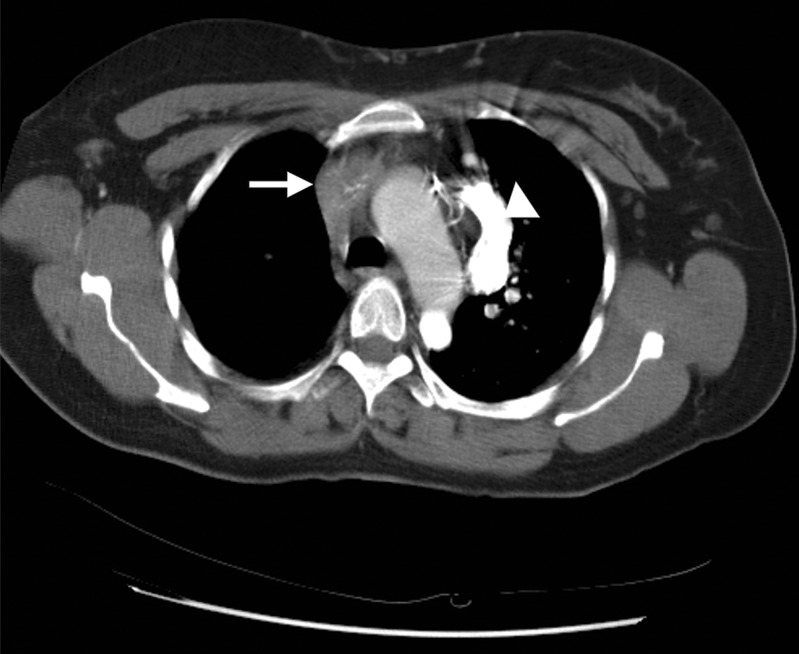
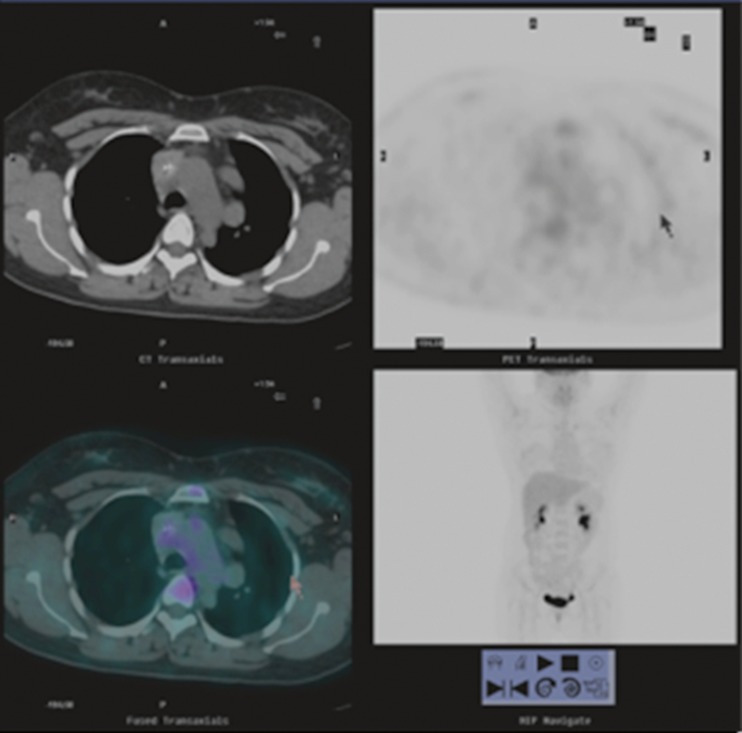
Fibrosing mediastinitis (FM), also known as granulomatous or sclerosing mediastinitis, is an uncommon but serious cause of chest symptoms. Due to an infectious or inflammatory challenge, production of collagen occurs in the confined space of the mediastinum. Collagen formation leads to compression of vital structures, resulting in cough, chest pain and dyspnea. The majority of cases of FM occur as a result of prior exposure to Histoplasma capsulatum. The following is a case of a previously healthy young woman who presented with a 3-month history of cough, chest pain and trouble breathing, and was subsequently found to have fibrosing mediastinitis. Fibrosing mediastinitis should be considered in the differential diagnosis of cough, chest pain and dyspnea, primarily when findings such as increased venous pressure are present on physical exam and hilar abnormalities are seen on chest radiograph. Clinical presentation, diagnosis and management of fibrosing mediastinitis are discussed.
Figures

Similar articles
-
Fibrosing mediastinitis mimicking sarcoidosis.Clin Respir J. 2015 Jan;9(1):125-8. doi: 10.1111/crj.12107. Epub 2014 May 15. Clin Respir J. 2015. PMID: 24405501
-
Pulmonary hypertension associated with combined fibrosing mediastinitis and bronchial anthracofibrosis: A retrospective analysis in a single Chinese hospital.Clin Respir J. 2018 Mar;12(3):1134-1140. doi: 10.1111/crj.12641. Epub 2017 May 11. Clin Respir J. 2018. PMID: 28419740
-
Ptosis and Miosis Associated with Fibrosing Mediastinitis.Am J Case Rep. 2021 Jan 12;22:e927556. doi: 10.12659/AJCR.927556. Am J Case Rep. 2021. PMID: 33431787 Free PMC article.
-
A review of endovascular stenting for superior vena cava syndrome in fibrosing mediastinitis.Vasc Med. 2020 Apr;25(2):174-183. doi: 10.1177/1358863X19884130. Epub 2019 Dec 5. Vasc Med. 2020. PMID: 31804157 Review.
-
Multimodality Imaging of Focal and Diffuse Fibrosing Mediastinitis.Radiographics. 2019 May-Jun;39(3):651-667. doi: 10.1148/rg.2019180143. Epub 2019 Apr 5. Radiographics. 2019. PMID: 30951437 Review.
Cited by
-
Diagnostic Snapshot: Acute Edema in the Oncology Patient.J Adv Pract Oncol. 2018 Sep-Oct;9(6):677-679. Epub 2018 Sep 1. J Adv Pract Oncol. 2018. PMID: 31186989 Free PMC article. Review. No abstract available.
-
Fibrosing mediastinitis with pulmonary hypertension as a complication of pulmonary vein stenosis: A case report and review of the literature.Medicine (Baltimore). 2018 Jan;97(4):e9694. doi: 10.1097/MD.0000000000009694. Medicine (Baltimore). 2018. PMID: 29369193 Free PMC article. Review.
-
Recurrent Pneumonia due to Fibrosing Mediastinitis in a Teenage Girl: A Case Report with Long-Term Follow-Up.Case Rep Pediatr. 2018 Mar 18;2018:3246929. doi: 10.1155/2018/3246929. eCollection 2018. Case Rep Pediatr. 2018. PMID: 29744231 Free PMC article.
-
Severe fibrosing mediastinitis with atypical presentation: Effective control with novel therapeutic approach.Ann Thorac Med. 2017 Jul-Sep;12(3):209-212. doi: 10.4103/atm.ATM_47_17. Ann Thorac Med. 2017. PMID: 28808494 Free PMC article.
-
Pulmonary Hypertension Complicating Fibrosing Mediastinitis.Medicine (Baltimore). 2015 Nov;94(44):e1800. doi: 10.1097/MD.0000000000001800. Medicine (Baltimore). 2015. PMID: 26554778 Free PMC article.
References
-
- NCHS. National Hospital Ambulatory Medical Care Survey. 2010 outpatient department summary tables. Available from: http://www.cdc.gov/nchs/data/ahcd/nhamcs_outpatient/2010_opd_web_tables.pdf Accessed 24 February 2013.
-
- NCHS. National Hospital Ambulatory Medical Care Survey: 2010 emergency department summary tables. Available from: http://www.cdc.gov/nchs/data/ahcd/nhamcs_emergency/2010_ed_web_tables.pdf Accessed 24 February 2013.
-
- NCHS. National Ambulatory Medical Care Survey: 2010 summary tables. Available from: http://www.cdc.gov/nchs/data/ahcd/nhamcs_summary/outpatieny/2010_opd_web... Accessed 24 February 2013.
-
- Loyd JE, Tillman BF, Atkinson JB, Desprez RM. Mediastinal fibrosis complicating histoplasmosis. Medicine. 1998;67:295–310. - PubMed
Publication types
MeSH terms
Supplementary concepts
LinkOut - more resources
Full Text Sources
Other Literature Sources
Medical
