. 2013 Dec;27(12):2366-75.
doi: 10.1038/leu.2013.194. Epub 2013 Jun 28.

Small-molecule multi-targeted kinase inhibitor RGB-286638 triggers P53-dependent and -independent anti-multiple myeloma activity through inhibition of transcriptional CDKs

Affiliations

Small-molecule multi-targeted kinase inhibitor RGB-286638 triggers P53-dependent and -independent anti-multiple myeloma activity through inhibition of transcriptional CDKs

D Cirstea et al. Leukemia. 2013 Dec.

Abstract

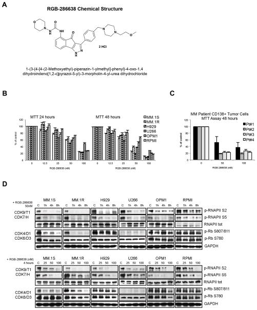
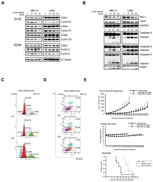
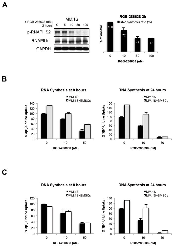
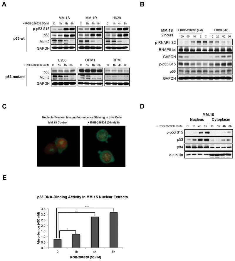
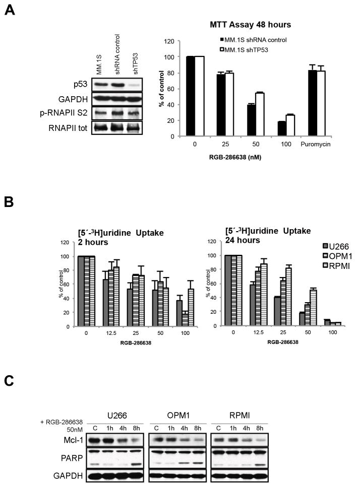
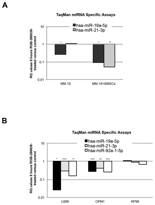
Small-molecule multi-targeted cyclin-dependent kinase (CDK) inhibitors (CDKIs) are of particular interest due to their potent antitumor activity independent of p53 gene alterations. P53 deletion is associated with a very poor prognosis in multiple myeloma (MM). In this regard, we tested the anti-MM activity of RGB-286638, an indenopyrazole-derived CDKI with Ki-nanomolar activity against transcriptional CDKs. We examined RGB-286638's mode-of-action in MM cell lines with wild-type (wt)-p53 and those expressing mutant p53. RGB-286638 treatment resulted in MM cytotoxicity in vitro associated with inhibition of MM tumor growth and prolonged survival in vivo. RGB-286638 displayed caspase-dependent apoptosis in both wt-p53 and mutant-p53 cells that was closely associated with the downregulation of RNA polymerase II phosphorylation and inhibition of transcription. RGB-286638 triggered p53 accumulation via nucleolar stress and loss of Mdm2, accompanied by induction of p53 DNA-binding activity. In addition, RGB-286638 mediated p53-independent activity, which was confirmed by cytotoxicity in p53-knockdown and p53-mutant cells. We also demonstrated downregulation of oncogenic miR-19, miR-92a-1 and miR-21. Our data provide the rationale for the development of transcriptional CDKIs as therapeutic agents, which activate p53 in competent cells, while circumventing p53 deficiency through alternative p53-independent cell death mechanisms in p53-mutant/deleted cells.


Conflict of interest statement

CONFLICT OF INTEREST

Hannes Loferer is the Agennix AG employee. Other authors have no relevant financial relationship(s) to disclose.

Figures

Figure 1
Figure 1. RGB-286638 Demonstrated Potent Activity Against Transcriptional CDKs in MM Cell Lines and Inhibited Myeloma Cell Growth in Vitro
(A) RGB-286638 chemical structure (left panel). Chemical name: 1-(3-{4-[4-(2-Methoxyethyl)-piperazin-1-ylmethyl]-phenyl}-4-oxo-1,4-dihydroindeno[1,2-c]pyrazol-5-yl)-3-morpholin-4-yl-urea dihydrochloride. Molecular formula: C29H37N7O4 2HCl. Molecular weight: 618.52. (B) Dose- and time-dependent cytotoxicity of RGB-286638 in MM cell lines. Expressing wild-type p53 (MM.1S, MM.1R, H929) or mutant-p53 (U266, OPM1, RPMI) cells were incubated with RGB-286638 (0–100nM), and viability was determined at 24 and 48 h using MTT assay. EC50 ranged between 20–70nM at 48h. (C) RGB-286638-mediated cytotoxicity in primary tumor cells from MM patients. Myeloma cells isolated from BM via CD138+ positive selection were cultured with RGB-286638 (0–100nM), and viability was assessed by MTT at 48h. RGB-286638 demonstrated comparable cytotoxicity to MM cell lines. Data represents means (+/−SD) of triplicate cultures. (D) RGB-286638 treatment results in transcriptional CDK inhibition. The indicated cell lines were exposed to RGB-286638 (0–100nM) 1, 4, and 8 h treatment. Whole lysates were subjected to western blotting and membranes probed for phosphorylated RNAPII at S2/S5 and Rb at S807/811/S780 sites using phosphospecific antibodies.
Figure 2
Figure 2. RGB-286638-Induced Cytotoxicity in Vitro Translates in Anti-MM Activity in Vivo
(A) RGB-286638 reduced the expression of cell cycle related proteins. MM.1S and U266 cells were treated with 50nM RGB-286638 for 2, 4, and 8 h, and whole cell extracts resolved by SDS-PAGE. Cell cycle-related proteins were assessed through probing membranes with the indicated antibodies. (B) Treatment with RGB-286638 reduced anti-apoptotic protein expression and triggered caspase and PARP activation. The expression of RNAPII anti-apoptotic Mcl-1 and XIAP transcripts was examined by western blot in MM.1S and U266 cells treated with 50nM RGB-286638 (0–8h). After 4, 8, and 24 h RGB-286638 (50nM) treatment, MM.1S and U266 cells were subjected to western blotting for detection of caspase 8, 9, 3 and PARP cleavage. (C) RGB-286638 affected cell cycle progression. Cell cycle progression was examined via flow cytometry in MM.1S cells stained with propidium iodide after exposure to media alone, or to 50nM RGB-286638 for 12 and 24 h. RGB-286638 treatment affected G1/S and G2/M cell cycle progression. (D) RGB-286638 induced early time point apoptosis. Annexin/PI staining and FACS analysis for apoptosis was performed on MM.1S cells cultured with or without 50nM RGB-286638 for 12 and 24 h. Viable cells are represented in the lower left quadrants. Increasing percentages of apoptotic cells after prolonged exposure to RGB-286638 treatment are shown in the lower and upper right quadrants. (E) RGB-286638 anti-MM activity in vivo: tumor growth, host weight, and Kaplan–Meier survival curves in xenografted with MM.1S cells mice. SCID mice were inoculated subcutaneously with 3 × 10⁶ MM.1S cells in 100 uL RPMI medium. Tumor-bearing mice were randomly assigned into 3 cohorts receiving daily IV tail vein injections for 5 consecutive days with 30 mg/kg or 40 mg/kg RGB-286638, or with control vehicle alone. RGB-286638 treatment resulted in tumor growth inhibition. Transient weight loss, with maximum body weight loss on day 5 (8.4%) after 30 mg/kg and on day 15 (9.9%) after 40mg/kg dosing, was followed by recovery in the following two weeks. Prolonged survival was observed in both 30 mg/kg and 40 mg/kg dosing groups.
Figure 3
Figure 3. RGB-286638 Inhibited Transcription
(A) RGB-286638 inhibited RNAPII-mediated transcription. MM.1S were incubated with RGB-286638 (0–100 nM) for 2 hours. Whole cell lysates were subjected to western blotting and analyzed using specific antibodies for p-RNAPII S2, RNAPII, and GAPDH. RNA synthesis was evaluated in MM.1S cells pre-incubated for one hour with the indicated concentrations of RGB-286638. [5′-3H]uridine was added, and after one hour incubation cells were assayed for radioactivity. Data represent means (+/−SD) of triplicate cultures. (B) RGB-286638 reduced BMSC-induced RNA synthesis in MM.1S cells. Cells were incubated with increasing concentrations of RGB-286638 for 8 and 24 h, alone or in co-culture with BMSCs; [5′-3H]uridine was added and RNA synthesis measured. Data represent means (+/−SD) of triplicate cultures. (C) RGB-286638 abrogated BMSC-induced DNA synthesis in MM.1S cells. MM.1S cells, in the presence or absence of BMSCs, were treated with increasing concentrations of RGB-286638 for 8 and 24 h; cell proliferation was then examined by [3H-TdR] uptake. Data represents means (+/−SD) of triplicate cultures.
Figure 4
Figure 4. RGB-286638 Triggered Stabilization and Activation of p53
(A) RGB-286638 induced p53 accumulation and reduced Mdm2 expression in wt-p53 MM cells. P53 levels remained unchanged in mutant-p53 MM cells, regardless of reduced Mdm2. Wt-p53 MM.1S, MM.1R, and H929, and mutant-p53 U266, OPM1, and RPMI cells were exposed to 50nM RGB-286638 for 0–8 h, and whole cell extracts then subjected to western blotting. Expression of p-p53 S15, p53, Mdm2, and GAPDH was determined using specific antibodies. (B) RGB-286638 triggered p53 accumulation prior to reducing Mdm2. p53 and Mdm2 expression were correlated with RNAPII inhibition in MM.1S cells cultured for 2h with control media, or with increasing concentrations of RGB-286638 (0–100nM) or DRB (0–60uM). Whole cell extracts were examined via western blotting for expression of p-RNAPII S2, RNAPII, p-p53 S15, p53, and GAPDH. (C) RGB-286638 induced nucleolar fragmentation. MM.1S cells incubated with RGB-286638 (50nM) or media alone for 3 hours were pelleted and re-suspended in 100 μL Dual Detection Reagent for 30 min. Viable cells were analyzed by immunofluorescence microscopy (60X magnification). (D) RGB-286638 triggered nuclear accumulation of p53. Nuclear and cytoplasmic extracts separated from MM.1S cells exposed to 50nM RGB-286638 for 0–8 h were analyzed by western blotting for expression of p-p53 S15 and p53 using specific antibodies. Expression of p84, or α-tubulin was used for loading control of nuclear or cytoplasmic proteins, respectively. (E) RGB-286638 increased p53 sequence specific DNA binding activity. P53/DNA binding activity was detected in nuclear extracts purified from MM.1S cells cultured with media, or RGB-286638 (50nM) for 1, 4, and 8 h. Data represents means (+/−SD) of triplicate cultures.
Figure 5
Figure 5. RGB-286638 Mediated p53-Independent Apoptosis in MM Cells
(A) RGB-286638 induced cytotoxicity in p53 knockdown MM.1S. P53 shRNA- or empty vector-transduced MM.1S were cultured with RGB-286638 (0–100nM) or 1.5ug/ml puromycin for 48h, and viability was assayed using MTT. Data represent means (+/−SD) of triplicate cultures. P53 and p-RNAPII S2 were examined by western blotting of whole cell lysates of parental MM.1S cells and MM.1S cells transduced with p53 shRNA or with empty vector. (B) RGB-286638 reduced transcription in mutant-p53 MM cell lines. U266, OPM1, and RPMI cells were treated for 2 and 24 h with RGB-286638 (0–100nM); [5′-3H]uridine was then added, and RNA synthesis measured. Data represent means (+/−SD) of triplicate cultures. (D) RGB-286638 triggered apoptosis in MM cell lines expressing mutant-p53. U266, OPM1, and RPMI cells incubated with or without 50nM RGB-286638 for 1, 4 and 8 h were examined by western blotting with anti-Mcl-1, -PARP, and - GAPDH antibodies.
Figure 6
Figure 6. RGB-286638 Effects on miRNA Expression
(A) RGB-286638 modulated miRNA expression in MM.1S cells alone and in culture with BMSCs. TaqMan® miR-21-3p and miR-19a-5p miRNA Specific Assays using RT-PCR were performed on aliquots of total RNA extracts from MM.1S cells cultured for 8h in control media or 50nM RGB-286638, with or without BMSCs. Data are presented as relative quantity (RQ) values in RGB-286638 treated versus untreated cells. The experiments were carried out in triplicates, (*) if p<0.05 and two (**) if p<0.01. (B) RGB-286638 suppressed the expression of the oncogenic miR-19a-5p, miR-21-3p, and miR-92a-1-5p in mutant-p53 MM cell lines. U266, OPM1, and RPMI cells were incubated in media alone or in 50nM RGB-286638 for 8h. Total RNA extracts were subjected to TaqMan Specific miRNA Assays for miR-21-3p, -19a-5p, and -92a-1-5p detection. Data is presented as RQ values of RGB-286638-treated versus -untreated cells. Experiments were carried out in triplicates, (*) if p<0.05 and two (**) if p<0.01.

Similar articles

Cited by

References

    1. Malumbres M, Harlow E, Hunt T, Hunter T, Lahti JM, Manning G, et al. Cyclin-dependent kinases: a family portrait. Nat Cell Biol. 2009;11(11):1275–6. - PMC - PubMed
    1. Malumbres M, Barbacid M. Cell cycle, CDKs and cancer: a changing paradigm. Nature reviews Cancer. 2009;9(3):153–66. - PubMed
    1. Pinhero R, Liaw P, Bertens K, Yankulov K. Three cyclin-dependent kinases preferentially phosphorylate different parts of the C-terminal domain of the large subunit of RNA polymerase II. European journal of biochemistry/FEBS. 2004;271(5):1004–14. - PubMed
    1. Romano G, Giordano A. Role of the cyclin-dependent kinase 9-related pathway in mammalian gene expression and human diseases. Cell cycle (Georgetown, Tex. 2008;7(23):3664–8. - PubMed
    1. Bergsagel PL, Kuehl WM. Molecular pathogenesis and a consequent classification of multiple myeloma. J Clin Oncol. 2005;23(26):6333–8. - PubMed

Publication types