

The role of MRI in diagnostic algorithm of cervicofacial vascular anomalies in children
- PMID: 23807878
- PMCID: PMC3693840
- DOI: 10.12659/PJR.883941
The role of MRI in diagnostic algorithm of cervicofacial vascular anomalies in children
Abstract
Background: Vascular anomalies are usually diagnosed through their clinical picture and history. The purpose of this study was to assess the role of MR imaging in initial assessment of cervicofacial vascular anomalies in children.
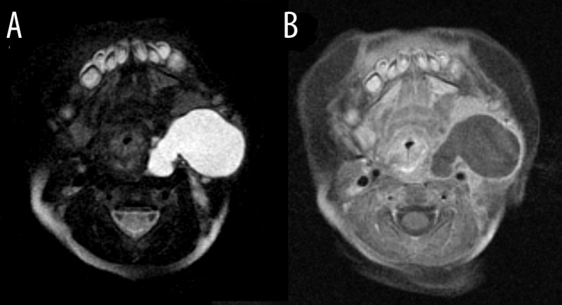
Material/methods: Twenty pediatric patients with vascular anomalies located in the cervicofacial region underwent MRI examination in our department. Images were evaluated for lesion detectability and its signal characteristics (on T1w, T2w images with fat suppression and contrast enhanced T1w sequences); the extent of the lesions and surrounding tissue involvement were also assessed.
Results: In the studied group MR images revealed all anomalies and provided information of their anatomic extent and invasion of surrounding anatomic structures. Nine hemangiomas and six venous malformations were found among studied patients. Two children had multiloculated lesions corresponding to lymphatic malformations. One examination visualized a lesion consisting mainly of dilated vascular channels with an apparent feeding artery, which was consistent with arteriovenous malformation. Two remaining lesions were mixed malformations. Nine patients had lesions limited to subcutaneous tissue. Two masses infiltrated bone structures. There was muscle involvement found in nine cases.
Conclusions: MR imaging is a well-established method for detection and monitoring of vascular anomalies in children. With ultrasound used mostly for initial diagnosis and additional flow assessment, angiography viewed as an invasive therapeutic method and computed tomography used only in specific situations due to its high irradiation dose, magnetic resonance is the best imaging method used in differential diagnosis and topographical characterization of vascular malformations and tumors of cervicofacial area in pediatric patients. Noninvasively and without irradiation, it enables evaluation of the extent and characteristics of lesions and planning proper therapeutic strategy.
Keywords: MRI; hemangioma; pediatrics; vascular malformations.
Figures


References
-
- Gelbert F, Riche MC, Reizine D, et al. MR imaging of head and neck vascular malformations. J Magn Reson Imaging. 1991;1(5):579–84. - PubMed
-
- Mulliken JB, Glowacki J. Hemangiomas and vascular malformations in infants and children: a classification based on endothelial characteristics. Plast Reconstr Surg. 1982;69(3):412–22. - PubMed
-
- Ernemann U, Kramer U, Miller S, et al. Current concepts in the classification, diagnosis and treatment of vascular anomalies. Eur J Radiol. 2010;75(1):2–11. - PubMed
-
- Donnelly LF, Adams DM, Bisset GS., III Vascular malformations and hemangiomas: a practical approach in a multidisciplinary clinic. AJR Am J Roentgenol. 2000;174(3):597–608. - PubMed
-
- Lee BB, Laredo J, Kim YW, et al. Congenital vascular malformations: general treatment principles. Phlebology. 2007;22(6):258–63. - PubMed
LinkOut - more resources
Full Text Sources
Other Literature Sources