

Clinical and radiological pictures of two newborn babies with manifestations of chondrodysplasia punctata and review of available literature
- PMID: 23807887
- PMCID: PMC3693839
- DOI: 10.12659/PJR.883947
Clinical and radiological pictures of two newborn babies with manifestations of chondrodysplasia punctata and review of available literature
Abstract
Background: Chondrodysplasia punctata (CDP) is a rare, heterogeneous congenital skeletal dysplasia, characterized by punctate or dot-like calcium deposits in cartilage observed on neonatal radiograms. A number of inborn metabolic diseases are associated with CDP, including peroxisomal and cholesterol biosynthesis dysfunction and other inborn errors of metabolism such as: mucolipidosis type II, mucopolysacharidosis type III, GM1 gangliosidosis. CDP is also related to disruption of vitamin K-dependent metabolism, causing secondary effects on the embryo, as well as fetal alcohol syndrome (FAS), chromosomal abnormalities that include trisomies 18 and 21, Turner syndrome.
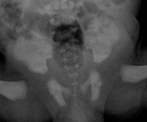
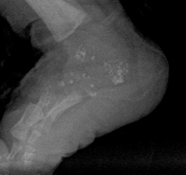
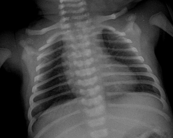
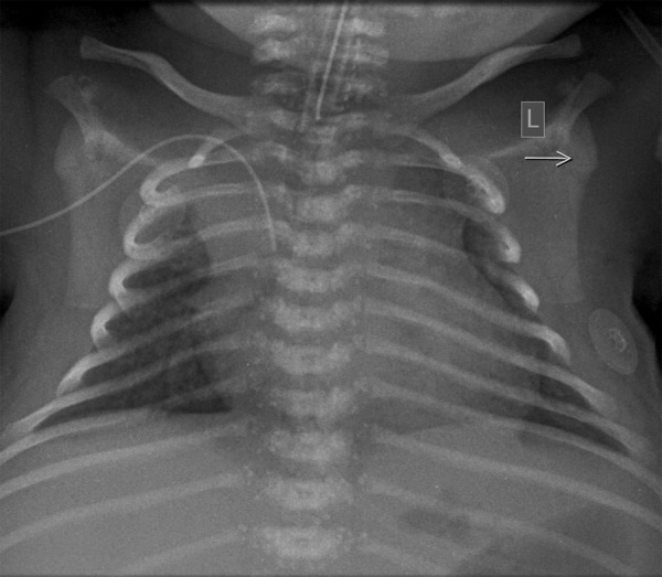
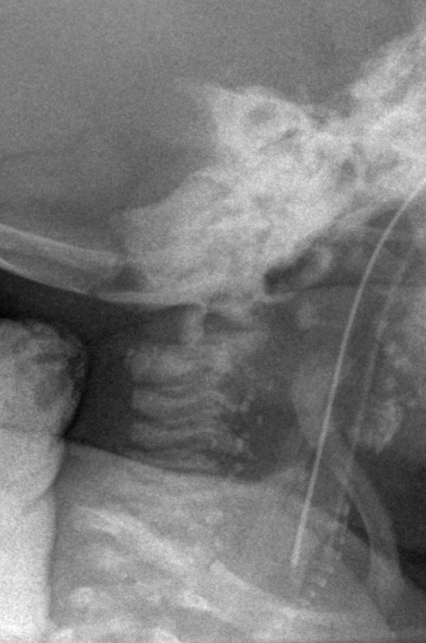
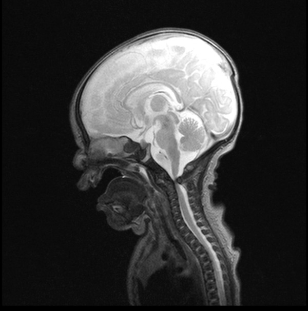
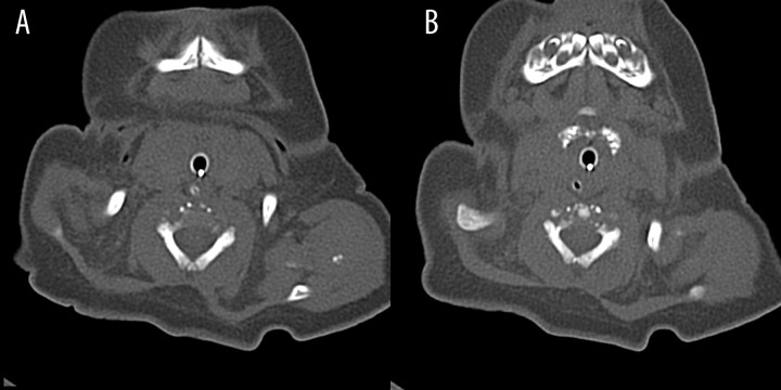
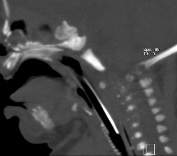
Case report: This article presents clinical data and diagnostic imaging findings of two newborn babies with chondrodysplasia punctata. Children presented with skeletal and cartilage anomalies, dysmorphic facial feature, muscles tone abnormalities, skin changes and breathing difficulties. One of the patients demonstrated critical stenosis of spinal canal with anterior subluxation of C1 vertebra relative to C2. The aim of this article is to present cases and briefly describe current knowledge on etiopathogenesis as well as radiological and clinical symptoms of diseases coexisting with CDP.
Conclusions: Radiological diagnostic imaging allows for visualization of punctate focal mineralization in bone epiphyses during neonatal age and infancy. Determining the etiology of chondrodysplasia punctata requires performing various basic as well as additional examinations, including genetic studies.
Keywords: chondrodysplasia punctata; congenital malformation; newborn; skeletal dysplasia.
Figures
Similar articles
-
A case of rhizomelic chondrodysplasia punctata in newborn.Case Rep Med. 2014;2014:879679. doi: 10.1155/2014/879679. Epub 2014 Mar 9. Case Rep Med. 2014. PMID: 24715923 Free PMC article.
-
Abnormal sterol metabolism in patients with Conradi-Hünermann-Happle syndrome and sporadic lethal chondrodysplasia punctata.Am J Med Genet. 1999 Mar 19;83(3):213-9. doi: 10.1002/(sici)1096-8628(19990319)83:3<213::aid-ajmg15>3.0.co;2-c. Am J Med Genet. 1999. PMID: 10096601
-
Chondrodysplasia punctata: a clinical diagnostic and radiological review.Clin Dysmorphol. 2008 Oct;17(4):229-41. doi: 10.1097/MCD.0b013e3282fdcc70. Clin Dysmorphol. 2008. PMID: 18978650 Review.
-
Management of Tracheobronchial Stenosis in Chondrodysplasia Punctata.Laryngoscope. 2024 Mar;134(3):1464-1468. doi: 10.1002/lary.30920. Epub 2023 Jul 31. Laryngoscope. 2024. PMID: 37522476
-
Fetal chondrodysplasia punctata associated with maternal autoimmune diseases: a review.Appl Clin Genet. 2018 Apr 20;11:31-44. doi: 10.2147/TACG.S150982. eCollection 2018. Appl Clin Genet. 2018. PMID: 29720879 Free PMC article. Review.
Cited by
-
X-linked dominant chondrodysplasia punctata due to novel mutation in EBP gene: A case report.Medicine (Baltimore). 2025 Apr 4;104(14):e42086. doi: 10.1097/MD.0000000000042086. Medicine (Baltimore). 2025. PMID: 40193659 Free PMC article.
-
Chondrodysplasia Punctata with Severe Airway Stenosis.Indian J Crit Care Med. 2018 Jul;22(7):552-554. doi: 10.4103/ijccm.IJCCM_105_18. Indian J Crit Care Med. 2018. PMID: 30111935 Free PMC article.
-
Rare Case of Rhizomelic Chondrodysplasia Punctata.J Orthop Case Rep. 2015 Jul-Sep;5(3):38-40. doi: 10.13107/jocr.2250-0685.303. J Orthop Case Rep. 2015. PMID: 27299065 Free PMC article.
-
Zebrafish models of skeletal dysplasia induced by cholesterol biosynthesis deficiency.Dis Model Mech. 2020 Jun 24;13(6):dmm042549. doi: 10.1242/dmm.042549. Dis Model Mech. 2020. PMID: 32430393 Free PMC article.
-
A case of rhizomelic chondrodysplasia punctata in newborn.Case Rep Med. 2014;2014:879679. doi: 10.1155/2014/879679. Epub 2014 Mar 9. Case Rep Med. 2014. PMID: 24715923 Free PMC article.
References
-
- Chitayat D, Keating S, Zand DJ, et al. Chondrodysplasia Punctata Associated With Maternal Autoimmune Disease: Expanding the Spectrum From Systemic Lupus Erythematosus (SLE) to Mixed Connective Tissue Disease (MCTD) and Scleroderma Report of Eight Cases. Am J Med Genet. 2008;Part A 146A:3038–53. - PubMed
-
- Irving MD, Chitty LS, Mansour S, et al. Chondrodysplasia punctata: a clinical diagnostic and radiological review. Clin Dysmorph. 2008;17:229–41. - PubMed
-
- Morrisom SC. Punctate epiphyses associated with Turner syndrome. Pediatr Radiol. 1999;29:478–80. - PubMed
-
- Leicher-Duber A, Schumacher R, Spranger J. Stippled epiphyses in fetal alcohol syndrome. Pediatr Radiol. 1990;20:369–70. - PubMed
-
- Alessandri, Jean-Luc D, Ramful, et al. Binder fenotype and brachytelephalanic chondrodysplasia punctata secondary to maternal vitamin K deficiency. Clin Dysmorph. 2010:85–87. - PubMed
Publication types
LinkOut - more resources
Full Text Sources
Other Literature Sources
Research Materials
Miscellaneous