. 2012:2012:215308.
doi: 10.6064/2012/215308.

B Cell in Autoimmune Diseases

Affiliations

B Cell in Autoimmune Diseases

Christiane S Hampe. Scientifica (Cairo). 2012.

Abstract

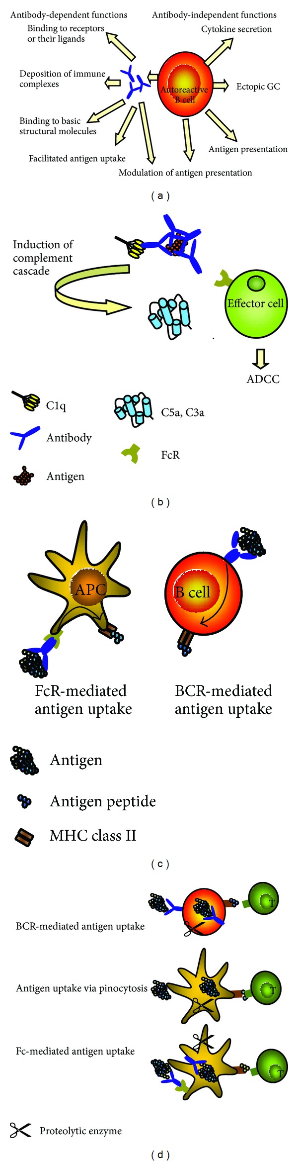
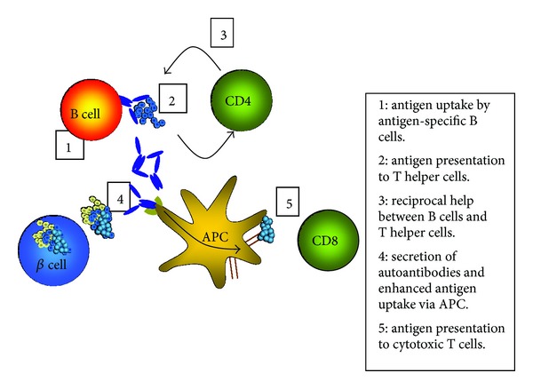
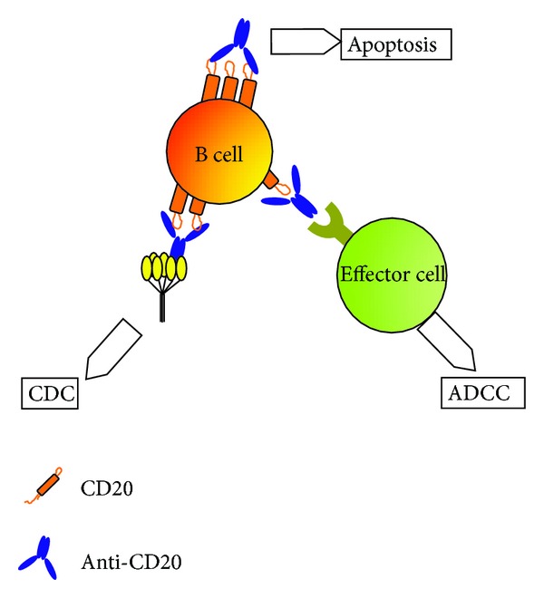
The role of B cells in autoimmune diseases involves different cellular functions, including the well-established secretion of autoantibodies, autoantigen presentation and ensuing reciprocal interactions with T cells, secretion of inflammatory cytokines, and the generation of ectopic germinal centers. Through these mechanisms B cells are involved both in autoimmune diseases that are traditionally viewed as antibody mediated and also in autoimmune diseases that are commonly classified as T cell mediated. This new understanding of the role of B cells opened up novel therapeutic options for the treatment of autoimmune diseases. This paper includes an overview of the different functions of B cells in autoimmunity; the involvement of B cells in systemic lupus erythematosus, rheumatoid arthritis, and type 1 diabetes; and current B-cell-based therapeutic treatments. We conclude with a discussion of novel therapies aimed at the selective targeting of pathogenic B cells.


Figures

Figure 1
Figure 1
(a) B cells in autoimmune diseases. B cells have antibody-dependent and antibody-independent pathogenic functions. Secreted autoantibodies specific to receptors or receptor ligands can activate or inhibit receptor functions. Deposited immune complexes can activate complement and effector cells. Autoantibodies can bind to basic structural molecules and interfere with the synthesis of structural elements and facilitate the uptake of antigen. Independent of antibody secretion B cells secrete proinflammatory cytokines, support the formation of ectopic GCs, and serve as antigen presenting cells. Both secreted autoantibodies and BCR on B cells can modulate the processing and presentation of antigen and thereby affect the nature of presented T-cell determinants. (b) Pathogenic effects of deposited immune complexes. The Fc portion of antibodies in immune complexes can be bound by C1q of the classical complement pathway, which eventually leads to the release of C5a and C3a. These anaphylatoxins promote release of proinflammatory cytokines and serve as chemoattractants for effector cells. Moreover they induce the upregulation of activating FcR on effector cells. Binding of the Fc portion of the antibodies to FcR leads to activation of effector cells and further release of proinflammatory cytokines and proteolytic enzymes, mediators of antibody-dependent cell-mediated cytotoxicity (ADCC). (c) Effect of antibodies and antigen-specific B cells on antigen uptake. Left panel: antigen bound by antibody is taken up via FcR on APCs such as dendritic cells or macrophages. After processing, antigen is presented on MHC molecules. This FcR-mediated antigen uptake is more efficient than antigen uptake by pinocytosis. Right panel: antigen binds to the BCR of antigen-specific B cells and is internalized. B cells are highly efficient APCs in situations of low antigen concentrations. (d) Effect of antibodies and antigen-specific B cells on antigen processing and presentation. BCR-mediated antigen uptake can influence antigen processing and the nature of MHC-displayed T-cell determinants. Likewise, antigen/antibody complexes are bound by the FcR of APCs and processed in a unique fashion dependent on the epitope specificity of the bound antibody. The BCR or antibody can shield certain protein determinants from the proteolytic attack in endocytic compartments (represented as scissors in this figure). Presentation of some determinants may thereby be suppressed, while others are boosted. Thereby cryptic pathogenic peptides may be presented and stimulate autoreactive T cells.
Figure 2
Figure 2
Model of pathogenic function of B cells in type 1 diabetes. Islet cell antigen released from the pancreatic beta cells is being taken up at low antigen concentrations by antigen-specific B cells, which present the antigen determinants to CD4+ T cells. T cells provide help to the B cells to eventually differentiate into antibody secreting plasma cells. Autoantibodies can now bind to the autoantigen and the resulting autoantibody/autoantigen complexes are efficiently taken up via FcR present on other APCs. This enhanced autoantigen uptake and presentation finally activates cytotoxic CD8+ T cells, which carry out the killing of the beta cells.
Figure 3
Figure 3
B-cell depletion with CD20 (Rituximab). Anti-CD20 mAb can direct the killing of B cells by antibody-dependent cytotoxicity (ADCC), complement-dependent cytotoxicity (CDC), or apoptosis. ADCC is triggered by the interaction between the Fc region of the antibody and the FcR on effector cells of the immune system. In CDC the Fc region is bound by the complement component C1q, which triggers a proteolytic cascade. Apoptosis occurs when CD20 molecules are cross-linked by anti-CD20 mAb in lipid rafts and activate signaling pathways leading to cell death.

Similar articles

Cited by

References

    1. Dandel M, Wallukat G, Potapov E, Hetzer R. Role of β1-adrenoceptor autoantibodies in the pathogenesis of dilated cardiomyopathy. Immunobiology. 2011;217(5):511–520. - PubMed
    1. Riemekasten G, Philippe A, Näther M, et al. Involvement of functional autoantibodies against vascular receptors in systemic sclerosis. Annals of the Rheumatic Diseases. 2011;70(3):530–536. - PubMed
    1. Xia Y, Kellems RE. Is preeclampsia an autoimmune disease? Clinical Immunology. 2009;133(1):1–12. - PMC - PubMed
    1. LaMarca BD, Gilbert J, Granger JP. Recent progress toward the understanding of the pathophysiology of hypertension during preeclampsia. Hypertension. 2008;51(4):982–988. - PMC - PubMed
    1. Wenzel K, Rajakumar A, Haase H, et al. Angiotensin II type 1 receptor antibodies and increased angiotensin II sensitivity in pregnant rats. Hypertension. 2011;58(1):77–84. - PMC - PubMed

LinkOut - more resources