. 2013 Apr 12;2(1):158.
doi: 10.1186/2193-1801-2-158. Print 2013 Dec.

The melanocyte photosensory system in the human skin

Affiliations

The melanocyte photosensory system in the human skin

Bhanu Iyengar. Springerplus. .

Abstract

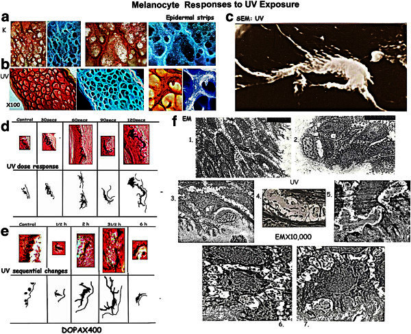
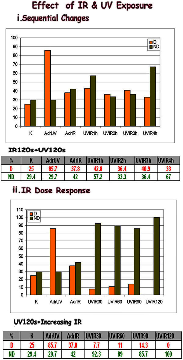
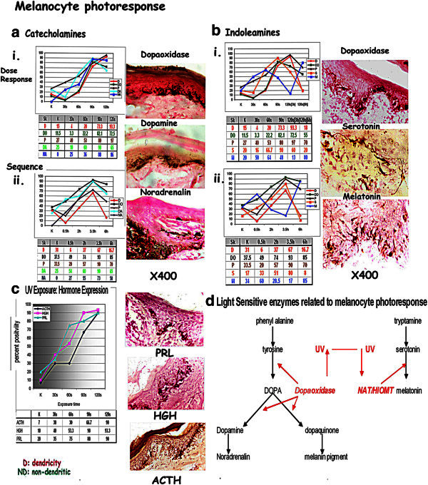
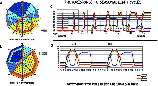
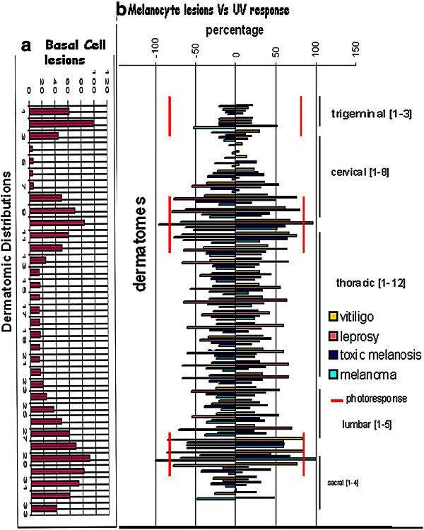
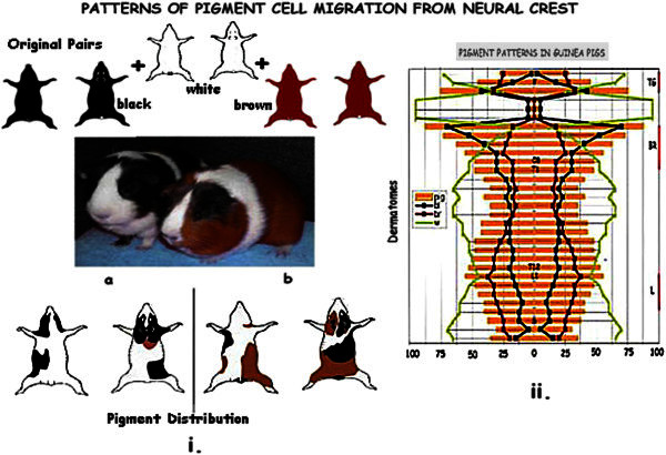
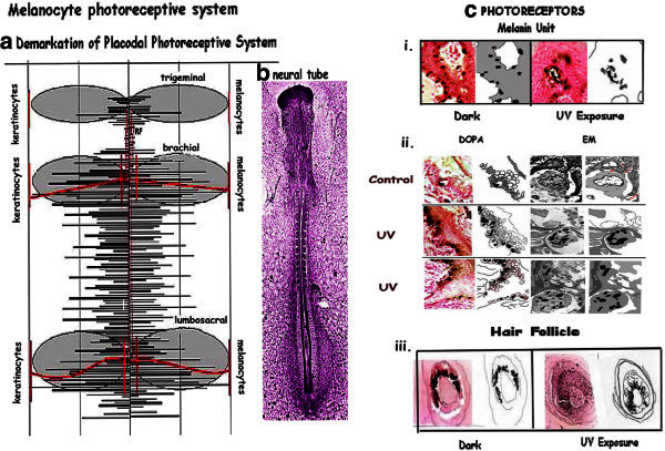
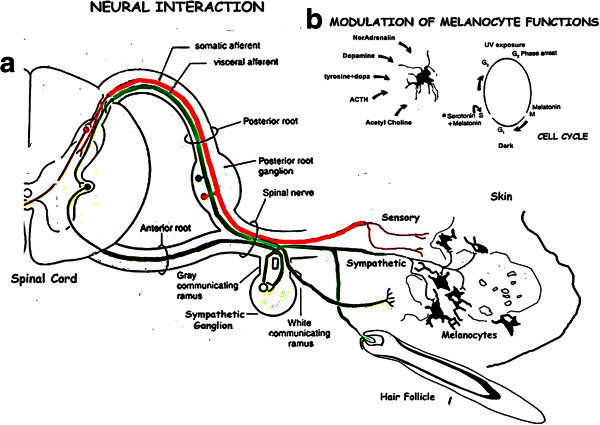
The pigment cells form the largest population of neural crest cells to migrate into the epidermis and hair follicle along each dermatomic area from the neural folds. The melanopsin system responsible for photoentrainment, was isolated from the photosensitive dermal melanophores of frogs Xenopus laevis responding to light. Melanocytes form a photoresponsive network which reads the environmental seasonal variations in the light cycles in the same manner. The present work was undertaken to study the organization of this system by: I. Experimental assessment of photoresponse and II. Evidence of an organized system of photoreception in the skin. Melanocytes, in whole skin organ cultures and epidermal strips, from margin of vitiligo in G2 phase show prominent dendricity, and express pigment, biogenic amines and hormones on UV exposure. The photoresponse depends on the photosensitive enzymes NAT/HIOMT and dopaoxidase. Melanocytes interact with adjacent keratinocytes, dermal capillaries, and nerve endings. The melanocyte network reads the diurnal and seasonal photophase by the melatonin/serotonin switch like the pineal. Sleep disorders and winter depression are corrected by phototherapy utilising this mechanism. Melanocytes showing photoactivity, aplasia, hypoplasia and hyperplasia, and interactive keratinocytes occupy the trigeminal, brachial and lumbosacral dermatomes, zones of high embryonic induction, forming an ectodermal placodal system. Melanin units and hair follicles serve as photoreceptors. Migration of active melanocytes to defined areas is evident in pigment patterns in guinea pigs. This study identifies defined photoreceptor melanocyte/epidermal domains which read the seasonal photophase and control the sleep waking cycle in response to the environmental light. I. Whole skin organ cultures, and epidermal strips from margin of vitiligo in G2 phase are exposed to UV and IR to study sequential and dose response of marginal melanocytes, using histochemistry, immunohistochemistry to assess pigment, biogenic amines and hormones on UV exposure. II. Dermatomic Distributions: Detailed maps of melanocyte photoresponse in 356 biopsies, lesions in 297 vitiligo, 100 melanosis, 165 melanomas 142 leprosy and 442 basal cell/keratinocytes lesions were assessed for patterns of dermatomic distribution. Embryonal melanocyte migration along dermatomes was assessed in 285 guinea pigs from an inbred colony having black, brown and white patches.

Keywords: Circadian; Photoperiod; Photoreceptor; Photosensitive enzymes; Placodal system; Seasonal light cycles.


Figures

Figure 1
Figure 1
Melanocyte Responses to UV exposure. [a] Epidermal strips: control: non dendritic melanocytes. [b] UV exposure: Dendritic melanocytes in G2 phase. [c] SEM: Dendritic melanocytes in 3D. [d]: Organ Cultures: UV dose response: Melanocyte dendricity increases with dose, maximum on 120 s UV exposure. [e]: Sequential changes: Dendricity increases sequentially on exposure to a pulse of 120 s UV, maximum being at 3½ h, the effect lasting for 3 h. [f]: Ultrastructure of epidermis: 1. Intercellular spaces between keratinocytes reach down to the basement membrane. 2/3. Melanocytes on the BM with surrounding keratinocytes. 3–7. UV exposure: Melanocyte dendritic processes extend into the intercellular spaces supported by desmosomes.
Figure 2
Figure 2
i: Melanocyte UVvsIR sequential changes: UV induced dendricity shows a flat curve on combined UV/IR exposure. ii: Melanocyte UVvsIR dose response: UV induced dendricity is abolished with increase in nondendricity, on increasing doses of IR. Non-dendricity increases to 92.3% with 30s IR, and 100% on 120 s IR, following an initial exposure to 120 s UV.
Figure 3
Figure 3
Melanocyte functions on UV exposure: [a] i-ii:Catecholamines:Catecholoxidase, pigment transfer, dopamine, and noradrenalin: increase with increasing dose of UV and sequentially on exposure to 120 s UV [b] i-ii:Indoleamines: UV dose response: Serotonin positivity increases with the dose of UV and sequentially to a 120 s pulse of UV to a peak at 3 h returning to control levels after 6 h. Melatonin decreases with increasing exposures of UV to13% after 120 s UV at 3 h followed by a rise to 85%, at 6 h incubation. [c]: Hormone expression: The melanocyte expresses ACTH: 80%, HGH: 85% and PRL: 93% 3 h after exposure to 120 s UV [d] Diagram depicting light sensitive enzymes NAT/HIOMT inhibited by light and catecholoxidase activated by light.
Figure 4
Figure 4
Response to light cycles: Dendricity, serotonin, and melatonin during [a] seasonal cycles and [b] the diurnal photophase. [c] Depicts the phase response curves, near the poles, measuring length of the photophase, with the serotonin/melatonin flux, during transit from winter, with short curves to summer with lengthening curves. [d] Correction of the serotonin/melatonin rhythm by phototherapy during long dark phase in winter.
Figure 5
Figure 5
Dermatomic maps to show favoured distributions of various lesions. [a] Basal Cell/melanocyte lesions; [b] Composite map of photoresponsive melanocytes; vitiligo, leprosy, melanosis, melanomas; showing preferential distribution in the trigeminal, brachial and lumbosacral distributions.
Figure 6
Figure 6
Melanocyte migration in guinea pigs derived from i. White, brown and black progenitors. ii. Pigment, indicating melanocyte migration, preferentially involve the trigeminal, brachial and lumbosacral distributions reflecting the active zones seen in the human material.
Figure 7
Figure 7
Melanocyte photoreceptive system: a. Dermatomic map outlining the placodal photoreceptive system shown as shaded areas. b. 40 h neural tube of chick embryo identifies areas of high inductive activity. c. Photoreceptors: i. Melanin Units: Non dendritic melanocytes on dark incubation and dendriticity on UV exposure. ii. Panel depicts melanin units on light and EM in controls and on UV exposure with dendrites extending towards the source of UV. iii. Hair follicle: Dark incubation shows non dendritic melanocytes in the hair bulb. UV exposure: Melanocytes show dendrites directed towards the shaft on UV exposure.
Figure 8
Figure 8
Composite diagram showing the schema of interactions and modulation of melanocyte functions. [a] Diagramatic representation of the sympathetic and sensory innervation, the possible pathways for entrainment of the seasonal photophase and light cycles through the melanocyte receptor complexes – melanin units and hair follicles, the cutaneous nerves being utilized as fiberoptic devises. [b] Modulation of the melanocyte cell cycle during the light and dark phases. Serotonin + Melatonin traverse and hold the melanocyte into G2 phase on UV exposure while Melatonin puts it through mitosis. NA, DA, ACTH and tyrosine + dopa enhance pigmentation while acetyl choline is inhibitory.

Similar articles

Cited by

References

    1. Altmeyer P, Holzman H. In: The relationship between α-MSH levels and coat color in white Camargue horse. Bagnara J, Klaus SN, Schartl M, editors. Tokyo: Pigment Cell.Univ. of Tokyo Press; 1985. pp. 159–163.
    1. Armstrong SM. Melatonin and circadian control in mammals. Experientia. 1989;45:932–938. doi: 10.1007/BF01953050. - DOI - PubMed
    1. Bellingham J, Foster RG. Opsins and mammalian photoentrainment. Cell Tissue Res. 2002;309:57–71. doi: 10.1007/s00441-002-0573-4. - DOI - PubMed
    1. Berson DM, Dunn FA. Phototransduction by retinal ganglion cells that set the circadian clock. Science. 2002;295:1070–3. doi: 10.1126/science.1067262. - DOI - PubMed
    1. Berson DM, Castrucci AM, Provencio I. Morphology and mosaics of melanopsin-expressing retinal ganglion cell types in mice. J Comp Neurol. 2010;518:2405–2522. doi: 10.1002/cne.22417. - DOI - PMC - PubMed

LinkOut - more resources