. 2013 Dec;49(6):912-22.
doi: 10.1165/rcmb.2013-0070OC.

Lysophosphatidic acid receptor-2 deficiency confers protection against bleomycin-induced lung injury and fibrosis in mice

Affiliations

Lysophosphatidic acid receptor-2 deficiency confers protection against bleomycin-induced lung injury and fibrosis in mice

Long Shuang Huang et al. Am J Respir Cell Mol Biol. 2013 Dec.

Abstract

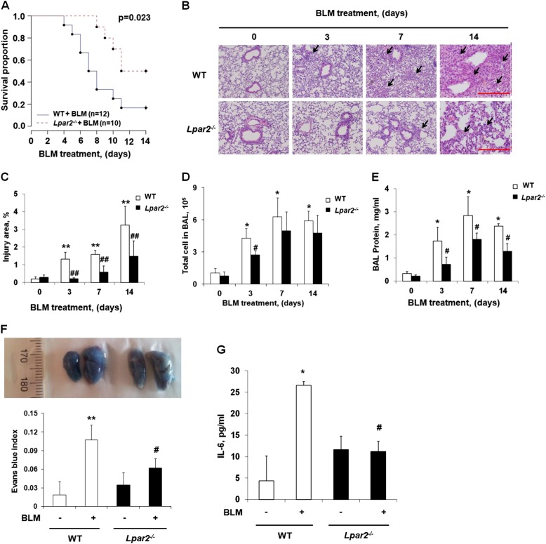
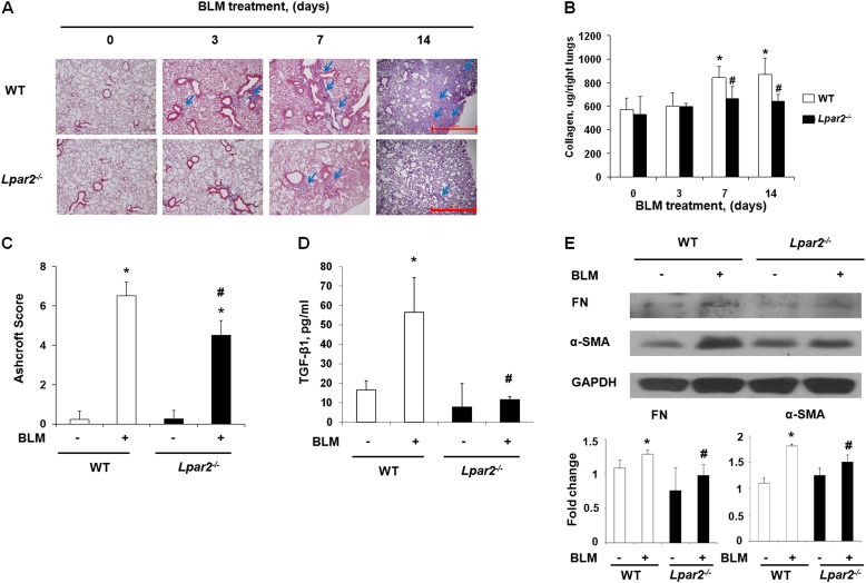
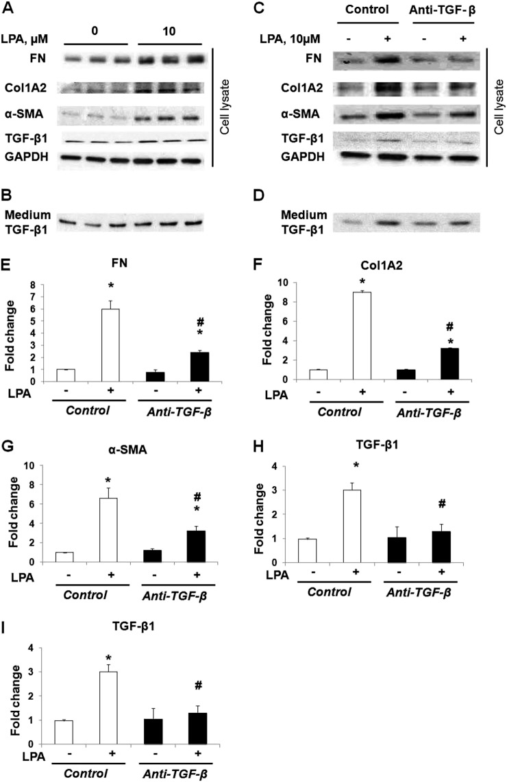
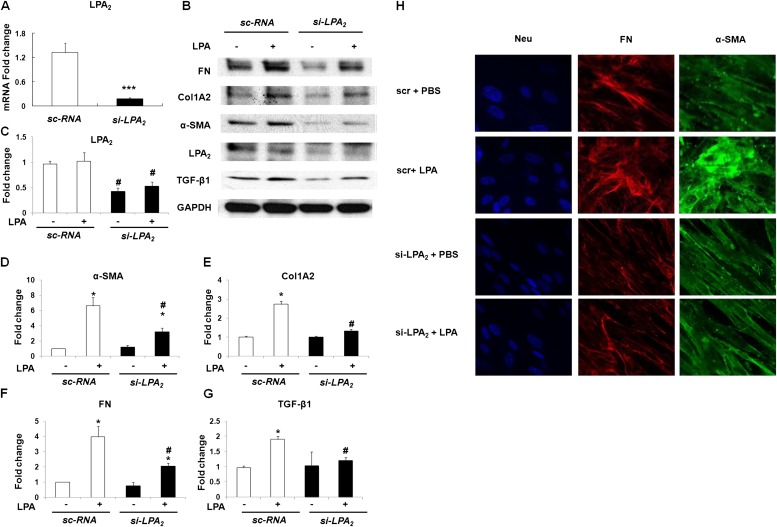
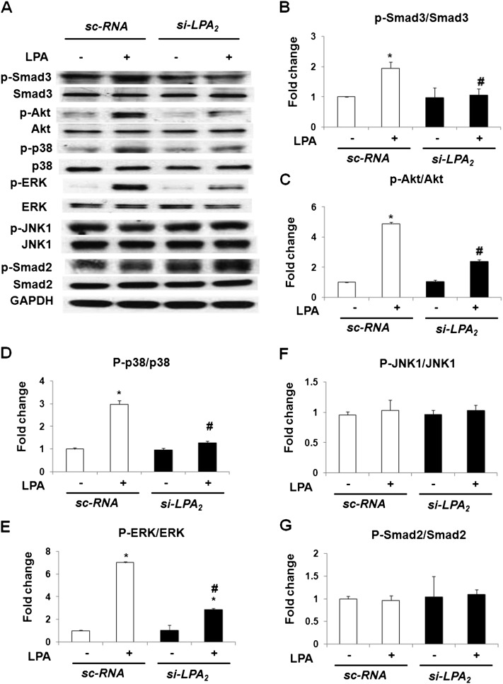
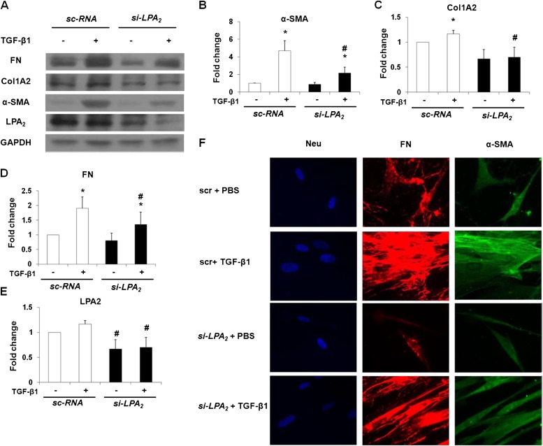
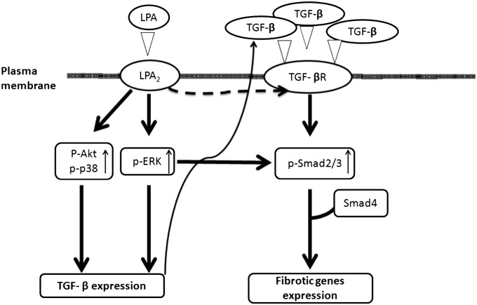
Idiopathic pulmonary fibrosis is a devastating disease characterized by alveolar epithelial cell injury, the accumulation of fibroblasts/myofibroblasts, and the deposition of extracellular matrix proteins. Lysophosphatidic acid (LPA) signaling through its G protein-coupled receptors is critical for its various biological functions. Recently, LPA and LPA receptor 1 were implicated in lung fibrogenesis. However, the role of other LPA receptors in fibrosis remains unclear. Here, we use a bleomycin-induced pulmonary fibrosis model to investigate the roles of LPA2 in pulmonary fibrogenesis. In the present study, we found that LPA2 knockout (Lpar2(-/-)) mice were protected against bleomycin-induced lung injury, fibrosis, and mortality, compared with wild-type control mice. Furthermore, LPA2 deficiency attenuated the bleomycin-induced expression of fibronectin (FN), α-smooth muscle actin (α-SMA), and collagen in lung tissue, as well as levels of IL-6, transforming growth factor-β (TGF-β), and total protein in bronchoalveolar lavage fluid. In human lung fibroblasts, the knockdown of LPA2 attenuated the LPA-induced expression of TGF-β1 and the differentiation of lung fibroblasts to myofibroblasts, resulting in the decreased expression of FN, α-SMA, and collagen, as well as decreased activation of extracellular regulated kinase 1/2, Akt, Smad3, and p38 mitogen-activated protein kinase. Moreover, the knockdown of LPA2 with small interfering RNA also mitigated the TGF-β1-induced differentiation of lung fibroblasts. In addition, LPA2 deficiency significantly attenuated the bleomycin-induced apoptosis of alveolar and bronchial epithelial cells in the mouse lung. Together, our data indicate that the knockdown of LPA2 attenuated bleomycin-induced lung injury and pulmonary fibrosis, and this may be related to an inhibition of the LPA-induced expression of TGF-β and the activation and differentiation of fibroblasts.


Figures

<i>Figure 1.</i>
Figure 1.
Lysophosphatidic acid receptor–2 (LPA2) deficiency protects against bleomycin (BLM)–induced lung injury, inflammation, and mortality in mice. Wild-type (WT) and LPA receptor–2 knockout (Lpar2−/−) mice (aged 8–10 wk, male) were anesthetized with 3 ml/kg of a mixture of ketamine (25 mg/kg) and 2.5 ml of xylazine, followed by an intratracheal injection of saline or bleomycin sulfate (2 U/kg, ∼ 0.04 U/animal) in saline in a total volume of 50 μl. Animals were killed for analysis on Days 0, 3, 7, or 14 (after bleomycin administration). Bronchoalveolar lavage (BAL) fluid was collected and analyzed, as described in Materials and Methods. Lungs were removed from the mice and their lobes were sectioned, embedded in paraffin, and cut into 5-μm sections for hematoxylin-and-eosin staining. (A) Survival of WT and Lpar2−/− mice challenged with bleomycin. (B) Representative hematoxylin-and-eosin staining of lung tissue obtained from bleomycin-challenged Lpar2−/− and WT mice (arrows show injured areas). Original magnification, ×10. Scale bar, 200 μm. (C) Quantitative analysis of injury area, expressed as the percentage of the total cross-sectional area. (D) Total infiltrated cell number. (E) Total protein levels in BAL fluid, expressed as means ± SEMs. (F) Evans blue dye extravasation assay for pulmonary vascular leakage. Briefly, 7 days after bleomycin challenge (2 U/kg body weight), mice were injected with Evans blue dye (20 mg/kg) intravenously for 3 hours. After perfusion, lungs were excised and imaged by a Canon (Tokyo, Japan) digital camera (top), followed by homogenization and analysis, as described in Materials and Methods. The Evans blue dye in lung-tissue supernatants and plasma was quantified and normalized to the plasma level (bottom). (G) IL-6 level in BAL fluid from WT and Lpar2−/− mice on Day 7 after bleomycin challenge. Data are expressed as means ± SEMs. *P < 0.05 and **P < 0.01, versus WT mice without BLM treatment. #P < 0.05 and ##P < 0.01, versus WT mice with BLM challenge at the same time point (n = 4–6 per group).
<i>Figure 2.</i>
Figure 2.
LPA2 deficiency attenuates bleomycin-induced pulmonary fibrosis in mice. Lpar2−/− or WT mice (male, aged 8–10 wk) received bleomycin (2 U/kg in 50 μl PBS, ∼ 0.04 U/animal) or PBS intratracheally, and were killed on Days 0, 3, 7, and 14 after bleomycin challenge. Lungs were removed, embedded in paraffin, and cut into 5-μm sections for staining. (A) Representative images of trichrome-stained lung sections were obtained from Lpar2−/− or WT mice, with or without bleomycin challenge (blue arrows show collagen deposition areas in blue). Original magnification, ×4. Scale bar, 1 mm. (B) Acid-soluble collagen in lung tissue. (C) Ashcroft score of lung sections from lung tissue obtained from Lpar2−/−or WT mice challenged with bleomycin on Days 0 and 14. (D) Transforming growth factor–β1 levels in BAL fluid obtained from Lpar2−/− or WT mice after bleomycin challenge on Day 14. (E) Protein levels of α–smooth muscle actin (α-SMA) and fibronectin (FN) in lung tissue from Lpar2−/− or WT mice after bleomycin challenge on Day 14. Data are expressed as means ± SEMs. *P < 0.05 versus wild-type mice without bleomycin treatment. #P < 0.05, versus WT mice after bleomycin treatment at the same time point (n = 4–6 per group). BLM, bleomycin; GAPDH, glyceraldehyde 3–phosphate dehydrogenase.
<i>Figure 3.</i>
Figure 3.
Lysophosphatidic acid (LPA) induces the activation and differentiation of lung fibroblasts via Transforming growth factor (TGF)-β1. Serum-starved (for 20 h) human lung fibroblasts (∼ 90% confluence) were pretreated with anti–TGF-β1 antibody or control IgG antibody (5 μg/ml, 1 h), and challenged with 18:1 LPA (10 μM) or vehicle for 48 hours, and cell lysates (20 μg protein) were subjected to SDS-PAGE and immunoblotting. The cell culture medium was concentrated, and TGF-β1 levels were analyzed by SDS-PAGE and immunoblotting. The intensity of the bands with anti-FN or anti–α-SMA, collagen 1A2 (Col1A2), and TGF-β1 antibody were quantified and normalized to GAPDH. LPA (10 μM) at 18:1 induced the expression of FN, α-SMA, Col1A2, and TGF-β1 in human lung fibroblasts (A) and the secretion of TGF-β1 into the culture medium (B). Effects of anti–TGF-β1 antibody on 18:1 LPA (10 μM)–induced expression of FN, α-SMA, Col1A2, and TGF-β1 in human lung fibroblasts (C) and TGF-β levels in culture medium (D). (E–I) Quantification of the expression of FN (E), Col1A2 (F), α-SMA (G), and TGF-β1 (H) induced by LPA in human lung fibroblasts, and the secreted TGF-β1 in culture medium (I). Data are expressed as means ± SEMs of three independent experiments. *P < 0.05, versus cells with control antibody but without LPA treatment. #P < 0.05, versus LPA-challenged cells with pretreatment of control antibody.
<i>Figure 4.</i>
Figure 4.
LPA2 small interfering RNA (siRNA) blocks the LPA-induced expression of transforming growth factor (TGF)-β, as well as the activation and differentiation of lung fibroblasts. Human lung fibroblasts (∼ 60% confluence) were treated with control scrambled siRNA (sc-RNA) or LPA2 siRNA (si-LPA2) (200 nM) for 24 hours, followed by serum starvation and treatment with 18:1 LPA (0 and 10 μM) for another 48 hours. Cell lysates (20 μg of protein) were subjected to SDS-PAGE and immunoblotting, as described in Materials and Methods. The intensity of the bands with anti-FN or anti–α-SMA, Col1A2, and TGF-β1 antibody were quantified and normalized to GAPDH. Data are expressed as the means ± SEMs of three independent experiments. (A) mRNA levels of LPA2 from human lung fibroblasts transfected with scrambled siRNA or LPA2 siRNA. ***P < 0.001, versus cells with sc-RNA treatment. (B) Immunoblotting to determine the expression of LPA2, FN, α-SMA, Col1A2, and TGF-β1. A representative immunoblot from three independent experiments is shown. (C–G) Quantification of the expression of LPA2 (C), α-SMA (D), Col1A2 (E), FN (F), and TGF-β1 (G) induced by LPA after LPA2 siRNA treatment. *P < 0.05 versus cells with sc-RNA but without LPA treatment. #P < 0.05 versus LPA-challenged cells with pretreatment of sc-RNA. (H) Human lung fibroblasts grown on eight-well slide chambers were transfected with scrambled or LPA2 siRNA for 24 hours, exposed to PBS or 18:1 LPA (10 μM) for 48 hours, washed, fixed, permeabilized, probed with anti-FN or anti–α-SMA antibodies, and examined by immunofluorescence microscopy, using a ×60 oil objective. The FN (red) and α-SMA (green) images showed matched cell fields for each condition. A representative image from three independent experiments is shown.
<i>Figure 5.</i>
Figure 5.
LPA2 siRNA attenuates the LPA-induced phosphorylation of Smad3, Akt, p38 mitogen-activated protein kinase (p38 MAPK), and extracellular regulated kinase (ERK) in human lung fibroblasts. Human lung fibroblasts (∼ 60% confluence) were treated with scrambled or LPA2 siRNA (200 nM) for 48 hours, followed by serum starvation and treatment with 18:1 LPA (10 μM) for another 15 minutes. (A) Cell lysates (20 μg of protein) were subjected to SDS-PAGE and immunoblotted with anti–p-Smad3, Smad3, p-Akt, Akt, p–p38 MAPK, p38 MAPK, p–c-Jun N-terminal kinase (JNK)–1, JNK1, p-Smad2, Smad2, p-ERK, ERK, and GAPDH antibodies. A representative immunoblot from three independent experiments is shown. (B–G) The intensity of each band was quantified using densitometry, and normalized to GAPDH. Data are expressed as the means ± SEMs of three independent experiments. *P < 0.05, versus cells without LPA treatment. #P < 0.05, versus scrambled siRNA–treated cells also treated with LPA.
<i>Figure 6.</i>
Figure 6.
LPA2 siRNA attenuates the Transforming growth factor (TGF)-β1–induced activation and differentiation of lung fibroblasts. Human lung fibroblasts (∼ 60% confluence) were treated with scrambled or LPA2 siRNA (200 nM) for 24 hours, followed by serum starvation and treatment with TGF-β1 (5 ng/ml) for another 48 hours. Cell lysates (20 μg protein) were subjected to SDS-PAGE and immunoblotting. The intensity of bands for anti-FN or anti–α-SMA and Col1A2 antibodies were quantified and normalized to GAPDH. Data are expressed as the means ± SEMs of three independent experiments. (A) Immunoblotting to determine relative expression levels of FN, α-SMA, Col1A2, and TGF-β1. A representative immunoblot from three independent experiments is shown. (B–E) Quantification of the expression of α-SMA (B), Col1A2 (C), FN (D), and LPA2 (E) induced by TGF-β1 after LPA2 siRNA treatment. *P < 0.05, versus cells without LPA treatment. #P < 0.05, versus scrambled siRNA-treated cells with LPA treatment. (F) Human lung fibroblasts grown on eight-well slide chambers were transfected with scrambled or LPA2 siRNA (for 24 h), exposed to PBS or TGF-β1 (for 48 h), washed, fixed, permeabilized, probed with anti-FN or anti–α-SMA antibodies, and examined according to immunofluorescence microscopy, using a ×60 oil objective. The FN (red) and α-SMA (green) images showed matched cell fields for each condition. A representative image from three independent experiments is shown.
<i>Figure 7.</i>
Figure 7.
Schematic diagram illustrates the effects of LPA and Transforming growth factor (TGF)-β on fibrotic gene expression, differentiation, and activation in human lung fibroblasts. LPA binding to LPA2 on the cell surface induces the activation of intracellular signaling pathways and triggers the expression of fibrotic genes (α-SMA, FN, Col1A2, and TGF-β). LPA induces TGF-β expression through the activation of Akt, p38, and ERK, and secreted TGF-β then signals through the TGF-β receptor (TGF-βR) and augments fibrogenesis. LPA2 is also required for the TGF-β signaling of Smad3 phosphorylation and expression of genes associated with fibrogenesis. However, the potential mechanisms involved in the cross-talk between LPA2 and the TGF-βR remain unclear.

Similar articles

Cited by

References

    1. King TE., Jr Update in pulmonary medicine. Ann Intern Med. 1998;129:806–812. - PubMed
    1. Raghu G, Freudenberger TD, Yang S, Curtis JR, Spada C, Hayes J, Sillery JK, Pope CE, II, Pellegrini CA. High prevalence of abnormal acid gastro-oesophageal reflux in idiopathic pulmonary fibrosis. Eur Respir J. 2006;27:136–142. - PubMed
    1. Cucoranu I, Clempus R, Dikalova A, Phelan PJ, Ariyan S, Dikalov S, Sorescu D. NAD(P)H oxidase 4 mediates transforming growth factor–beta1–induced differentiation of cardiac fibroblasts into myofibroblasts. Circ Res. 2005;97:900–907. - PubMed
    1. Sappino AP, Schurch W, Gabbiani G. Differentiation repertoire of fibroblastic cells: expression of cytoskeletal proteins as marker of phenotypic modulations. Lab Invest. 1990;63:144–161. - PubMed
    1. Phan SH. The myofibroblast in pulmonary fibrosis. Chest. 2002;122(Suppl. 6):286S–289S. - PubMed

Publication types

MeSH terms