

Review
doi: 10.1021/cr300015c.
Epub 2013 Jun 28.
Arsenic binding to proteins
Affiliations
- PMID: 23808632
- PMCID: PMC3797521
- DOI: 10.1021/cr300015c
Item in Clipboard
Review
Arsenic binding to proteins
Chem Rev.
.
No abstract available
Figures
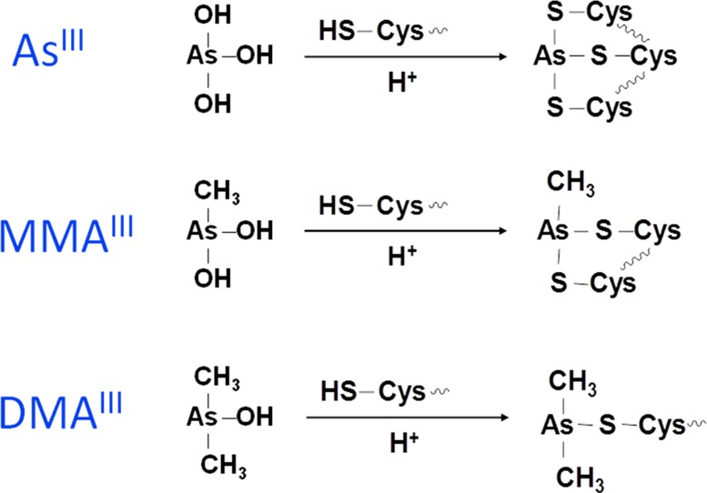
Binding of inorganic arsenite (iAsIII), monomethylarsonous
acid (MMAIII), and dimethylarsinous acid (DMAIII) to cysteines in a protein.
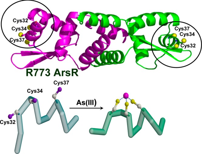
Binding of iAsIII to the ArsR repressor from E. coli plasmid R773 (P15905) results in conformational
change of the repressor. iAsIII binds to Cys32, Cys34,
and Cys37 of the ArsR repressor. Unwinding the helix disrupts DNA
binding, resulting in dissociation of the repressor from the operator
site. Dissociation of the repressor induces gene expression. (Adapted
from ref (65).)
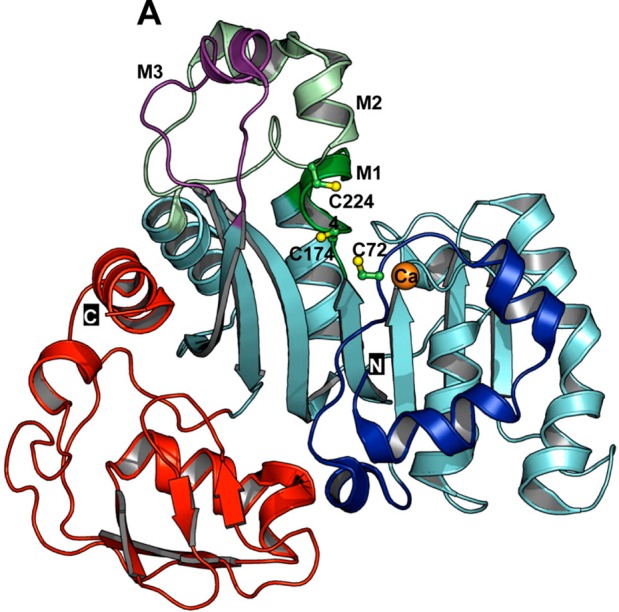
Ribbon representation of ligand-free AS3MT (CmArsM) from
the thermophilic
eukaryotic alga C. merolae. N and C
indicate the N- and C-terminal domains and are colored blue and red,
respectively. Cysteine residues are shown as balls and sticks and
colored green (carbon) and yellow (sulfur). Three cysteine residues,
C72, C174, and C224, are believed to be involved in arsenic binding.
(Reprinted with permission from ref (75). Copyright 2012 American Chemical Society.)
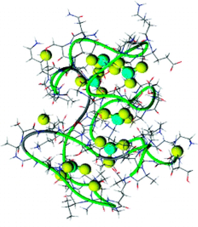
A model depicting six arsenic atoms bound
to 18 cysteines in metallothionein.
(Reprinted with permission from ref (155). Copyright 2008 American Chemical Society.)
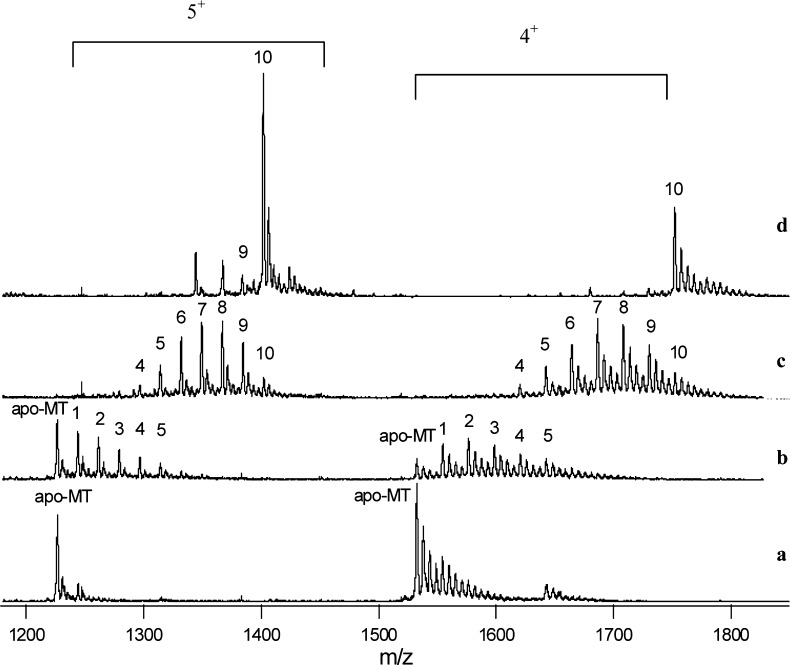
Mass spectra showing the number of monomethylarsonous
acid (MMAIII) molecules bound to metallothionein (MT).
The solutions
contained 7 μM metallothionein (rMT-IIa) and increasing concentrations
of MMAIII. The concentration ratios of MMAIII to MT in the solutions were (a) 1:5, (b) 1:1, (c) 5:1, and (d) 50:1.
The ions carrying 5+ and 4+ charges are shown.
The numbers on the peaks represent the number of MMAIII bound to the MT molecule. For example, peak 6 represents MT-(CH3As)6. A maximum of 10 MMAIII molecules
were bound to a single MT that contained 20 cysteines. (Adapted from
ref (151).)
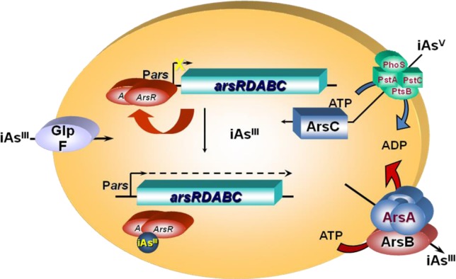
Schematic representation of arsenic interaction
with the arsRDABC
operon and related enzymes in E. coli. ArsR is an AsIII-responsive transcriptional repressor
that binds to the ars promoter, repressing transcription. Binding
of iAsIII to ArsR results in dissociation of the repressor
from the DNA and hence gene expression. iAsV is taken up
by the phosphate transporter, while iAsIII is taken up
by the aquaglyceroporin GlpF. iAsV is reduced to iAsIII by ArsC. Intracellular iAsIII is extruded from
the cells by ArsB alone or by the ArsAB ATPase. (Modified from ref (163).)
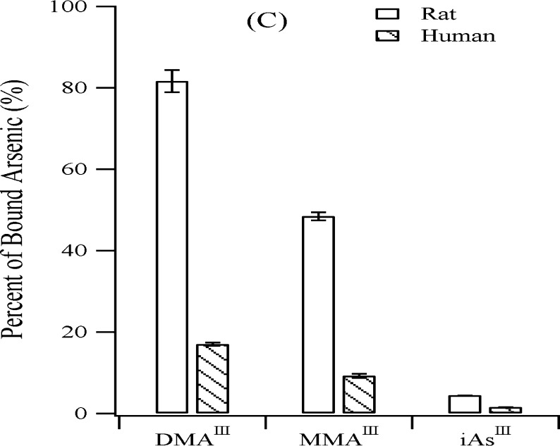
Comparison
of rat hemoblobin (rHb) and human hemoblobin (hHb) binding
to three trivalent arsenicals (iAsIII, MMAIII, and DMAIII). Higher percentages of the trivalent arsenicals
are bound to rHb than to hHb. (Reprinted with permission from ref (169). Copyright 2004 American
Chemical Society.)
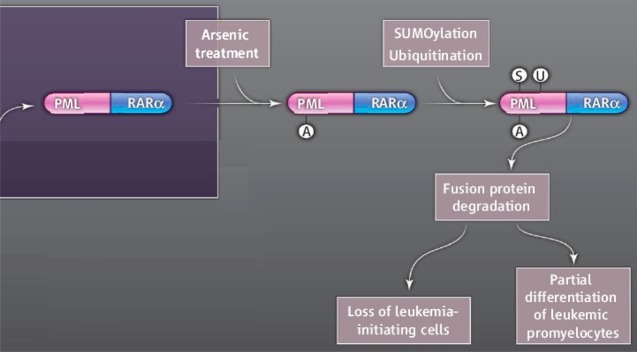
A proposed mechanism for the treatment
of acute promyelocytic leukemia
(APL) using inorgaic arsenic. Binding of arsenic to the promyelocytic
leukemia (PML) retinoic acid receptor alpha (RARα) fusion protein
triggers SUMOylation and ubiquitination and ultimately leads to the
degradation of the oncoprotein and cell death. (Reprinted with permission
from ref (66) and Kogan,
S.C. 10.1126/science.1189198.
Copyright 2010 American Association for the Advancement of Science.)
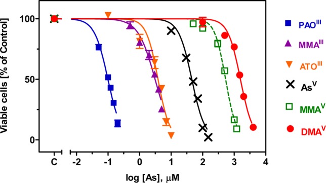
Toxicity of six arsenic compounds to HL-60 cells
after a 48-h incubation.
(Adapted from ref (200).)
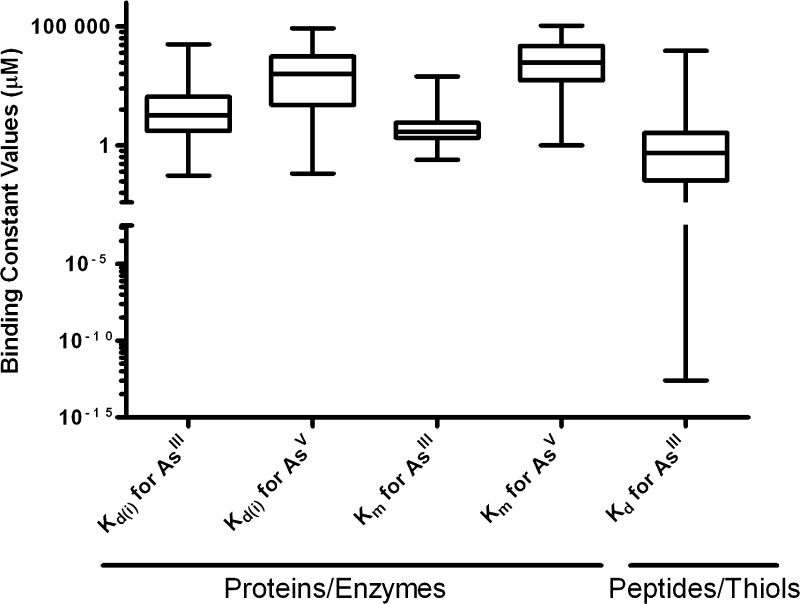
A comparison
of binding affinity of trivalent and pentavalent arsenicals
to proteins.
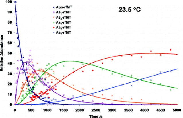
Kinetic data
showing the relative abundance of the various arsenic–metallothionein
species detected using electrospray mass spectrometry. The solution
was analyzed following reaction of 9 μM apo-rfMT with 108 μM
arsenite at 23.5 °C. The experimental data have a relative standard
error of 7%. (Reprinted with permission from ref (153).)
References
-
- Cullen W. R.Is Arsenic an Aphrodisiac? The Sociochemistry of an Element; RSC Publishing: Cambridge, U.K., 2008; 412 pp.
-
- Oremland R. S.; Stolz J. F. Science 2003, 300, 939. - PubMed
-
- Nordstrom D. K. Science 2002, 296, 2143. - PubMed
-
- Jain C. K.; Ali I. Water Res. 2000, 34, 4304.
-
- Bhattacharya P.; Welch A. H.; Stollenwerk K. G.; McLaughlin M. J.; Bundschuh J.; Panaullah G. Sci. Total Environ. 2007, 379, 109. - PubMed
Publication types
MeSH terms
Substances
Grants and funding
LinkOut - more resources
Full Text Sources
Other Literature Sources
Medical
