. 2013 Sep;53(8):1250-61.
doi: 10.1111/head.12152. Epub 2013 Jun 28.

pH-evoked dural afferent signaling is mediated by ASIC3 and is sensitized by mast cell mediators

Affiliations

pH-evoked dural afferent signaling is mediated by ASIC3 and is sensitized by mast cell mediators

Jin Yan et al. Headache. 2013 Sep.

Abstract

Background: Prior studies have shown that decreased meningeal pH activates dural afferents via opening of acid-sensing ion channels (ASICs), suggesting one pathophysiological mechanism for the generation of headaches. The studies described here further examined the ASIC subtype mediating pH-induced dural-afferent activation and examined whether sensitization influences pH responses.

Objective: Given the potential importance of meningeal mast cells to headache, the goal of this study was to evaluate dural afferent responses to pH following sensitization with mast cell mediators.

Methods: Cutaneous allodynia was measured in rats following stimulation of the dura with decreased pH alone or in combination with mast cell mediators. Trigeminal ganglion neurons retrogradely labeled from the dura were stained with an ASIC3 antibody using immunohistochemistry. Current and action potentials evoked by changes in pH alone or in combination with mast cell mediators were measured in retrogradely labeled dural afferents using patch-clamp electrophysiology.

Results: pH-sensitive dural afferents generated currents in response to the ASIC3 activator 2-guanidine-4-methylquinazoline (GMQ), approximately 80% of these neurons express ASIC3 protein, and pH-evoked behavioral responses were inhibited by the ASIC3 blocker APETx2. Following exposure to mast cell mediators, dural afferents exhibited increased pH-evoked excitability, and cutaneous allodynia was observed at higher pH than with pH stimuli alone.

Conclusions: These data indicate that the predominant ASIC subtype responding to decreased meningeal pH is ASIC3. Additionally, they demonstrate that in the presence of inflammation, dural afferents respond to even smaller decreases in pH providing further support for the ability of small pH changes within the meninges to initiate afferent input leading to headache.

Keywords: ASIC3; dural afferent; meninges.


Figures

Figure 1
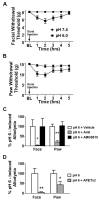
Figure 1. Application of pH 6.0 SIF solution to the dura elicited cutaneous allodynia via activation of ASIC3 subunit
Withdrawal thresholds to tactile stimuli applied to the face (A) and the hindpaws (B) were measured in rats before and immediately after dural application of pH 6.0 SIF (n = 11) or pH 7.4 SIF (n = 13). (C) Application of pH 6.0 solution was given with either vehicle (1% DMSO, white bar, n = 13), amiloride (100 nmol, grey bar, n = 12) or AMG-9810 (10 nmol, black bar, n = 10). Withdrawal thresholds to tactile stimuli were measured for 5 hrs and data were converted to area over the time-effect curve and normalized as a percentage of the pH 6.0 – treated group to allow for multiple comparisons. Comparisons among several treatment groups were performed by one-way ANOVA followed by post hoc Dunnett’s test. Coapplication of amiloride significantly abolished behavioral signs of tactile allodynia of the face and hindpaw (*p < 0.05, **p < 0.01). Cotreatment with AMG-9810 failed to prevent development of behavioral signs of tactile allodynia of the face or hindpaw. (D) Application of pH 6.0 solution was given alone (white bar, n = 13) or with the selective ASIC3 antagonist APETx2 (200 pmol, grey bar, n = 13). Coapplication of APETx2 (200 pmol) significantly abolished behavioral signs of tactile allodynia of the face and hindpaw (*p < 0.05, **p < 0.01).
Figure 2
Figure 2. Retrogradely-labeled dural afferents express ASIC3 immunoreactivity
Images are representative sections of trigeminal ganglia taken from rats in which fluorogold was applied to the dura 7 days prior. Immunolabeling with an ASIC3-specific primary antibody and Alexa Fluor 555-conjugated secondary antibody is shown in red in (A). Retrogradely-labeled dural afferents (fluorogold-positive neurons) are shown in blue in (B). An overlay of (A) and (B) is shown in (C). Neurons that appear purple are dural afferents that express ASIC3 immunoreactivity.
Figure 3
Figure 3. pH-evoked responses were mediated through ASIC3 subunits in vitro
GMQ-evoked currents in pH-sensitive dural afferents. Representative traces illustrating acid-(pH 6.0) and GMQ-induced currents in a dural afferent.
Figure 4
Figure 4. Application of pH to the dura dose dependently elicited cutaneous allodynia
(A) Withdrawal thresholds to tactile stimuli applied to the face and the hindpaws were measured in rats before and immediately after dural application of pH 6.4, pH 6.6, pH 6.8, and pH 7.4 solutions. (B) Data were converted to area over the time-effect curve to allow for multiple comparisons. pH dose-dependently decreased the withdrawal threshold both in the face and the hindpaws. Significant (*p < 0.05) differences among means for each group were determined by one-way ANOVA followed by Dunnett’s post hoc test.
Figure 5
Figure 5. Animals exhibited enhanced withdrawal responses to pH changes following coapplication of sub-threshold mast cell mediators
M indicates sub-threshold mast cell mediators, which were composed of Histamine (200 μM), Serotonin (200 μM), AC55541 (10 μM) and Iloprost (20 μM). Data were converted to area over the time-effect curve to allow for multiple comparisons. Significant (**p < 0.01) differences among means for each group were determined by one-way ANOVA followed by Dunnett’s post hoc test. Application of sub-threshold mast cell mediators (black bar, n = 9) by itself did not cause significant changes in facial and hindpaw withdrawal thresholds compared with SIF administration (white bar, n = 11). (A) Coapplication of sub-threshold mast cell mediators and pH 6.6 solution caused a significant decrease in facial withdrawal threshold (red bar, n = 12) compared with pH 7.4 application, whereas application of pH 6.6 solution by itself did not (red bar, n = 13). (B) Coapplication of sub-threshold mast cell mediators and pH 6.8 solution caused a significant decrease in hindpaw withdrawal threshold (blue bar, n = 14) compared with pH 7.4 application, whereas application of pH 6.8 solution by itself did not (blue bar, n = 14).
Figure 6
Figure 6. Dural afferents exhibited enhanced sensitivity to pH-induced action potential firing following application of mast cell mediators
Mast cell mediators were composed of Histamine (100 μM), Serotonin (10 μM), AC55541 (10 μM), Iloprost (1 μM). (A) The percentage of neurons firing action potentials was calculated before and after a 5 min application of mast cell mediators. For each pH tested, the percentage of neurons firing action potentials increased following application of mast cell mediators. n = 23. (B) Number of spikes was counted from the same dural afferent before and after 5 mins application of mast cell mediators. Application of mast cell mediators significantly increased the number of spikes for each pH tested (paired t-test, **p < 0.01 for pH 6.9, *p < 0.05 for pH 6.8 and pH 7.0) (C) Application of mast cell mediators led to the transient and persistent firing of action potential at pH 7.0 and 6.9 in two representative dural afferents.

Comment in

References

    1. Goadsby PJ, Lipton RB, Ferrari MD. Migraine--current understanding and treatment. N Engl J Med. 2002;346:257–270. - PubMed
    1. Yan J, Edelmayer RM, Wei X, Felice MD, Porreca F, Dussor G. Dural afferents express acid-sensing ion channels: A role for decreased meningeal pH in migraine headache. Pain. 2011;152:106–113. - PMC - PubMed
    1. Wemmie JA, Price MP, Welsh MJ. Acid-sensing ion channels: advances, questions and therapeutic opportunities. Trends Neurosci. 2006;29:578–586. - PubMed
    1. Lingueglia E. Acid-sensing ion channels in sensory perception. J Biol Chem. 2007;282:17325–17329. - PubMed
    1. Benson CJ, Xie J, Wemmie JA, et al. Heteromultimers of DEG/ENaC subunits form H+-gated channels in mouse sensory neurons. PNAS. 2002;99:2338–2343. - PMC - PubMed

Publication types

Substances

LinkOut - more resources