Review
. 2013 Jun;11 Suppl 1(Suppl 1):46-66.
doi: 10.1111/jth.12253.

Evolutionary origins of the blood vascular system and endothelium

Affiliations
Review

Evolutionary origins of the blood vascular system and endothelium

R Monahan-Earley et al. J Thromb Haemost. 2013 Jun.

Abstract

Every biological trait requires both a proximate and evolutionary explanation. The field of vascular biology is focused primarily on proximate mechanisms in health and disease. Comparatively little attention has been given to the evolutionary basis of the cardiovascular system. Here, we employ a comparative approach to review the phylogenetic history of the blood vascular system and endothelium. In addition to drawing on the published literature, we provide primary ultrastructural data related to the lobster, earthworm, amphioxus, and hagfish. Existing evidence suggests that the blood vascular system first appeared in an ancestor of the triploblasts over 600 million years ago, as a means to overcome the time-distance constraints of diffusion. The endothelium evolved in an ancestral vertebrate some 540-510 million years ago to optimize flow dynamics and barrier function, and/or to localize immune and coagulation functions. Finally, we emphasize that endothelial heterogeneity evolved as a core feature of the endothelium from the outset, reflecting its role in meeting the diverse needs of body tissues.

Keywords: biological evolution; blood vessels; endothelium; heart; phylogeny.


Figures

Fig. 1
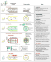
Fig. 1. Metazoan phylogenetic tree
Adapted and modified from Juliano CE, Swartz SZ, Wessel GM. A conserved germline multipotency program. Development. 2010; 137:4113-26.
Fig. 2
Fig. 2
Schematic of different circulatory systems.
Fig. 3
Fig. 3. Blood vascular system of the lobster
A, Schematic of the blood vascular system of the lobster. a, artery. B, Open view of the dorsal thorax showing the single-chambered heart suspended in the pericardial sinus by alary ligaments. C, Electron micrograph of the heart showing a cardiomyocyte (cm) filled with cross-sections of thick and thin microfibrils (consistent with myosin and actin) and mitochondria (*). The inner surface of the heart chamber is covered by a thin basal lamina. There is no endocardial lining. A hemocyte (h) is shown in the heart chamber. D, One-micron transverse histological section of a dorsal abdominal artery stained with Giemsa. E, Electron micrograph of the dorsal abdominal artery shows the wall consisting of extracellular matrix composed of a dense meshwork of collagen fibrils. There is no endothelial lining. F, Photomicrograph of a swimmeret of a lobster that has been injected with Evans blue dye in the dorsal abdominal artery. Note the branching network of vessels ending in plumes of extravasated dye in the interstitial space. Panel A is adapted McLaughlin PA. Internal Anatomy. In LH Mantel (Ed.), The biology of crustacea: Internal anatomy and physiological regualtion. New York, NY: Academic Press, 1983.
Fig. 4
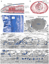
Fig. 4. Blood vascular system of the earthworm
A, Schematic of the blood vascular system of the earthworm. In this case, all vessels are colored red since there is no specialized respiratory organ where blood is oxygenated and because there is no central heart that divides vessels into afferent channels (veins) and efferent channels (arteries). B, Transverse histological section through the body of the earthworm shows the dorsal vessel (arrow) on the dorsal side of the gut. The section was stained with H&E. C, One-micron histological section stained with Giemsa shows two small blood vessels in the body wall. Ep, epidermis; mu, circular muscle layer. D, Electron micrograph of a small blood vessel surrounded by a continuous layer of myoepithelial cells. The lumen is filled with hemoglobin particles. E, Higher power electron micrograph shows a blood vessel lined by several myoepithelial cells. The cells are connected by specialized lateral borders. They contain numerous thick myofilaments (arrows) consistent with myosin that are oriented circumferentially around the vessel. A well-formed basal lamina (*) separates the myoepithelial cells from the lumen of the blood vessel. The fact that the hemoglobin particles are retained in the lumen indicates that the basal lamina forms an effective barrier. F, A similar electron micrograph to E, but shows the presence of an amoebocyte in the blood vessel lumen adjacent to the myoepithlial lining. Panel A is adapted from Brusca R.C., Brusca G.J. Invertebrates. Sunderland, MA: Sinauer Associates, 2003.
Fig. 5
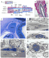
Fig. 5. Blood vascular system of amphioxus
A, Schematic of the blood vascular system of amphioxus. B, Transverse histological section through the pharynx shows the paired dorsal aortae (arrows) lying on either side of and ventral to the notochord (NC). The hepatic vein is on the dorsal surface of the liver (also referred to as the hepatic cecum). The section was stained with H&E. P, pharynx. C, One-micron histological section stained with Giemsa shows a discontinuous coverage of dorsal aorta with amoebocytes (arrows). D, Electron micrograph of the contractile hepatic vein shows the lumen delimited by parts of three myoepithelial cells, which contain myofilaments (*) in their basal portion. a, amoebocyte. E, Electron micrograph of a skeletal vessel in the gill surrounded by skeletal rod (skr). The skeletal rod, in turn is surrounded by atrial epithelial cells (ae) and lateral ciliated cells (lcc). Occasional chromaffin-like cells (with membrane-delimited electron dense granules) are scattered between the atrial epithelial cells. Note the presence of amoebocytes (a) on the luminal surface of the blood vessel wall. F, Electron micrograph shows an amoebocyte “clinging” to the luminal surface of the dorsal aorta. The cell contains a well-developed Golgi apparatus (g), numerous vesicles, and granules containing tubular structures (arrows). Note that the amoebocyte has no underling basal lamina. Rather it is applied to the connective tissue matrix. G, Higher magnification of an amoebocyte from dorsal aorta shows two tubule-filled granules.
Fig. 6
Fig. 6. Blood vascular system of the hagfish
A, Schematic of the blood vascular system of hagfish. Top, Transverse section through mid-region shows several features that are typical of other vertebrates, including the arrangement of myomeres, neural tube, and aorta. Features that are unique to hagfish include the large subcutaneous sinus between skin and skeletal muscle, the retention of the notochord in the adult, and the presence of slime glands on the ventrolateral surface. Bottom, schematic of hagfish circulation. The gills are in series with the systemic circulation. Blood enters the subcutaneous sinus via skeletal muscle capillaries and renters the systemic circulation via accessory hearts (the caudal and cardinal hearts). The portal heart pumps blood from the intestinal vasculature into the systemic heart via the common portal vein. B, Photomicrograph of ventral aorta and two (of a total of 12) gills in an animal that has been injected with Evans blue dye through the heart. C, Electron micrograph of the heart shows electron-lucent endothelial cells (EC) overlying a thick extracellular matrix containing a chromaffin-like cell, and a cardiomyocyte (cm) with electron-lucent cytoplasm, and well-preserved mitochondria and muscle filaments. D, Electron micrograph of the dermis shows a microvessel in cross-section containing two nucleated red blood cells. The blood vessel is lined by a continuous layer of endothelial cells. A melanocyte is seen on the left side of the vessel. E, Electron micrograph of a kidney glomerulus shows podocyte foot processes abutting a well formed basal lamina. On the other side of the basal lamina is an endothelial cell (EC) facing the lumen of a glomerular capillary. The endothelial cell contains many vesicles, vacuoles and tubular structures. F, Electron micrograph of a liver sinusoid shows a large gap in a single endothelial cell (E1, double-headed arrow), well-preserved attachments between two endothelial cells (EC1 and EC2), and a continuum of proteinaceous material from the lumen to extravascular space (Space of Disse). EC2 is a second endothelial cell. Panel A is reprinted with permission from Cheruvu PK, Gale D, Dvorak AM, Haig D, Aird WC. Hagfish: a model for early endothelium. In W Aird (Ed.), Endothelial Biomedcine. Cambridge, UK; New York: Cambridge University Press, 2007. Panels C, D and F are reprinted with permission from Yano K, Gale D, Massberg S, Cheruvu PK, Monahan-Earley R, Morgan ES, Haig D, von Andrian UH, Dvorak AM, Aird WC. Phenotypic heterogeneity is an evolutionarily conserved feature of the endothelium. Blood. 2007;109:613-5.
Fig. 7
Fig. 7. Phylogenetic perspective of the blood vascular system
Blood vascular system types and major propulsive organs are shown for representative extant phyla. The phylogenetic tree is schematic only, and the timelines are not drawn to scale. EC, endothelial cell; mya, mullion years ago.

Similar articles

Cited by

References

    1. Nesse RM, Bergstrom CT, Ellison PT, Flier JS, Gluckman P, Govindaraju DR, Niethammer D, Omenn GS, Perlman RL, Schwartz MD, Thomas MG, Stearns SC, Valle D. Evolution in health and medicine Sackler colloquium: Making evolutionary biology a basic science for medicine. Proc Natl Acad Sci U S A. 2010;107(Suppl 1):1800–7. doi: 10.1073/pnas.0906224106. 0906224106 [pii] - DOI - PMC - PubMed
    1. Tinbergen N. On aims and methods in ethology. Z Tierpsychol. 1963;20:410–33.
    1. Brusca RC, Brusca GJ. Invertebrates. Sunderland, Mass: Sinauer Associates; 2003.
    1. Ruppert EE, Carle KJ. Morphology of metazoan circulatory systems. Zoomorphology. 1983;103:193–208.
    1. Farmer CG. Evolution of the vertebrate cardio-pulmonary system. Annu Rev Physiol. 1999;61:573–92. doi: 10.1146/annurev.physiol.61.1.573. - DOI - PubMed

Publication types