

Impact of Hepatitis C Virus (HCV) infection on biomolecular markers influencing the pathogenesis of bladder cancer
- PMID: 23809295
- PMCID: PMC3704792
- DOI: 10.1186/1750-9378-8-24
Impact of Hepatitis C Virus (HCV) infection on biomolecular markers influencing the pathogenesis of bladder cancer
Abstract
Objective: The present study was designed to determine the possible impact of hepatitis C virus (HCV) infection on the expression of telomerase (TERT), retinoblastoma (RB1), E2F3, TP53, CDKN1A (p21) and fibroblast growth factor receptor- 3 (FGFR3) genes in patients with bladder cancer (BC).
Materials and methods: 100 patients with bladder cancer (15 female and 85 male) were divided into 2 groups; Group I: 50 HCV negative subjects (age range 36-79), and Group II: 50 HCV positive subjects (age range 42-80). Expressions of the telomerase, retinoblastoma (Rb), E2F3, TP53 and FGFR3 genes were tested by immunohistochemistry and real time PCR in tumour tissues and healthy bladder tissues. Also, telomerase activity was assessed by telomeric repeats amplification protocol (TRAP).
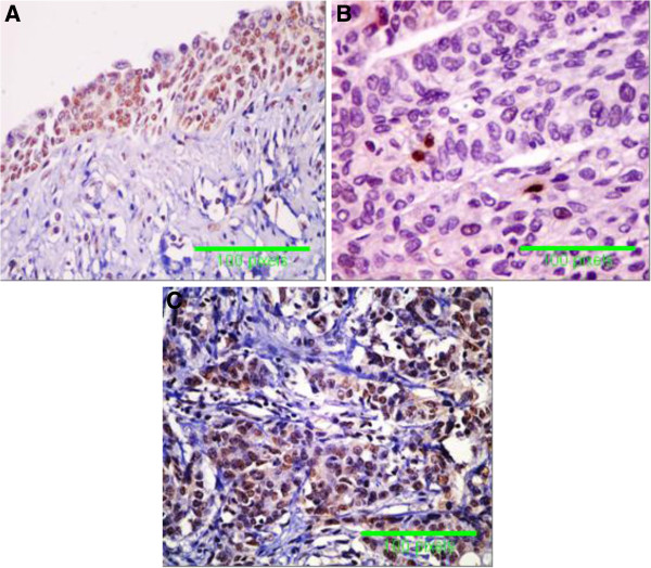
Results: Bladder tumors associated with HCV infection were of high grade and invasive squamous cell carcinomas (SCCs). Expressions of hTERT, Rb, E2F3, TP53 and FGFR3 as well as telomerase activity were significantly higher in bladder tissues of HCV-infected patients compared with bladder tissues of non infected patients (p<0.05). On the contrary, CDKN1A (p21) expression was significantly lower in bladder tissues of HCV-infected patients compared to bladder tissues of non infected patients (p<0.05).
Conclusion: The expressions of hTERT, Rb, E2F3, TP53 and FGFR3 as well as the activity of telomerase were significantly high in malignant bladder tissues associated with HCV infection. On the other hand, CDKN1A (p21) expression was low in bladder tissues of HCV-infected subjects. Moreover, there was a positive correlation between HCV infection and expression of telomerase, E2F3, TP53 and FGFR3. There was a negative correlation between HCV infection and expression of Rb and p21.
Figures


Similar articles
-
Detection of telomerase status by semiquantitative and in situ assays, and by real-time reverse transcription-polymerase chain reaction (telomerase reverse transcriptase) assay in bladder carcinomas.BJU Int. 2003 Apr;91(6):567-72. doi: 10.1046/j.1464-410x.2003.04117.x. BJU Int. 2003. PMID: 12656916
-
Evaluation of Fibroblast Growth Factor Receptor 3 (FGFR3) and Tumor Protein P53 (TP53) as Independent Prognostic Biomarkers in High-Grade Non-muscle Invasive Bladder Cancer.Cureus. 2024 Jul 31;16(7):e65816. doi: 10.7759/cureus.65816. eCollection 2024 Jul. Cureus. 2024. PMID: 39219882 Free PMC article.
-
[Detection of telomerase activity by semi-quantitative and in situ assays and quantification of hTERT expression in bladder carcinomas].Prog Urol. 2003 Apr;13(2):238-45. Prog Urol. 2003. PMID: 12765058 French.
-
Molecular mechanism of hepatocarcinogenesis.J Gastroenterol Hepatol. 1997 Oct;12(9-10):S309-13. doi: 10.1111/j.1440-1746.1997.tb00514.x. J Gastroenterol Hepatol. 1997. PMID: 9407351 Review.
-
Expression of telomerase genes in thyroid carcinoma.Int J Oncol. 2002 Aug;21(2):265-72. Int J Oncol. 2002. PMID: 12118320 Review.
Cited by
-
The role of Kaposi's sarcoma-associated herpesvirus infection in the proliferation of human bladder cancer cells.Tumour Biol. 2016 Feb;37(2):2587-96. doi: 10.1007/s13277-015-4096-5. Epub 2015 Sep 21. Tumour Biol. 2016. PMID: 26392109
-
Proteomic snapshot of saliva samples predicts new pathways implicated in SARS-CoV-2 pathogenesis.Clin Proteomics. 2024 May 22;21(1):37. doi: 10.1186/s12014-024-09482-9. Clin Proteomics. 2024. PMID: 38778280 Free PMC article.
-
The association between hepatitis C virus infection and renal cell cancer, prostate cancer, and bladder cancer: a systematic review and meta-analysis.Sci Rep. 2021 May 25;11(1):10833. doi: 10.1038/s41598-021-90404-2. Sci Rep. 2021. PMID: 34035396 Free PMC article.
References
-
- Khaled H. Systematic management of bladder cancer in Egypt: revisited. J Egypt Natl Canc Inst. 2005;17:127–131. - PubMed
-
- Sandberg AA, Berger CS. Review of chromosome studies in urological tumors II. Cytogenetics and molecular genetics of bladder cancer. J Urol. 1994;151:545–560. - PubMed
-
- Carroll PR. In: General urology. 14. Tanagho EA, Mc Aninch JW, editor. Philadelphia: Prentice-Hall International Inc; 1995. Urothelial carcinoma: cancers of the bladder ureter & renal pelvis; pp. 353–372.
LinkOut - more resources
Full Text Sources
Other Literature Sources
Research Materials
Miscellaneous
