. 2013 Sep;183(3):881-92.
doi: 10.1016/j.ajpath.2013.05.007. Epub 2013 Jun 26.

Exserohilum infections associated with contaminated steroid injections: a clinicopathologic review of 40 cases

Collaborators, Affiliations

Exserohilum infections associated with contaminated steroid injections: a clinicopathologic review of 40 cases

Jana M Ritter et al. Am J Pathol. 2013 Sep.

Abstract

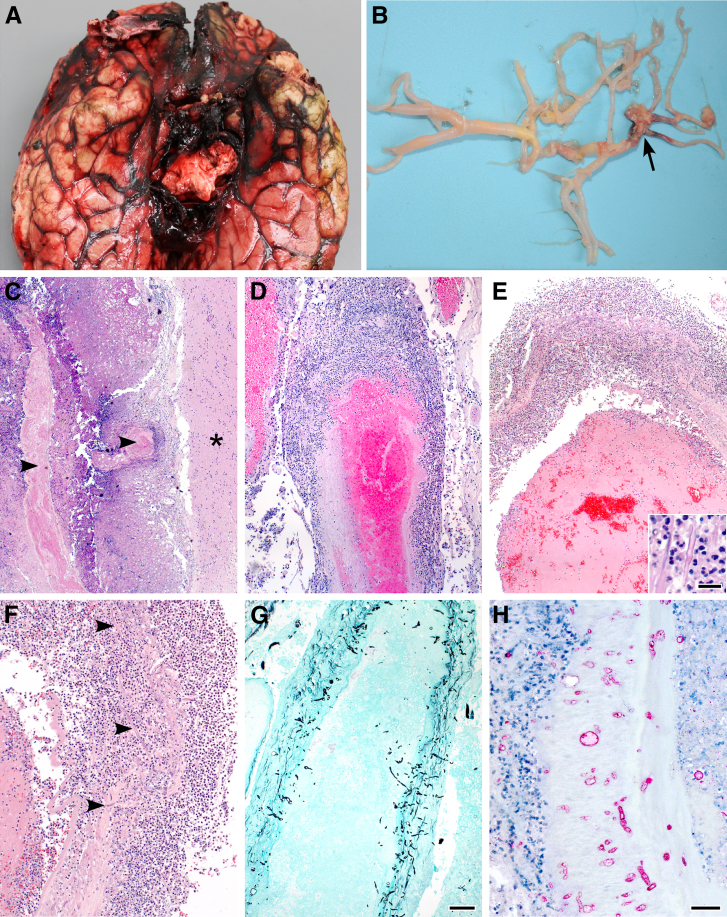
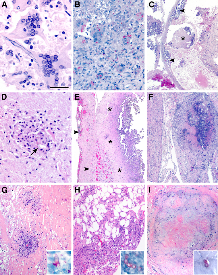
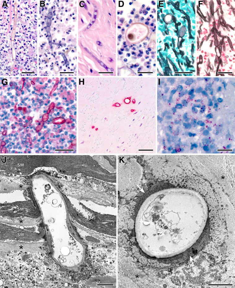
September 2012 marked the beginning of the largest reported outbreak of infections associated with epidural and intra-articular injections. Contamination of methylprednisolone acetate with the black mold, Exserohilum rostratum, was the primary cause of the outbreak, with >13,000 persons exposed to the potentially contaminated drug, 741 confirmed drug-related infections, and 55 deaths. Fatal meningitis and localized epidural, paraspinal, and peripheral joint infections occurred. Tissues from 40 laboratory-confirmed cases representing these various clinical entities were evaluated by histopathological analysis, special stains, and IHC to characterize the pathological features and investigate the pathogenesis of infection, and to evaluate methods for detection of Exserohilum in formalin-fixed, paraffin-embedded (FFPE) tissues. Fatal cases had necrosuppurative to granulomatous meningitis and vasculitis, with thrombi and abundant angioinvasive fungi, with extensive involvement of the basilar arterial circulation of the brain. IHC was a highly sensitive method for detection of fungus in FFPE tissues, demonstrating both hyphal forms and granular fungal antigens, and PCR identified Exserohilum in FFPE and fresh tissues. Our findings suggest a pathogenesis for meningitis involving fungal penetration into the cerebrospinal fluid at the injection site, with transport through cerebrospinal fluid to the basal cisterns and subsequent invasion of the basilar arteries. Further studies are needed to characterize Exserohilum and investigate the potential effects of underlying host factors and steroid administration on the pathogenesis of infection.


Figures

Figure 1
Figure 1
Most frequent gross and histopathological features of acute fatal Exserohilum meningitis. A: Hemorrhage around base of brain. B: Cerebral artery thrombus (arrow), as part of the circle of Willis. C: Expansion of meninges by necrosuppurative infiltrate and vascular thrombosis (arrowheads). The asterisk indicates brain parenchyma. D: Infiltration of a large meningeal arterial wall by neutrophils. E: Infiltration and destruction of the middle cerebral arterial wall by neutrophils (inset) and a large luminal thrombus. F: Fungal elements (arrowheads) admixed with the mural inflammatory infiltrate, higher magnification of same section as E. G: GMS stain highlights numerous fungal hyphae within a large arterial wall. H: Polyfungal IHC shows many fungi unaccompanied by inflammation in the tunica media of a large cerebral artery. H&E staining (CF); GMS staining (G); polyfungal immunoalkaline phosphatase staining, naphthol fast red substrate with hematoxylin counterstain (H). Original magnification: ×6.75 (C); ×12.5 (D and G); ×12.5 (E); ×158 (E, inset); ×25 (F); ×50 (H). Scale bars: 10 μm (E); 100 μm (G); 40 μm (H).
Figure 2
Figure 2
Additional features of Exserohilum meningitis (AD) and features of injection site pathology (EI). A: Multinucleated giant cells with intracytoplasmic fungal elements in a patient with granulomatous meningitis. B: Polyfungal IHC shows abundant fungal fragments within giant cells. C: Dissection of a basilar arterial wall by granulomatous infiltrates (arrowheads), with aneurysm formation (asterisk) as the result of accumulation of inflammatory debris within the tunica media. D: A single fungal fragment (arrow) within a granuloma in the deep pontine parenchyma. E: Disruption of the dura by chronic inflammation and neovascularization (arrowheads); necrosuppurative exudate subjacent to the dura (asterisks). F: Necrotizing granulomatous cauda equina neuritis associated with lumbar epidural steroid injection. G: Paraspinal collagenous tissue with necrosis and multiple foci of inflammation. Inset: A fragment of a fungal hypha within a focus of inflammation (Polyfungal IHC). H: Epidural adipose tissue with lymphohistiocytic infiltrates. Inset: Chain of fungal hyphae amid the inflammation (Polyfungal IHC). I: Necrotizing granuloma within a hip joint synovial biopsy specimen. Inset: Fungal hyphae in the central area of necrosis (Polyfungal IHC). H&E staining (A and CI); polyfungal immunoalkaline phosphatase staining, naphthol fast red substrate with hematoxylin counterstain (B and insets of G–I). Original magnification: ×158 (A); ×50 (B and D); ×6.75 (C and F); ×12.5 (E and I); ×25 (G and H). Scale bar = 30 μm (A).
Figure 3
Figure 3
Exserohilum morphological characteristics, detection by IHC, and ultrastructural features of arteritis. A: Pigmented, septate, branching fungal hyphae. B: Chains of yeast-like structures. C: Conidial forms. D: Large spore-like structures. E: GMS stain shows septate fungal hyphae with bulbous dilations. F: FM stain shows septate, branching hyphae of irregular width. G: IHC detection of Exserohilum amid an intense suppurative infiltrate. H: Lack of inflammatory response to Exserohilum in dense avascular connective tissue at paraspinal injection site. I: Detection of granular fungal antigen by IHC in the absence of fungal hyphae. J: Exserohilum penetrating multiple layers of smooth muscle (SM) and collagen (asterisks) in the tunica media of the basilar artery. K: Dissolution of collagen (asterisks) with replacement by electron-dense material (white arrows) surrounding an Exserohilum hypha in the adventitia of a large artery. H&E staining (AD); GMS staining (E); FM staining (F); polyfungal immunoalkaline phosphatase staining, naphthol fast red substrate with hematoxylin counterstain (GI); and thin-section electron microscopy, 4% uranyl acetate and lead citrate stain (J and K). Original magnification: ×100 (A–C and E–I); ×158 (D). Scale bars: 30 μm (A and B); 20 μm (CF and I); 40 μm (G and H); 2 μm (J and K).

Comment in

Similar articles

Cited by

References

    1. Carrino J.A., Morrison W.B., Parker L., Schweitzer M.E., Levin D.C., Sunshine J.H. Spinal injection procedures: volume, provider distribution, and reimbursement in the U.S. medicare population from 1993 to 1999. Radiology. 2002;225:723–729. - PubMed
    1. Windsor R.E., Storm S., Sugar R. Prevention and management of complications resulting from common spinal injections. Pain Physician. 2003;6:473–483. - PubMed
    1. Derby R., Lee S.H., Kim B.J., Chen Y., Seo K.S. Complications following cervical epidural steroid injections by expert interventionalists in 2003. Pain Physician. 2004;7:445–449. - PubMed
    1. Kabbara A., Rosenberg S.K., Untal C. Methicillin-resistant Staphylococcus aureus epidural abscess after transforaminal epidural steroid injection. Pain Physician. 2004;7:269–272. - PubMed
    1. Gaul C., Neundörfer B., Winterholler M. Iatrogenic (para-) spinal abscesses and meningitis following injection therapy for low back pain. Pain. 2005;116:407–410. - PubMed

MeSH terms