Review
. 2013:87:1-35.
doi: 10.1016/B978-0-12-407698-3.00001-6.

Mechanisms of reovirus bloodstream dissemination

Affiliations
Review

Mechanisms of reovirus bloodstream dissemination

Karl W Boehme et al. Adv Virus Res. 2013.

Abstract

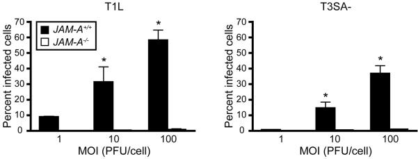
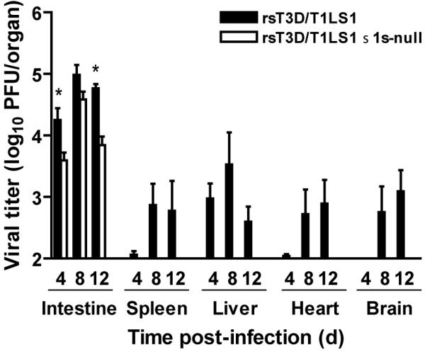
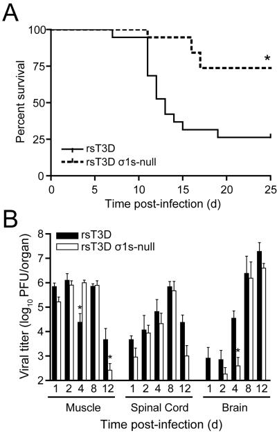
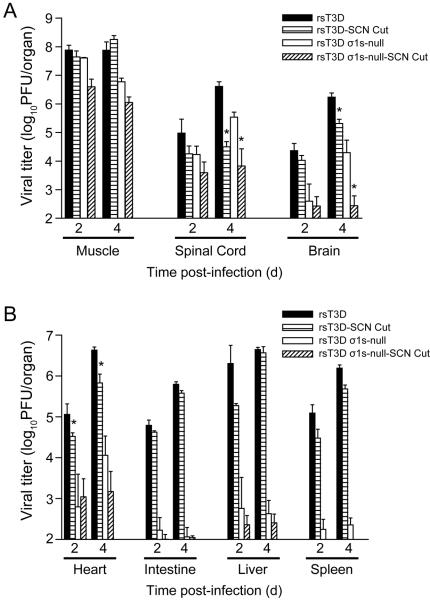
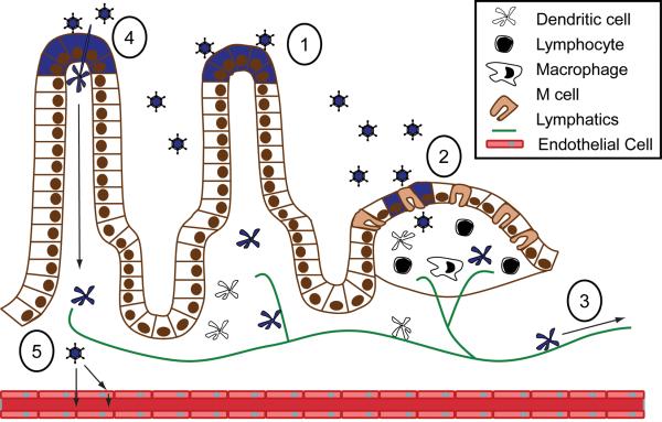
Many viruses cause disease within an infected host after spread from an initial portal of entry to sites of secondary replication. Viruses can disseminate via the bloodstream or through nerves. Mammalian orthoreoviruses (reoviruses) are neurotropic viruses that use both bloodborne and neural pathways to spread systemically within their hosts to cause disease. Using a robust mouse model and a dynamic reverse genetics system, we have identified a viral receptor and a viral nonstructural protein that are essential for hematogenous reovirus dissemination. Junctional adhesion molecule-A (JAM-A) is a member of the immunoglobulin superfamily expressed in tight junctions and on hematopoietic cells that serves as a receptor for all reovirus serotypes. Expression of JAM-A is required for infection of endothelial cells and development of viremia in mice, suggesting that release of virus into the bloodstream from infected endothelial cells requires JAM-A. Nonstructural protein σ1s is implicated in cell cycle arrest and apoptosis in reovirus-infected cells but is completely dispensable for reovirus replication in cultured cells. Surprisingly, a recombinant σ1s-null reovirus strain fails to spread hematogenously in infected mice, suggesting that σ1s facilitates apoptosis of reovirus-infected intestinal epithelial cells. It is possible that apoptotic bodies formed as a consequence of σ1s expression lead to reovirus uptake by dendritic cells for subsequent delivery to the mesenteric lymph node and the blood. Thus, both host and viral factors are required for efficient hematogenous dissemination of reovirus. Understanding mechanisms of reovirus bloodborne spread may shed light on how microbial pathogens invade the bloodstream to disseminate and cause disease in infected hosts.


Figures

Figure 1
Figure 1
The reovirus virion. (A) Cryo-electron micrograph image reconstruction of a reovirus virion. Outer-capsid protein σ3 (blue), is the initial target for virion disassembly in infected cells. Pentameric λ2 protein (yellow) forms an insertion pedestal for σ1, which is the viral attachment protein. Copyright © American Society for Microbiology, Journal of Virology, vol. 75, 2001, pages 146625–6634, doi: 10.1128/JVI.75.14.6625-6634.2001 (Nason et al., 2001). (B) Schematic of a reovirus virion. Reovirus particles are formed from two concentric protein shells, the outer capsid and core. The core contains the viral genome, which consists of ten dsRNA segments. Reovirus also encodes nonstructural proteins, σNS, μNS, μNSC, and σ1s.
Figure 2
Figure 2. Structure of σ1 and JAM-A
(A) Full-length model of attachment protein σ1 bound to JAM-A. A model of full-length σ1 extending from the virion is shown as a ribbon drawing, with the known structure of the C-terminus (Chappell et al., 2002) in tricolor and the predicted structure of the N-terminus in grey. Arrows indicate predicted regions of flexibility. A model of full-length JAM-A is shown in green as a ribbon drawing of the known structure of the extracellular domain (Prota et al., 2003) and a schematic representation of the transmembrane and intracellular domains. For clarity, only two JAM-A monomers are shown bound to σ1. Adapted from Kirchner et al. (2008, Fig. 1). (B) Structure of human JAM-A D1 and D2 domains. Ribbon drawings of a JAM-A homodimer, with one monomer shown in yellow and the other in green. Two orthogonal views are displayed. Adapted from Prota et al. (2003). Copyright (2003) National Academy of Sciences, USA.
Figure 3
Figure 3. JAM-A is required for hematogenous reovirus dissemination
(A) Newborn JAM-A+/+ and JAM-A−/− mice were inoculated perorally with 106 PFU of strain T1L. At days 4, 8, and 12 after inoculation, mice were euthanized, organs were resected, and viral titers were determined by plaque assay. Results are expressed as mean viral titers for 6 animals for each time point. Error bars indicate SD. *, P < 0.005 by Student's t test. When all values are less than the limit of detection (spleen, liver, heart, and brain in JAM-A−/− mice), a Student's t test P value cannot be calculated. (B) Newborn JAM-A+/+ and JAM-A−/− mice were inoculated perorally with 104 PFU of strain T3SA-. At days 4, 8, and 12 after inoculation, mice were euthanized, organs were resected, and viral titers were determined by plaque assay. Results are expressed as mean viral titers for 6–13 animals for each time point. Error bars indicate SD. *, P < 0.05 by Student's t test in comparison to JAM-A−/− mice.
Figure 4
Figure 4. JAM-A is required for reovirus viremia
Newborn JAM-A+/+ and JAM-A−/− mice were inoculated perorally with 108 PFU of T1L. At days 1, 2, 4, and 6 after inoculation, mice were euthanized, mesenteric lymph node (MLN), blood, and spleen were collected, and viral titers were determined by plaque assay. Results are expressed as mean viral titers for 3–8 animals for each time point. Error bars indicate SD.
Figure 5
Figure 5. JAM-A is required efficient reovirus infection of endothelial cells
JAMA+/+ and JAM-A−/− primary endothelial cells were adsorbed with T1L or T3SA- at MOIs of 1, 10, and 100 PFU/cell and incubated for 20 h. The percentage of infected cells was quantified by dividing the number of cells exhibiting reovirus staining by the total number of cell nuclei exhibiting DAPI staining in whole 96 wells for triplicate experiments. Wells contained between 200 and 1600 nuclei. Error bars indicate SD. *, P < 0.05 as determined by Student's t test in comparison to JAM-A−/− endothelial cells inoculated at the same MOI.
Figure 6
Figure 6. The σ1s protein is required for systemic reovirus dissemination following peroral inoculation
Newborn C57/BL6 mice were inoculated perorally with 104 PFU of wild-type or σ1s-null reovirus. At days 4, 8, and 12 post-inoculation, viral titers in the organs shown were determined by plaque assay. Error bars indicate SEM. *, P < 0.05 as determined by Mann-Whitney test in comparison to wild-type virus. When all values are less than the limit of detection, a Mann-Whitney test P value cannot be calculated. Adapted from Boehme et al. (2009). Copyright (2009) National Academy of Sciences, USA.
Figure 7
Figure 7
The σ1s protein enhances reovirus virulence following intramuscular inoculation. (A) Newborn C57/BL6 mice were inoculated in the left hindlimb with 106 PFU of wild-type or σ1s-null T3 reovirus. Mice (n = 19 for each virus strain) were monitored for survival for 25 days. *, P < 0.001 as determined by log-rank test in comparison to wild-type T3 reovirus. (B) The σ1s protein is not required for reovirus spread by neural routes. Newborn C57/BL6 mice were inoculated in the left hindlimb with 106 PFU of wild-type or σ1s-null T3 reovirus. At days 1, 2, 4, 8, and 12 post-inoculation, mice were euthanized, hindlimb muscle, spinal cord, and brain were resected, and viral titers were determined by plaque assay. Results are expressed as mean viral titers for 6–9 animals for each time point. Error bars indicate SEM. *, P < 0.05 as determined by Mann-Whitney test in comparison to wild-type T3 reovirus. Copyright © American Society for Microbiology, Journal of Virology, vol. 85, 2011, pages 11781 1790, doi: 10.1128/JVI.02289-10.
Figure 8
Figure 8. Reovirus disseminates to the CNS by hematogenous and neural routes
The left sciatic nerve of newborn C57/BL6 mice was sectioned prior to inoculation in the left hindlimb with 106 PFU of wild-type or σ1s-null T3 reovirus. In parallel, mice in which the left sciatic nerve was not sectioned were inoculated in the left hindlimb with 106 PFU of wild-type or σ1s-null T3 reovirus. At days 2 and 4 post-inoculation, mice were euthanized, (A) hindlimb muscle, spinal cord, and brain and (B) heart, intestine, liver, and spleen were resected, and viral titers were determined by plaque assay. Results are expressed as mean viral titers for 6 animals for each time point. Error bars indicate SEM. *, P < 0.05 as determined by Mann-Whitney test in comparison to animals in which the sciatic nerve was not sectioned. Copyright © American Society for Microbiology, Journal of Virology, vol. 85, 2011, pages 11781 1790, doi: 10.1128/JVI.02289-10.
Figure 9
Figure 9. Model of reovirus hematogenous spread from the intestine
(1) Following peroral inoculation, reovirus infects intestinal epithelial cells (2) and is taken up by lymphoid cells in the Peyer patch. (3) Infected dendritic cells or lymphocytes carry reovirus from the Peyer patch through the lymphatics and finally to the blood. (4) Phagocytic cells that extend processes into the lumen of the intestine also might be infected for subsequent transport of virus through the lymphatics. (5) Reovirus may enter directly into the blood by passing between endothelial cells or via release into the bloodstream from infected cells.

Similar articles

Cited by

References

    1. Adair RA, Roulstone V, Scott KJ, Morgan R, Nuovo GJ, Fuller M, Beirne D, West EJ, Jennings VA, Rose A, Kyula J, Fraser S, Dave R, Anthoney DA, Merrick A, Prestwich R, Aldouri A, Donnelly O, Pandha H, Coffey M, Selby P, Vile R, Toogood G, Harrington K, Melcher AA. Cell carriage, delivery, and selective replication of an oncolytic virus in tumor in patients. Science translational medicine. 2012;4:138ra177. - PMC - PubMed
    1. Amerongen HM, Wilson GAR, Fields BN, Neutra MR. Proteolytic processing of reovirus is required for adherence to intestinal M cells. J. Virol. 1994;68:8428–8432. - PMC - PubMed
    1. Antar AAR, Konopka JL, Campbell JA, Henry RA, Perdigoto AL, Carter BD, Pozzi A, Abel TW, Dermody TS. Junctional adhesion molecule-A is required for hematogenous dissemination of reovirus. Cell Host Microbe. 2009;5:59–71. - PMC - PubMed
    1. Armstrong GD, Paul RW, Lee PW. Studies on reovirus receptors of L cells: virus binding characteristics and comparison with reovirus receptors of erythrocytes. Virology. 1984;138:37–48. - PubMed
    1. Barratt-Boyes SM, MacLachlan NJ. Dynamics of viral spread in bluetongue virus infected calves. Vet. Microbiol. 1994;40:361–371. - PubMed

Publication types

LinkOut - more resources