

Normal standards for computer-ECG programs for prognostically and diagnostically important ECG variables derived from a large ethnically diverse female cohort: the Women's Health Initiative (WHI)
- PMID: 23809992
- PMCID: PMC3825808
- DOI: 10.1016/j.jelectrocard.2013.05.136
Normal standards for computer-ECG programs for prognostically and diagnostically important ECG variables derived from a large ethnically diverse female cohort: the Women's Health Initiative (WHI)
Abstract
Background: Substantial new information has emerged recently about the prognostic value for a variety of new ECG variables. The objective of the present study was to establish reference standards for these novel risk predictors in a large, ethnically diverse cohort of healthy women from the Women's Health Initiative (WHI) study.
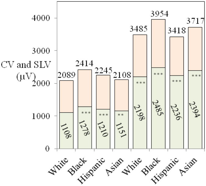
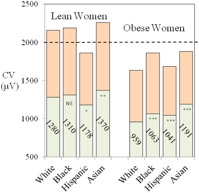
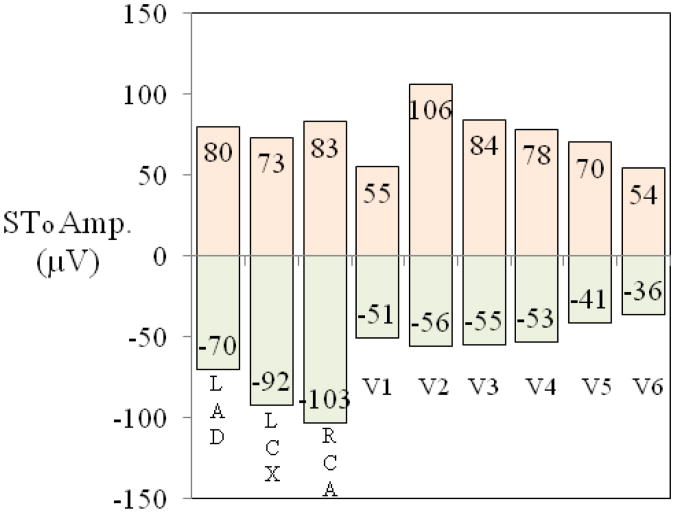
Methods and results: The study population consisted of 36,299 healthy women. Racial differences in rate-adjusted QT end (QT(ea)) and QT peak (QT(pa)) intervals as linear functions of RR were small, leading to the conclusion that 450 and 390 ms are applicable as thresholds for prolonged and shortened QT(ea) and similarly, 365 and 295 ms for prolonged and shortened QT(pa), respectively. As a threshold for increased dispersion of global repolarization (T(peak)T(end) interval), 110 ms was established for white and Hispanic women and 120 ms for African-American and Asian women. ST elevation and depression values for the monitoring leads of each person with limb electrodes at Mason-Likar positions and chest leads at level of V1 and V2 were first computed from standard leads using lead transformation coefficients derived from 892 body surface maps, and subsequently normal standards were determined for the monitoring leads, including vessel-specific bipolar left anterior descending, left circumflex artery and right coronary artery leads. The results support the choice 150 μV as a tentative threshold for abnormal ST-onset elevation for all monitoring leads. Body mass index (BMI) had a profound effect on Cornell voltage and Sokolow-Lyon voltage in all racial groups and their utility for left ventricular hypertrophy classification remains open.
Conclusions: Common thresholds for all racial groups are applicable for QT(ea), and QT(pa) intervals and ST elevation. Race-specific normal standards are required for many other ECG parameters.
Keywords: Electrocardiogram; Monitoring; Normal standards; QT; ST; TpTe.
Copyright © 2013 Elsevier Inc. All rights reserved.
Figures
References
-
- Rautaharju PM, Kooperberg C, Larson JC, LaCroix A. Electrocardiographic abnormalities that predict coronary heart disease events and mortality in postmenopausal women: the Women's Health Initiative. Circulation. 2006;113:473. - PubMed
-
- Rautaharju PM, Prineas RJ, Wood J, et al. Electrocardiographic predictors of new-onset heart failure in men and in women free of coronary heart disease (from the Atherosclerosis in Communities [ARIC] Study) Am J Cardiol. 2007;100:1437. - PubMed
-
- Aro AL, Anttonen O, Tikkanen JT, et al. Intraventricular conduction delay in a standard 12-lead electrocardiogram as a predictor of mortality in the general population. Circulation Arrhythm Electrophysiol. 2011;4:704. - PubMed
-
- Kurl S, Mäkikallio TH, Rautaharju P, et al. Duration of QRS complex in resting electrocardiogram is a predictor of sudden cardiac death in men. Circulation. 2012;125:2588. - PubMed
Publication types
MeSH terms
Grants and funding
LinkOut - more resources
Full Text Sources
Other Literature Sources
Medical
Research Materials
