. 2013 Jul 11;93(1):150-7.
doi: 10.1016/j.ajhg.2013.05.023. Epub 2013 Jun 27.

SHORT syndrome with partial lipodystrophy due to impaired phosphatidylinositol 3 kinase signaling

Affiliations

SHORT syndrome with partial lipodystrophy due to impaired phosphatidylinositol 3 kinase signaling

Kishan Kumar Chudasama et al. Am J Hum Genet. .

Abstract

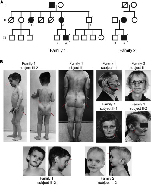
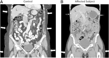
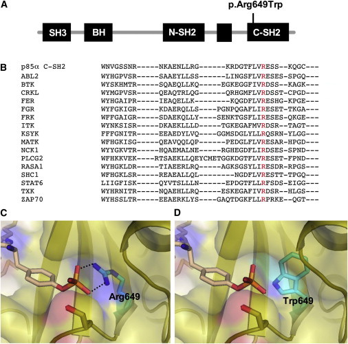
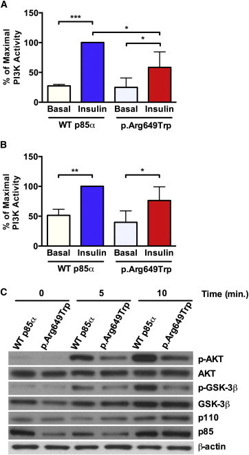
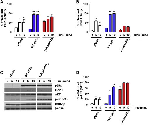
The phosphatidylinositol 3 kinase (PI3K) pathway regulates fundamental cellular processes such as metabolism, proliferation, and survival. A central component in this pathway is the p85α regulatory subunit, encoded by PIK3R1. Using whole-exome sequencing, we identified a heterozygous PIK3R1 mutation (c.1945C>T [p.Arg649Trp]) in two unrelated families affected by partial lipodystrophy, low body mass index, short stature, progeroid face, and Rieger anomaly (SHORT syndrome). This mutation led to impaired interaction between p85α and IRS-1 and reduced AKT-mediated insulin signaling in fibroblasts from affected subjects and in reconstituted Pik3r1-knockout preadipocytes. Normal PI3K activity is critical for adipose differentiation and insulin signaling; the mutated PIK3R1 therefore provides a unique link among lipodystrophy, growth, and insulin signaling.


Figures

Figure 1
Figure 1
Pedigrees and Photos of Affected Family Members (A) Pedigrees of Norwegian family 1 and German family 2. Filled symbols represent affected family members, and open symbols represent healthy individuals. DNA from subject I-1 of family 1 was unavailable for testing. The five other affected family members were investigated and had the mutation. Healthy subjects II-3 and III-1 of family 1 and subject III-1 of family 2 did not have the mutation. Circular symbols represent females, and squares represent males. Arrows represent the probands of each family. Stars indicate those individuals from whom DNA was available. (B) The photos reveal the key clinical signs. Partial lipodystrophy is especially noticeable in the face and the buttocks, and dysmorphic features, including a progeroid face with hypotrichosis (thin hair), frontal bossing, wide and deep-set eyes, thin ala nasi, a beaked nose, downturned lateral corners of the mouth, and large ears, are evident. Red arrows point to the typical features. Photos are printed with permission from John Wiley & Sons and Wolters Kluwer Health.
Figure 2
Figure 2
CT Scans Coronal reconstruction of contrast-enhanced CT scans of the abdomen and proximal lower extremities of subject III-2 of family 1 (B) and a 79-year-old healthy female control (A). In the affected person (B), the amount of subcutaneous fat (arrows) is strikingly sparse in the upper abdomen and lower thorax (closed arrows), whereas it seems normal in the buttocks and thighs (filled arrows). In the control (A), there is proportional distribution of subcutaneous fat (arrows) in the thorax, abdomen, and pelvis. The affected individual had normal liver attenuation (62 versus 8 Hounsfield units in a subject with fatty liver), indicating no signs of hepatic steatosis (not shown). The CT scans were performed according to standard procedures.
Figure 3
Figure 3
Overview of the p85 Protein with the p.Arg649Trp Substitution and SH2 Sequences of Various Proteins Related to p85 (A) Schematic illustration of p85α, the different domains, and the position of the p.Arg649Trp substitution. (B) Multiple-sequence alignment of SH2 domains from the indicated human proteins was performed with the Clustal Omega sequence-alignment tool and demonstrated absolute conservation of the arginine residue (red color). (C and D) Illustration that the p.Arg649Trp-containing protein impairs binding with the phosphotyrosine phosphate moiety. The panels show the substrate-binding pocket of the p85α SH2 domain (Protein Data Bank ID 1H90) with important residues for recognition of the phosphotyrosine of a platelet-derived growth-factor-receptor peptide (pYMPMS). (C) Arg649 in the wild-type (WT) p85α SH2 domain is critical for the formation of a bidentate interaction with the phosphotyrosine phosphate. Represented by black dotted lines, the distances between the amino group of Arg649 and the phosphate group of pTyr are 2.7 Å (top) and 2.8 Å (bottom). (D) Introduction of the p.Arg649Trp substitution causes loss of this key interaction for substrate recognition.
Figure 4
Figure 4
The p85α p.Arg649Trp Substitution Leads to Impaired Insulin Signaling in Primary Human Fibroblasts (A and B) PI3K activity was evaluated in (A) anti-phosphotyrosine (PY) or (B) anti-IRS-1 immunoprecipitates in WT (normal) and p.Arg649Trp human fibroblast cell lines before and after insulin stimulation (10 mM) for 10 min. Data are expressed as a percentage of maximal stimulation, and p values were calculated by ordinary one-way ANOVA testing (p < 0.05, ∗∗∗p < 0.001) (n = 3, data are represented as mean ± SEM). (C) Immunoblot analysis of lysates obtained from WT (normal) and p.Arg649Trp fibroblasts before and after insulin stimulation was performed with the indicated antibodies. Representative immunoblots are shown.
Figure 5
Figure 5
Assessment of PI3K Pathway Activation in WT and p.Arg649Trp-Reconstituted Cell Lines (A and B) PI3K activity was measured in (A) anti-phosphotyrosine or (B) anti-IRS-1 immunoprecipitates before and after insulin stimulation (10 mM) for 10 min. (C) Immunoblot analysis using the indicated antibodies was performed on cell lysates obtained from cell lines at the indicated time points after insulin stimulation (10 mM). Representative immunoblots are shown. (D) Quantification of AKT phosphorylation (S473) was performed with ImageJ. Data are expressed as a percentage of maximal stimulation, and p values were calculated by ordinary one-way ANOVA testing (p < 0.05, ∗∗p < 0.01) (n = 3, data are represented as mean ± SEM).

References

    1. Aubin D., Gagnon A., Sorisky A. Phosphoinositide 3-kinase is required for human adipocyte differentiation in culture. Int J Obes (Lond) 2005;29:1006–1009. - PubMed
    1. Engelman J.A., Luo J., Cantley L.C. The evolution of phosphatidylinositol 3-kinases as regulators of growth and metabolism. Nat. Rev. Genet. 2006;7:606–619. - PubMed
    1. Ueki K., Fruman D.A., Yballe C.M., Fasshauer M., Klein J., Asano T., Cantley L.C., Kahn C.R. Positive and negative roles of p85 alpha and p85 beta regulatory subunits of phosphoinositide 3-kinase in insulin signaling. J. Biol. Chem. 2003;278:48453–48466. - PubMed
    1. Miki H., Yamauchi T., Suzuki R., Komeda K., Tsuchida A., Kubota N., Terauchi Y., Kamon J., Kaburagi Y., Matsui J. Essential role of insulin receptor substrate 1 (IRS-1) and IRS-2 in adipocyte differentiation. Mol. Cell. Biol. 2001;21:2521–2532. - PMC - PubMed
    1. Taniguchi C.M., Emanuelli B., Kahn C.R. Critical nodes in signalling pathways: insights into insulin action. Nat. Rev. Mol. Cell Biol. 2006;7:85–96. - PubMed

Publication types

MeSH terms

Substances

Supplementary concepts