. 2013 Nov;34(11):2585-603.
doi: 10.1016/j.neurobiolaging.2013.05.026. Epub 2013 Jun 28.

Clearance of the mutant androgen receptor in motoneuronal models of spinal and bulbar muscular atrophy

Affiliations

Clearance of the mutant androgen receptor in motoneuronal models of spinal and bulbar muscular atrophy

Paola Rusmini et al. Neurobiol Aging. 2013 Nov.

Abstract

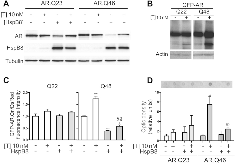
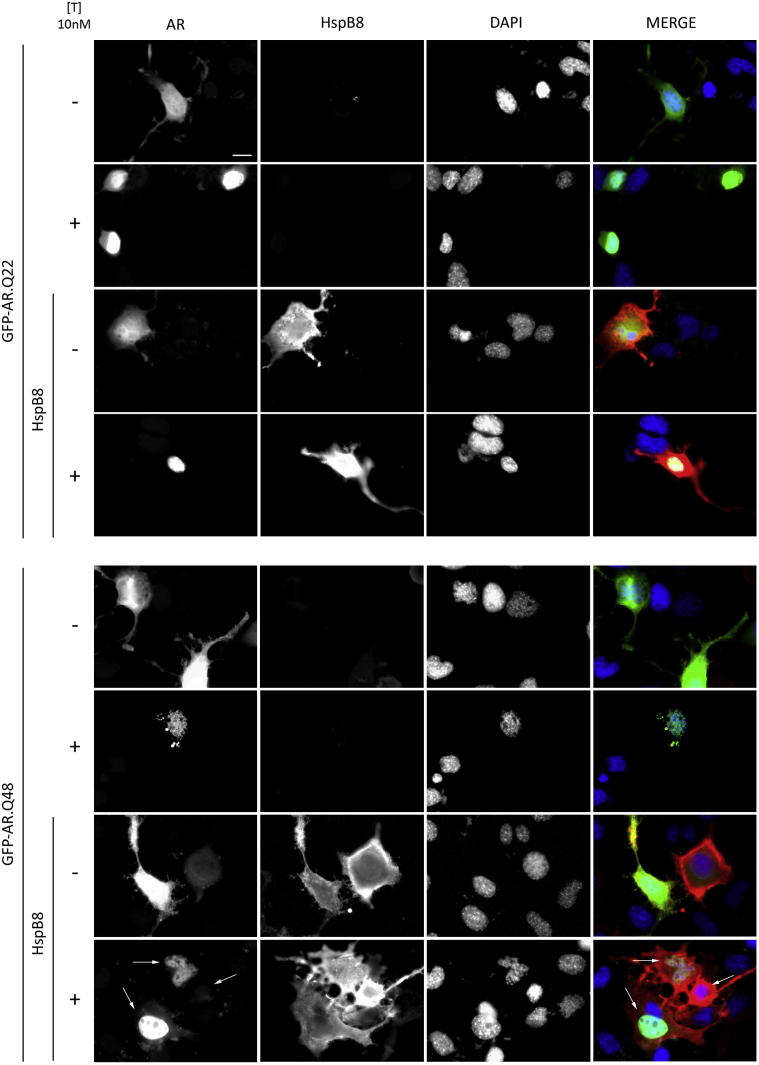
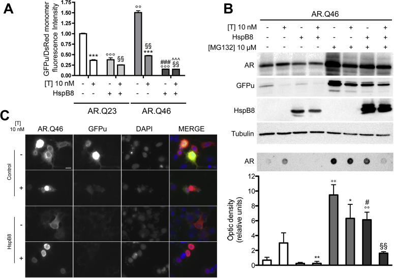
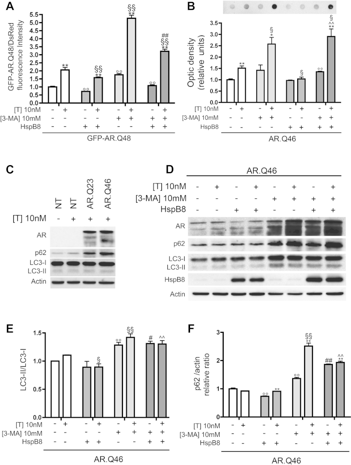
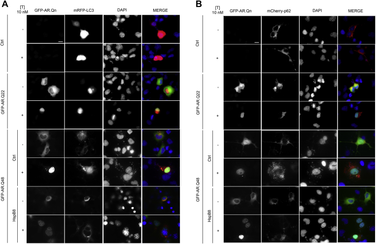
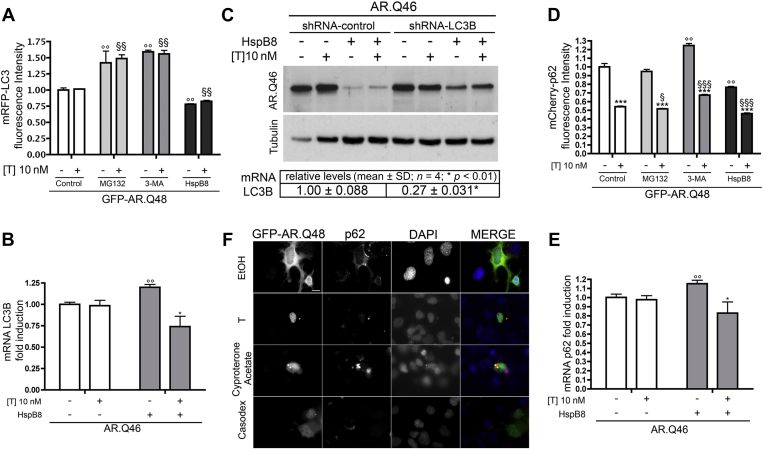
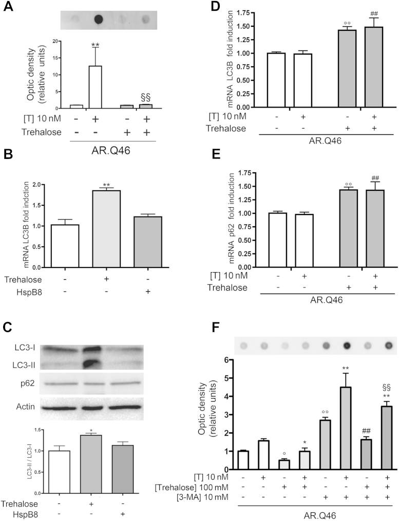
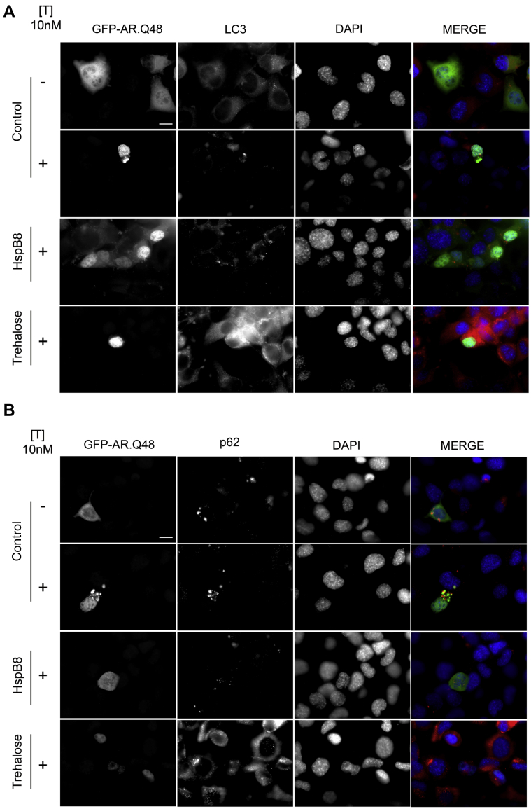
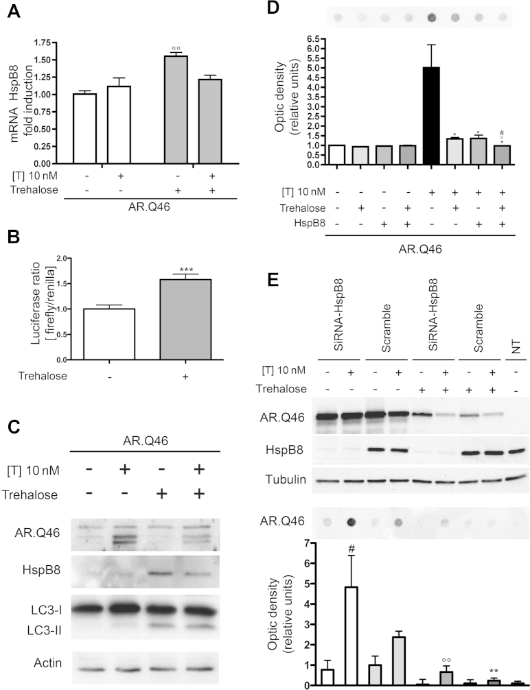
Spinal and bulbar muscular atrophy (SBMA) is an X-linked motoneuron disease caused by an abnormal expansion of a tandem CAG repeat in exon 1 of the androgen receptor (AR) gene that results in an abnormally long polyglutamine tract (polyQ) in the AR protein. As a result, the mutant AR (ARpolyQ) misfolds, forming cytoplasmic and nuclear aggregates in the affected neurons. Neurotoxicity only appears to be associated with the formation of nuclear aggregates. Thus, improved ARpolyQ cytoplasmic clearance, which indirectly decreases ARpolyQ nuclear accumulation, has beneficial effects on affected motoneurons. In addition, increased ARpolyQ clearance contributes to maintenance of motoneuron proteostasis and viability, preventing the blockage of the proteasome and autophagy pathways that might play a role in the neuropathy in SBMA. The expression of heat shock protein B8 (HspB8), a member of the small heat shock protein family, is highly induced in surviving motoneurons of patients affected by motoneuron diseases, where it seems to participate in the stress response aimed at cell protection. We report here that HspB8 facilitates the autophagic removal of misfolded aggregating species of ARpolyQ. In addition, though HspB8 does not influence p62 and LC3 (two key autophagic molecules) expression, it does prevent p62 bodies formation, and restores the normal autophagic flux in these cells. Interestingly, trehalose, a well-known autophagy stimulator, induces HspB8 expression, suggesting that HspB8 might act as one of the molecular mediators of the proautophagic activity of trehalose. Collectively, these data support the hypothesis that treatments aimed at restoring a normal autophagic flux that result in the more efficient clearance of mutant ARpolyQ might produce beneficial effects in SBMA patients.

Keywords: Androgen receptor; CAG repeat; Chaperones; HspB8; Motoneuron; Motoneuron disease; Neurodegeneration; Polyglutamine; Protein misfolding; Spinal and bulbar muscular atrophy.


Figures

Fig. 1
Fig. 1
Effect of heat shock protein (Hsp)B8 on wild type (wt) and mutant androgen receptor (AR) gene with an abnormally long polyglutamine tract (ARpolyQ) aggregation and clearance. (A) Western blot analysis on cell lysates of NSC34 expressing wt AR (AR.Q23) or mutant ARpolyQ (AR.Q46), pCDNA3 or HspB8 in the absence (−T) or in the presence (+T) of 10 nM of T for 48 hours. α-tubulin was used to normalize protein loading. (B) Western blot analysis performed on NSC34 cells transfected with GFP-AR.Q22 or GFP-AR.Q48, in the absence (−T) or in the presence (+T) of 10 nM of T for 48 hours. β-actin was used to normalize protein loading. (C) Cytofluorimetric analysis performed on NSC34 expressing DsRed monomer, GFP-AR.Q22, or GFP-AR.Q48, pCDNA3, or HspB8 in the absence (−T) or in the presence (+T) of 10 nM of T for 48 hours (* p < 0.05 vs. T-untreated controls; ** p < 0.01 vs. T-untreated controls; °° p < 0.01 vs. GFP-AR.Q48−T; §§ p < 0.01 vs. GFP-AR.Q48+T). (D) Filter retardation assay performed on NSC34 cells transfected with AR.Q23 or AR.Q46, pCDNA3, or HspB8 in the absence (−T) or in the presence (+T) of 10 nM of T for 48 hours (** p < 0.01 vs. T-untreated controls; §§ p < 0.01 vs. AR.Q46+T). Abbreviations: T, testosterone; pCDNA3, empty vector.
Fig. 2
Fig. 2
Effect of heat shock protein (Hsp)B8 on aggregation and intracellular distribution of wild type (wt) and mutant androgen receptor (AR) gene with an abnormally long polyglutamine tract (ARpolyQ). High resolution immunofluorescence microscopy analysis (magnification ×63) on NSC34 cells transfected with GFP-AR.Q22 or GFP-ARQ.48 (green), pCDNA3 or HspB8 (red) in the absence (−T) or in the presence (+T) of 10 nM of T for 48 hours. Nuclei were stained with DAPI (blue) (scale bar, 10 μm). Abbreviations: DAPI, 4′,6-diamidino-2-phenylindole; pCDNA3, empty vector; T, testosterone.
Fig. 3
Fig. 3
.The antiaggregation/prodegradative activity of heat shock protein (Hsp)B8 on androgen receptor (AR) gene with an abnormally long polyglutamine tract (ARpolyQ) does not require the ubiquitin proteasome system. (A) Cytofluorimetric analysis performed on NSC34 cells expressing GFPu, DsRed monomer, AR.Q23 or AR.Q46, pCDNA3, or HspB8 in the absence (−T) or in the presence (+T) of 10 nM of T for 48 hours (*** p < 0.001 vs. T-untreated controls; °°° p < 0.001 vs. AR.Q23−T; §§ p < 0.01 vs. AR.Q23+T; ### p < 0.001 vs. AR.Q46−T; ˆˆˆ p < 0.001 vs. AR.Q46+T; °° p < 0.01 vs. AR.Q23−T). (B) Upper insets, Western blot analysis, and lower insets, filter retardation assay, on cell lysates of NSC34 cells expressing GFPu, AR.Q46, pCDNA3, or HspB8 in the absence (−T) or in the presence (+T) of 10 nM of T for 48 hours, in basal condition or after treatment with 10 μM of MG132 for 24 hours. α-Tubulin was used to normalize protein loading. (°° p < 0.01 vs. T-untreated controls; ** p < 0.01 vs. T-treated controls; * p < 0.05 vs. T-treated controls; # p < 0.05 vs. −T/+MG132; §§ p < 0.01 vs. +T/+MG132). (C) High resolution immunofluorescence analysis of NSC34 cells transfected with GFPu, AR.Q46, pCDNA3, or HspB8 in the absence (−T) or in the presence (+T) of 10 nM of T for 48 hours. Nuclei were stained with DAPI (blue). Images were obtained at magnification ×63 (scale bar, 10 μm). Abbreviations: DAPI, 4,6-diamidino-2-phenylindole; GFPu, green fluorescent protein fused to a constitutive degron signal, CL1; MG132, Z-leu-leu-leu-al; pCDNA3, empty vector; T, testosterone.
Fig. 4
Fig. 4
Active autophagy is required to mediate the antiaggregation and/or prodegradative activity of heat shock protein (Hsp)B8 on the androgen receptor (AR) gene with an abnormally long polyglutamine tract (ARpolyQ). (A) Cytofluorimetric analysis performed on NSC34 cells expressing DsRed monomer, GFP-AR.Q48, pCDNA3, or HspB8 in the absence (−T) or in the presence (+T) of 10 nM of T for 48 hours, in basal condition or after treatment with 10 mM of 3-methyladenine (3-MA) for 24 hours (** p < 0.01 vs. T-untreated controls; °° p < 0.01 vs. GFP-AR.Q48−T; §§ p < 0.01 vs. GFP-AR.Q48+T; ## p < 0.01 vs. GFP-AR.Q48+T+HspB8). (B) Filter retardation assay performed on NSC34 cells transfected with AR.Q46, pCDNA3, or HspB8 in the absence (−T) or in the presence (+T) of 10 nM of T for 48 hours, in basal condition or after treatment with 10 mM of 3-MA for 24 hours (* p < 0.05 vs. T-untreated controls; ** p < 0.01 vs. T-untreated controls; °° p < 0.01 vs. AR.Q46−T; § p < 0.05 vs. AR.Q46+T; ˆˆ p < 0.01 vs. AR.Q46+T+HspB8). (C) Western blot analysis on cell lysates of NSC34 cells untrasfected (NT) or expressing AR.Q23 or AR.Q46, in the absence (−T) or in the presence (+T) of 10 nM of T for 48 hours. β-actin was used to normalize protein loading. (D) Western blot analysis on cell lysates of NSC34 cells expressing AR.Q46, pCDNA3 or HspB8 in the absence (−T) or in the presence (+T) of 10 nM of T for 48 hours, in basal condition or after treatment with 10 mM of 3-MA for 24 hours. β-actin was used to normalize protein loading. (E) The histogram represents a quantitative evaluation of LC3-II/LC3-I ratio protein level carried out by densitometric scanning of the blots (3 replicates) (°° p < 0.01 vs. AR.Q46−T; § < 0.05 vs. AR.Q46+T; §§ p < 0.01 vs. AR.Q46+T; # p < 0.05 vs. AR.Q46−T+HspB8; ˆˆ p < 0.005 vs. AR.Q46+T+HspB8). (F) The histogram represents a quantitative evaluation of p62 protein level normalized on actin carried out by densitometric scanning of the blots (3 replicates) (°° p < 0.01 vs. AR.Q46−T; §§ p < 0.01 vs. AR.Q46+T; ** p < 0.01 vs. T-untreated controls; ## p < 0.01 vs. AR.Q46−T+HspB8; ˆˆ p < 0.01 vs. AR.Q46+T+HspB8). Abbreviations: pCDNA3, empty vector; T, testosterone.
Fig. 5
Fig. 5
Autophagy is activated by the presence of androgen receptor (AR) gene with an abnormally long polyglutamine tract (ARpolyQ), but not by heat shock protein (Hsp)B8. (A) High-resolution fluorescence microscopy analysis (magnification ×63) on NSC34 cells expressing mRFP-LC3, and pCDNA3, GFP-AR.Q22 or GFP-AR.Q48 (−/+HspB8), in the absence (−T) or in the presence (+T) of 10 nM of T. Nuclei were stained with DAPI (blue) (scale bar, 10 μm). (B) High-resolution fluorescence microscopy analysis (magnification ×63) on NSC34 cells expressing mCherry-p62 and pCDNA3, GFP-AR.Q22 or GFP-AR.Q48 (−/+HspB8) in the absence (−T) or in the presence (+T) of 10 nM of T. Nuclei were stained with DAPI (blue) (scale bar, 10 μm). Abbreviations: Ctrl, control; DAPI, 4′,6-diamidino-2-phenylindole; GFP, green fluorescent protein; mRFP-LC3, monomeric red fluorescent protein; pCDNA3, empty vector; T, testosterone.
Fig. 6
Fig. 6
Autophagic flux is impaired by the mutant androgen receptor (AR) gene with an abnormally long polyglutamine tract (ARpolyQ) and restored by heat shock protein (Hsp)B8. (A) Cytofluorimetric analysis performed on NSC34 cells expressing mRFP-LC3, GFP-AR.Q48, pCDNA3, or HspB8 in the absence (−T) or in the presence (+T) of 10 nM of T for 48 hours, in basal condition or after treatment with 10 μM of MG132 or 10 mM of 3-methyladenine (3-MA) for 24 hours (°° p < 0.01 vs. GFP-AR.Q48−T; §§ p < 0.01 vs. GFP-AR.Q48+T). (B) Real-time polymerase chain reaction (PCR) on LC3B mRNA expression levels on NSC34 cells expressing AR.Q46, pCDNA3, or HspB8 in the absence (−T) or in the presence (+T) of 10 nM of T for 48 hours (°° p < 0.01 vs. AR.Q46−T; * p < 0.05 vs. AR.Q46+T). (C) Top insets, Western blot analysis of cell lysates of NSC34 cells expressing AR.Q46, pCDNA3, or HspB8 and shRNA against LC3B or shRNA scrambled control in the absence (−T) or in the presence (+T) of 10 nM of T for 48 hours. α-Tubulin was used to normalize protein loading. Bottom inset, real time PCR on LC3B mRNA levels on NSC34 cells expressing shRNA-LC3B or shRNA-control. Data (expressed as fold changes) have been normalized to the amount of GAPDH mRNA, and are expressed as relative to the levels determined in shRNA-control transfected cells, which are taken as internal reference. Data are mean ± SD of 4 independent replicates. (D) Cytofluorimetric analysis performed on NSC34 expressing mCherry-p62, GFP-AR.Q48, pCDNA3, or HspB8 in the absence (−T) or in the presence (+T) of 10 nM of T for 48 hours, in basal condition or after treatment with 10 μM of MG132 or 10 mM of 3-MA for 24 hours (*** p < 0.001 vs. T-untreated controls; °° p < 0.01 vs. GFP-AR.Q48−T; § p < 0.05 vs. GFP-AR.Q48+T; §§§ p < 0.001 vs. GFP-AR.Q48+T). (E) Real-time PCR on p62 mRNA expression levels on NSC34 cells expressing AR.Q46, pCDNA3, or HspB8 in the absence (−T) or in the presence (+T) of 10 nM of T for 48 hours (°° p < 0.01 vs. AR.Q46−T; * p < 0.05 vs. AR.Q46−T+HspB8). (F) High resolution immunofluorescence analysis on NSC34 cells transfected with GFP-AR.Q48 in the absence (−T) or in the presence (+T) of 10 nM of T or 100 nM of cyproterone acetate or 100 nM of casodex for 48 hours. Nuclei were stained with DAPI (blue). Images were obtained at magnification ×63 (scale bar, 10 μm). Abbreviations: DAPI, 4′,6-diamidino-2-phenylindole; GAPDH, glyceraldehyde-3-phosphate dehydrogenase; MG132, Z-leu-leu-leu-al; mRFP-LC3, monomeric red fluorescent protein-LC3; mRNA, messenger RNA; pCDNA3, empty vector; shRNA, short hairpin RNA; T, testosterone.
Fig. 7
Fig. 7
Trehalose induces the clearance of mutant androgen receptor (AR) gene with an abnormally long polyglutamine tract (ARpolyQ), via autophagy activation. (A) Filter retardation assay performed on NSC34 cells transfected with AR.Q46 in the absence (−T) or in the presence (+T) of 10 nM of T for 48 hours, in basal condition or after treatment with 100 mM of trehalose for 48 hours (** p < 0.01 vs. T-untreated control; §§ p < 0.01 vs. AR.Q46+T). (B) Real-time polymerase chain reaction (PCR) on LC3B mRNA expression levels on NSC34 cells expressing pCDNA3 or heat shock protein (Hsp)B8 in basal condition or after treatment with 100 mM of trehalose for 48 hours (** p < 0.01 vs. untreated control). (C) Upper insets, Western blot analysis of cell lysates of NSC34 expressing pCDNA3 or HspB8 in basal condition or after treatment with 100 mM of trehalose for 48 hours. β-actin was used to normalize protein loading. Lower inset, the histogram represents a quantitative evaluation of LC3-II/LC3-I ratio protein level carried out by densitometric scanning of the blots (3 replicates) (* p < 0.05 vs. untreated control). (D) Real-time PCR on LC3B mRNA expression levels on NSC34 cells expressing AR.Q46 in the absence (−T) or in the presence (+T) of 10 nM of T for 48 hours, in basal condition or after treatment with 100 mM of trehalose for 48 hours (°° p < 0.01 vs. AR.Q46−T; ## p < 0.01 vs. AR.Q46+T). (E) Real-time PCR of p62 mRNA expression levels on NSC34 cells expressing AR.Q46 in the absence (−T) or in the presence (+T) of 10 nM of T for 48 hours, in basal condition or after the treatment with 100 mM of trehalose for 48 hours (°° p < 0.01 vs. AR.Q46−T; ## p < 0.01 vs. AR.Q46+T). (F) Filter retardation assay performed on cell lysates of NSC34 cells expressing AR.Q46, in the absence (−T) or in the presence (+T) of 10 nM of T for 48 hours, in basal condition or after treatment with 10 mM of 3-MA or 100 mM of trehalose for 24 hours. (° p < 0.05 and °° p < 0.01 vs. AR.Q46−T; * p < 0.05 and ** p < 0.01 vs. AR.Q46+T; ## p < 0.01 vs. AR.Q46−T/+trehalose; §§ p < 0.01 vs. AR.Q46+T/+trehalose). Abbreviations: mRNA, messenger RNA; pCDNA3, empty vector; T, testosterone.
Fig. 8
Fig. 8
Heat shock protein (Hsp)B8 and trehalose exert different activities on the autophagic removal of androgen receptor (AR) gene with an abnormally long polyglutamine tract (ARpolyQ). (A) and (B) High resolution immunofluorescence analysis of NSC34 cells transfected with GFP-AR.Q48, pCDNA3, or HspB8 in the absence (−T) or in the presence (+T) of 10 nM of T for 48 hours, in basal condition or after treatment with 100 mM of trehalose for 48 hours. Nuclei were stained with DAPI (blue). Images obtained at magnification ×63 (scale bar, 10 μm). Abbreviations: DAPI, 4,6-diamidino-2-phenylindole; pCDNA3, empty vector; T, testosterone.
Fig. 9
Fig. 9
Trehalose induces heat shock protein (Hsp)B8 expression and both act in the autophagic clearance of mutant androgen receptor (AR) gene with an abnormally long polyglutamine tract (ARpolyQ). (A) Real-time polymerase chain reaction of HspB8 mRNA expression levels on NSC34 cells expressing AR.Q46 in the absence (−T) or in the presence (+T) of 10 nM of T for 48 hours, in basal condition or after treatment with 100 mM of trehalose for 48 hours (°° p < 0.01 vs. AR.Q46−T). (B) Transcriptional activity assay of HspB8 promoter performed on NSC34 cells transfected with promB8 and pRL-TK, in basal condition or after treatment with 100 mM of trehalose for 48 hours (*** p < 0.001 vs. untreated control). (C) Western blot analysis on cell lysates of NSC34 cells expressing AR.Q46 in the absence (−T) or in the presence (+T) of 10 nM of T for 48 hours, in basal condition or after treatment with 100 mM of trehalose for 48 hours. β-actin was used to normalize protein loading. (D) Filter retardation assay performed on NSC34 cells transfected with AR.Q46, pCDNA3, or HspB8, in the absence (−T) or in the presence (+T) of 10 nM of T for 48 hours, in basal condition or after treatment with 100 mM of trehalose for 48 hours (* p < 0.05 vs. AR.Q46+T; ° p < 0.05 vs. AR.Q46+T+trehalose; # p < 0.05 vs. AR.Q46+T+HspB8). (E) Upper insets, Western blot analysis, and lower insets, filter retardation assay, on cell lysates of NSC34 cotransfected with AR.Q46 and a small interfering RNA for endogenous HspB8 (SiRNA-HspB8) or its negative control (scramble). Cells were analyzed in the absence (−T) or in the presence (+T) of 10 nM of T for 48 hours, in basal condition or after treatment with 100 mM of trehalose for 48 hours. α-Tubulin was used to normalize protein loading (# p < 0.05 vs. scramble+T, °° p < 0.01 vs. scramble+T; °° p < 0.01 vs. SiRNA-HspB8+T). Abbreviations: mRNA, messenger RNA; NT, untransfected cells; pCDNA3, empty vector; pRL-TK, renilla luciferase expression vector; promB8, firefly luciferase controlled by HspB8 promoter; SiRNA, small interfering RNA; T, testosterone.

References

    1. Adachi H., Katsuno M., Minamiyama M., Sang C., Pagoulatos G., Angelidis C., Kusakabe M., Yoshiki A., Kobayashi Y., Doyu M., Sobue G. Heat shock protein 70 chaperone overexpression ameliorates phenotypes of the spinal and bulbar muscular atrophy transgenic mouse model by reducing nuclear-localized mutant androgen receptor protein. J. Neurosci. 2003;23:2203–2211. - PMC - PubMed
    1. Adachi H., Katsuno M., Minamiyama M., Waza M., Sang C., Nakagomi Y., Kobayashi Y., Tanaka F., Doyu M., Inukai A., Yoshida M., Hashizume Y., Sobue G. Widespread nuclear and cytoplasmic accumulation of mutant androgen receptor in SBMA patients. Brain Res. 2005;128:659–670. - PubMed
    1. Adachi H., Waza M., Tokui K., Katsuno M., Minamiyama M., Tanaka F., Doyu M., Sobue G. CHIP overexpression reduces mutant androgen receptor protein and ameliorates phenotypes of the spinal and bulbar muscular atrophy transgenic mouse model. J. Neurosci. 2007;27:5115–5126. - PMC - PubMed
    1. Anagnostou G., Akbar M.T., Paul P., Angelinetta C., Steiner T.J., de Belleroche J. Vesicle associated membrane protein B (VAPB) is decreased in ALS spinal cord. Neurobiol. Aging. 2010;31:969–985. - PubMed
    1. Arndt V., Dick N., Tawo R., Dreiseidler M., Wenzel D., Hesse M., Furst D.O., Saftig P., Saint R., Fleischmann B.K., Hoch M., Hohfeld J. Chaperone-assisted selective autophagy is essential for muscle maintenance. Curr. Biol. 2010;20:143–148. - PubMed

Publication types

MeSH terms