. 2013 Aug:44:21-33.
doi: 10.1016/j.jaut.2013.06.003. Epub 2013 Jun 28.

Breakdown of immune privilege and spontaneous autoimmunity in mice expressing a transgenic T cell receptor specific for a retinal autoantigen

Affiliations

Breakdown of immune privilege and spontaneous autoimmunity in mice expressing a transgenic T cell receptor specific for a retinal autoantigen

Reiko Horai et al. J Autoimmun. 2013 Aug.

Abstract

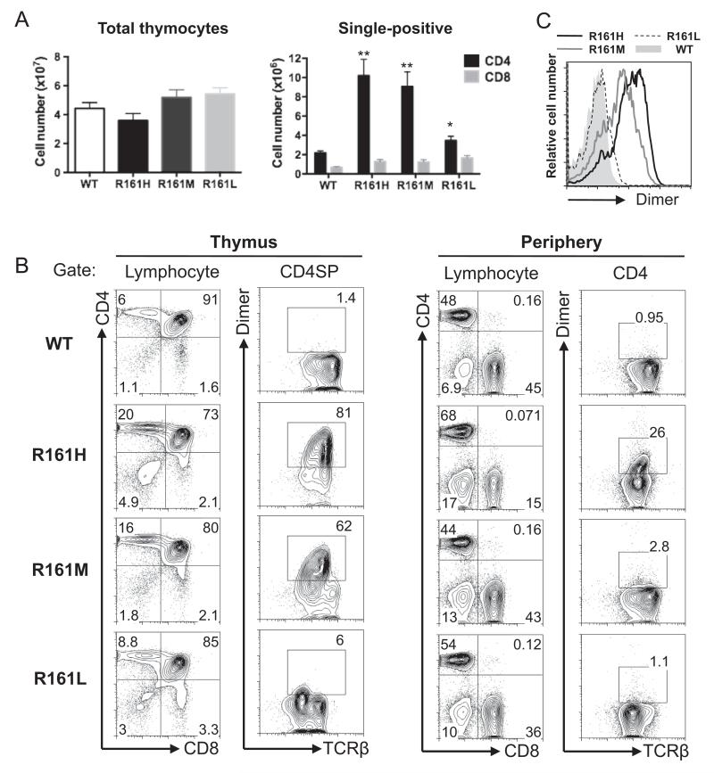
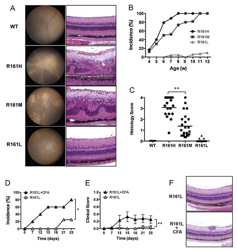
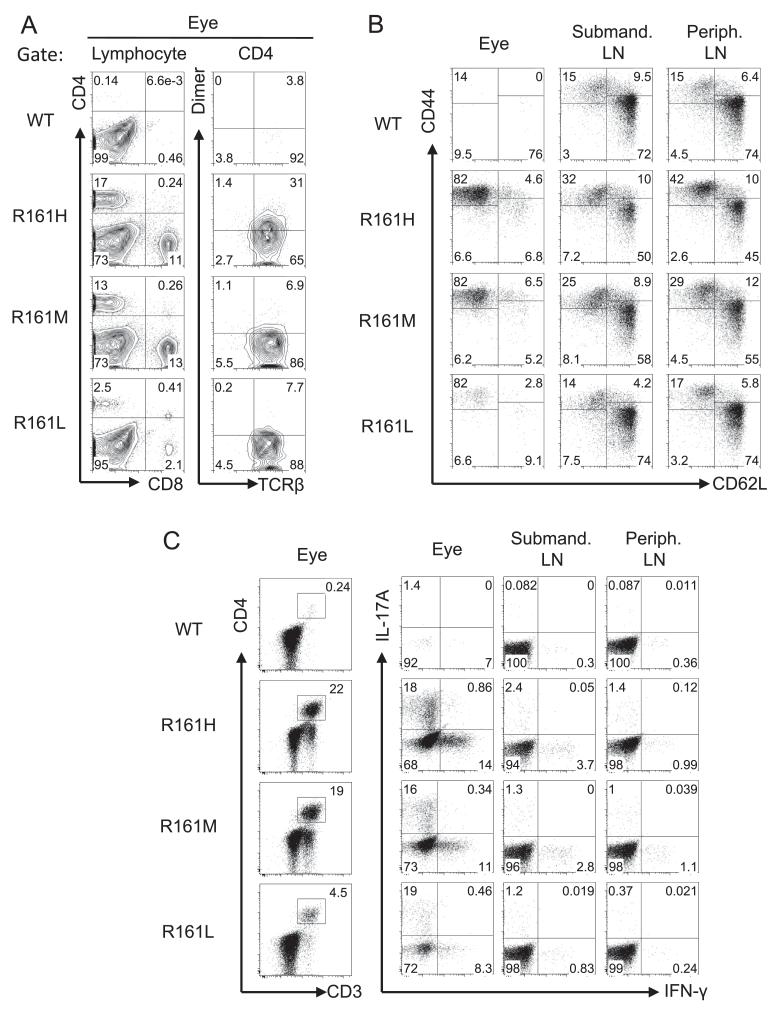
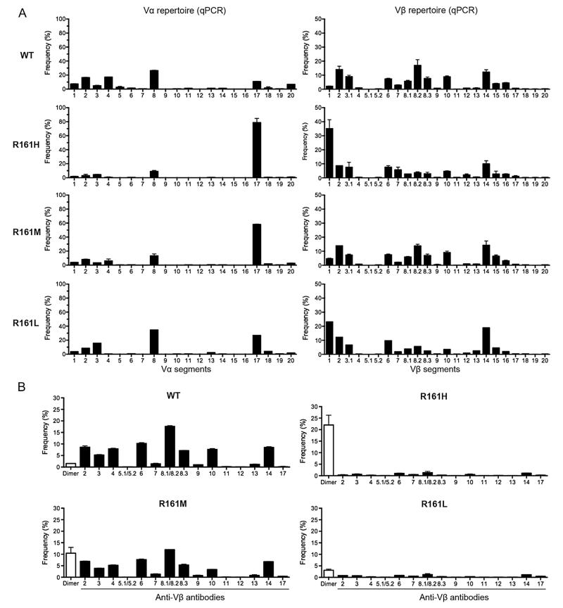
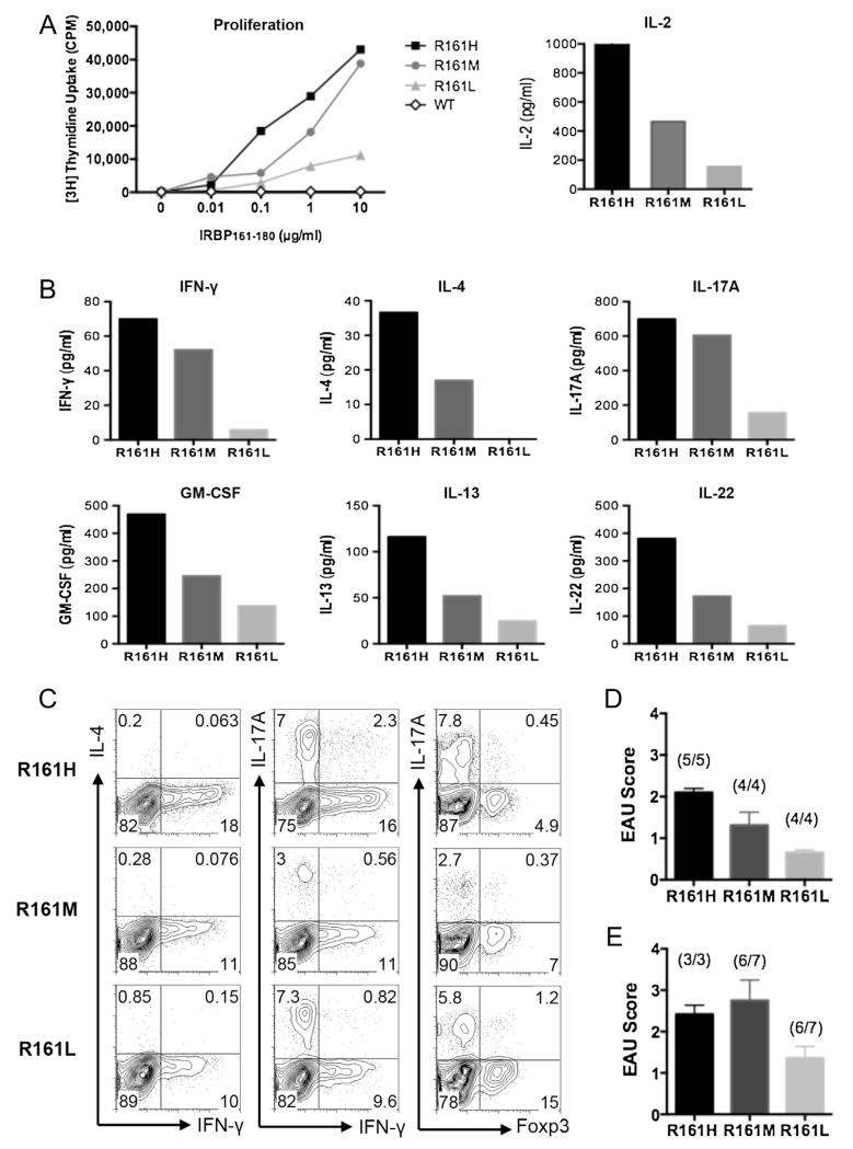
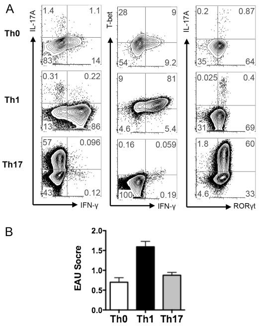
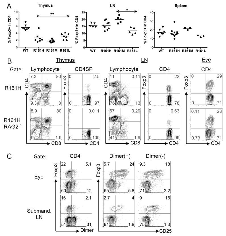
Despite presence of circulating retina-specific T cells in healthy individuals, ocular immune privilege usually averts development of autoimmune uveitis. To study the breakdown of immune privilege and development of disease, we generated transgenic (Tg) mice that express a T cell receptor (TCR) specific for interphotoreceptor retinoid-binding protein (IRBP), which serves as an autoimmune target in uveitis induced by immunization. Three lines of TCR Tg mice, with different levels of expression of the transgenic R161 TCR and different proportions of IRBP-specific CD4⁺ T cells in their peripheral repertoire, were successfully established. Importantly, two of the lines rapidly developed spontaneous uveitis, reaching 100% incidence by 2 and 3 months of age, respectively, whereas the third appeared "poised" and only developed appreciable disease upon immune perturbation. Susceptibility roughly paralleled expression of the R161 TCR. In all three lines, peripheral CD4⁺ T cells displayed a naïve phenotype, but proliferated in vitro in response to IRBP and elicited uveitis upon adoptive transfer. In contrast, CD4⁺ T cells infiltrating uveitic eyes mostly showed an effector/memory phenotype, and included Th1, Th17 as well as T regulatory cells that appeared to have been peripherally converted from conventional CD4⁺ T cells rather than thymically derived. Thus, R161 mice provide a new and valuable model of spontaneous autoimmune disease that circumvents the limitations of active immunization and adjuvants, and allows to study basic mechanisms involved in maintenance and breakdown of immune homeostasis affecting immunologically privileged sites such as the eye.

Keywords: AIRE; Ag; Autoimmune uveitis; CFA; EAU; HEL; IRBP; Immune privilege; PMA; RAG; SP; Spontaneous disease; T cell receptor; TCR; TCR transgenic mouse; WT; antigen; autoimmune regulator; complete Freund's adjuvant; experimental autoimmune uveitis; hen egg lysozyme; interphotoreceptor retinoid binding protein; phorbol myristate acetate; recombination activating gene; single positive; wild type.


Figures

Fig. 1
Fig. 1
Generation of IRBP-specific TCR transgenic (R161) mice. A. Total thymocyte and CD4/CD8 single positive (SP) cell counts (mean ± SEM) in WT and R161 lines between 8 and 15 weeks of age. The data was averaged from several experiments using total of 7–12 mice in each Tg line. **p < 0.001, *p < 0.05. No significance was detected in total thymocyte or CD8 SP numbers of R161 lines compared to that of WT. B. Lymphocyte profiles of thymus (left) and peripheral T cells (right) of WT and R161 lines. IRBP-specific T cells were detected within the CD4+ population by the IRBP161-180/IAr-Ig dimer reagent (Dimer) and TCRβ expression as gated. C. The expression levels of IRBP-specific TCR (Dimer-binding) in the thymus CD4 SP population were overlaid and compared between R161 lines in the histograms. The data is representative of at least 3 experiments using 2–3 mice per line.
Fig. 2
Fig. 2
Development of spontaneous uveitis in R161 lines. A. Fundus images (left) and histology pictures of retina (right, original mag. ×200) of R161 lines. WT shows normal fundus and histology. R161H and R161M show moderate to severe inflammatory lesions e.g. retinitis and choroiditis (see Suppl. Fig. 2 for detailed histopathology of R161H), whereas R161L shows mild lesions with few cellular infiltrates in the vitreous. B. Incidence of spontaneous uveitis determined by weekly fundus examination. Numbers of mice evaluated per time points; R161H (N = 11–40), R161M (N = 15–24), R161L (N10–22). C. Individual histology scores (symbols) and the average (line) evaluated by histology between 8 and 16 weeks of age. **p < 0.0001 by unpaired t-test. D–F. R161L mice (N = 5) were injected s.c. with 0.2 ml CFA (containing 250 ug M. tuberculosis). Shown is disease incidence (D) and clinical score (E) by fundus examination compared to age-matched R161L littermates without injection (N = 6). *p < 0.05 and **p < 0.0001 by 2-way ANOVA. F. Histology picture of R161L retina at day 25 post-injection of CFA and its control.
Fig. 3
Fig. 3
Ocular infiltrating lymphocytes display an activated/memory phenotype in R161 lines. A. Infiltrating lymphocytes in uveitic eyes included CD4+ and CD8+ T cells in all R161 lines. Substantial proportions of these CD4+ T cells expressed IRBP-specific TCRs. B. Naïve (CD62LhiCD44lo) and memory (CD62LloCD44hi) profiles of eye-infiltrating CD4+ T cells and lymph node (LN) CD4+ T cells. C. CD3/CD4 profiles (left) and IFN-γ and IL-17A production (right) from eye-infiltrating cells and LN cells following ex vivo PMA and ionomycin stimulation in the presence of Brefeldin A. CD3/CD4 plots are gated on lymphocytes by size and IFN-γ/IL-17A plots are gated on CD3+CD4+CD8 T cells. Peripheral (non-draining) LN are pooled inguinal, axillary and brachial LN.
Fig. 4
Fig. 4
Skewed TCR repertoire in R161 mouse lines. A. Vα and Vβ TCR repertoire. CD4+ T cells were sorted from lymph nodes and each Vα and Vβ TCR mRNA expression was measured by real-time quantitative PCR (q-PCR). Relative frequency of each Vα or Vβ TCR was calculated as described in Materials and methods. B. Cell surface Vβ expression analysis by flow cytometry with indicated anti-Vβ mAbs (black bars, Vβ2-14, 17) or with IRBP-specific dimer (Dimer) that binds to transgenic TCR (white bar). Frequency (%) was determined in the CD4+ gate.
Fig. 5
Fig. 5
Antigen-specific responses of R161 CD4+ T cells. A. Left: Proliferation of purified peripheral CD4+ T cells from R161 lines to various concentrations of specific Ag IRBP161–180 in the presence of irradiated (3000 rad) splenic APC from syngeneic WT mice. Right: IL-2 secretion at 24 h stimulation with 1 μg/ml IRBP161–180 peptide. B. Inflammatory cytokine secretion in the culture supernatant at 48 h stimulation with 1μg/ml IRBP161–180 peptide. Data is representative of at least 3 independent experiments. WT supernatant did not contain detectable cytokines. C. Flow cytometric intracellular cytokine analysis of in vitro activated R161 lymph node T cells with 1μg/ml IRBP161-180 peptide at 72 h followed by PMA and ionomycin pulse in the presence of Brefeldin A. D. Adoptive transfer of 1 × 10⁶ in vitro activated R161 T cells into B10.RIII-RAG2−/− recipients. EAU was evaluated by histopathology at day 10 of the transfer. E. Adoptive transfer of 1 × 10⁶ sorted naïve (CD62LhiCD44lo) R161 CD4+ T cells into B10.RIII-RAG2−/− recipients. EAU was evaluated by histopathology on day 12 (R161H) or day 14 (R161M, L) after adoptive transfer. The incidence (number of mice develop disease/total mice) is indicated above the bar of each group.
Fig. 6
Fig. 6
Th0, Th1 and Th17 R161H TCR Tg cells are all pathogenic in naïve recipients. A. Polarization of R161H T cells in non-skewing (Th0), Th1 or Th17 conditions. Total lymph node cells were stimulated with 1μg/ml IRBP161–180 for 3 days. B. Adoptive transfer of Th0, Th1 and Th17 cells in WT recipient mice. Eyes were collected for EAU scoring by histopathology on day 11–14 of the transfers. Shown are pooled data from 4 independent experiments for Th0 and 2 independent experiments for Th1 and Th17 conditions.
Fig. 7
Fig. 7
Foxp3 expression in the thymus, lymphoid tissues and uveitic eyes of R161 mice. A. Percent of Foxp3+ cells in the CD4 population in the thymus, lymph nodes and spleen from WT and R161 mice was determined by flow cytometry. **p < 0.005, *p < 0.05 compared to WT. B. R161H mice on the RAG2−/− background do not develop Foxp3+ Treg cells in the thymus. C. Uveitic eyes of R161H contain Foxp3+ Treg cells, including IRBP-specific Tregs.

References

    1. Streilein JW. Ocular immune privilege: the eye takes a dim but practical view of immunity and inflammation. J Leukoc Biol. 2003;74:179–85. - PubMed
    1. Caspi RR. Ocular autoimmunity: the price of privilege? Immunol Rev. 2006;213:23–35. - PubMed
    1. Nussenblatt RB, Whitcup SM. Uveitis: fundamentals and clinical practice. 3rd ed. Elsevier; Philadelphia, PA: 2004. Mosby.
    1. Gritz DC, Wong IG. Incidence and prevalence of uveitis in Northern California; the Northern California Epidemiology of Uveitis Study. Ophthalmology. 2004;111:491–500. - PubMed
    1. Durrani OM, Meads CA, Murray PI. Uveitis: a potentially blinding disease. Ophthalmologica. 2004;218:223–36. - PubMed

Publication types

MeSH terms