. 2013 Sep 17:248:571-84.
doi: 10.1016/j.neuroscience.2013.06.042. Epub 2013 Jun 28.

Long-term effects of cocaine experience on neuroplasticity in the nucleus accumbens core of addiction-prone rats

Affiliations

Long-term effects of cocaine experience on neuroplasticity in the nucleus accumbens core of addiction-prone rats

M Waselus et al. Neuroscience. .

Abstract

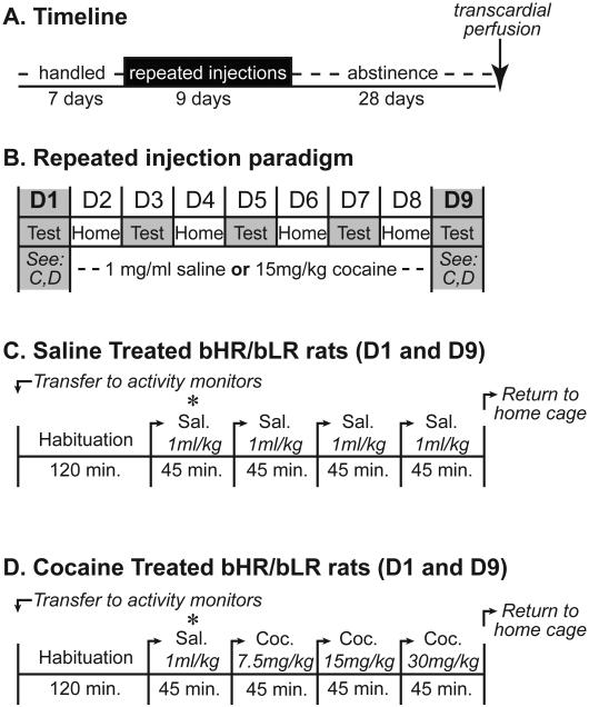
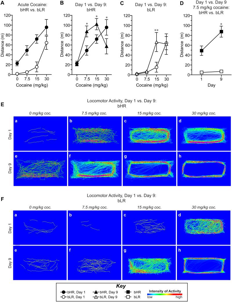
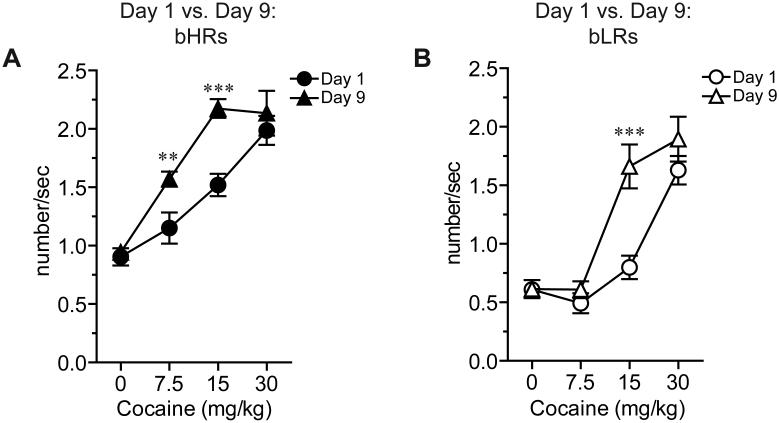
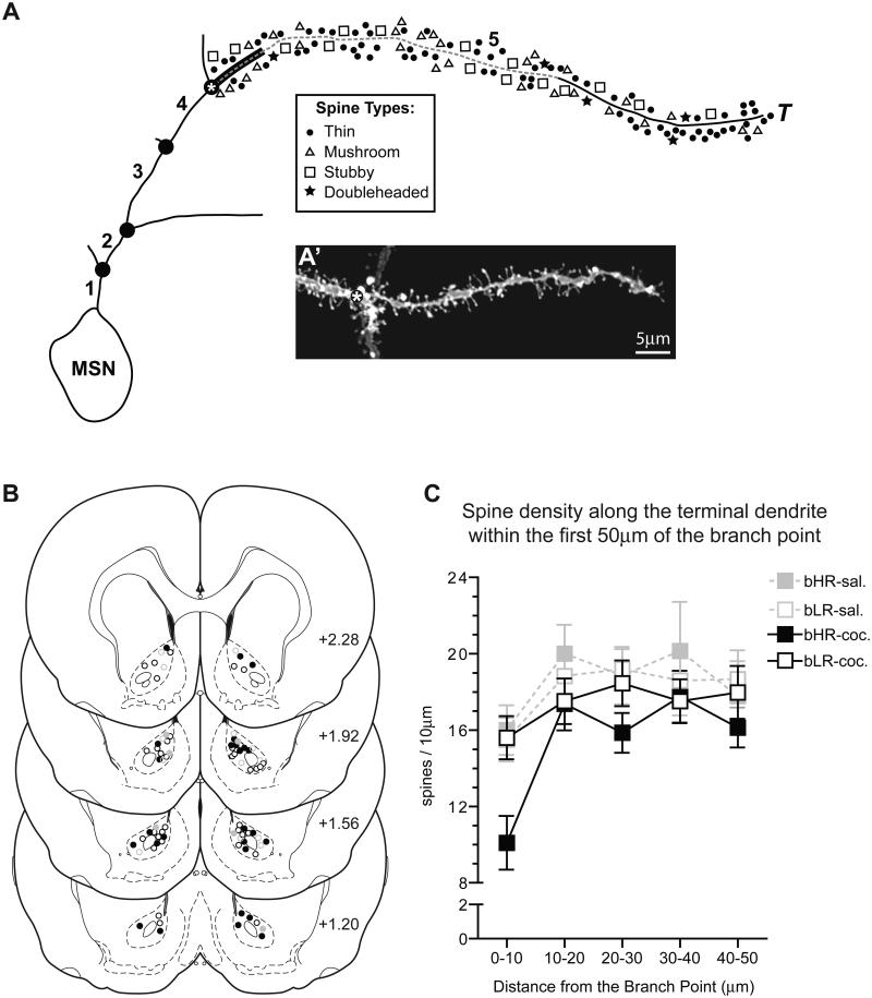
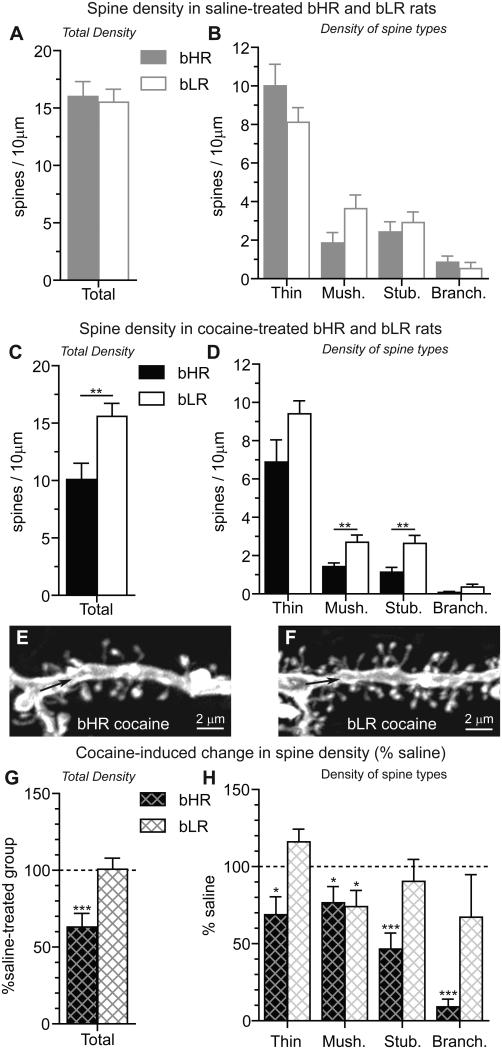
Repeated exposure to drugs of abuse is associated with structural plasticity in brain reward pathways. Rats selectively bred for locomotor response to novelty differ on a number of neurobehavioral dimensions relevant to addiction. This unique genetic animal model was used here to examine both pre-existing differences and long-term consequences of repeated cocaine treatment on structural plasticity. Selectively bred high-responder (bHR) and low-responder (bLR) rats received repeated saline or cocaine injections for 9 consecutive days. Escalating doses of cocaine (7.5, 15 and 30 mg/kg) were administered on the first (day 1) and last (day 9) days of treatment and a single injection of the intermediate dose (15 mg/kg) was given on days 2-8. Motor activity in response to escalating doses of cocaine was compared on the first and last days of treatment to assess the acute and sensitized response to the drug. Following prolonged cocaine abstinence (28 days), spine density was examined on terminal dendrites of medium spiny neurons in the nucleus accumbens core. Relative to bLRs, bHRs exhibited increased psychomotor activation in response to both the acute and repeated effects of cocaine. There were no differences in spine density between bHR and bLR rats under basal conditions or following repeated saline treatment. However, spine density differed markedly between these two lines following prolonged cocaine abstinence. All spine types were decreased in cocaine-treated bHRs, while only mushroom spines were decreased in bLRs that received cocaine. Changes in spine density occurred specifically near the branch point of terminal dendrites. These findings indicate that structural plasticity associated with prolonged cocaine abstinence varies markedly in two selected strains of rats that vary on numerous traits relevant to addiction. Thus, genetic factors that contribute to individual variation in the behavioral response to cocaine also influence cocaine-induced structural plasticity.

Keywords: addiction; cocaine; dendrites; prolonged abstinence; psychomotor sensitization; spine density.


Figures

Figure 1
Figure 1. Timeline for repeated cocaine treatment
A) Rats were handled for 7 days and then administered repeated injections of saline or cocaine for 9 consecutive days. For the next 28 days, rats remained cocaine abstinent until transcardial perfusion and brain extraction. B) Repeated injections of saline (C) or escalating doses of cocaine (D) were administered on the first (day 1; D1) and last (day 9; D9) days of treatment; single injections of saline or cocaine were given on the intermediate days (D2-D8). On days 2-8, injections were associated with the activity boxes (“test”; days 3,5,7) or home cages (“home”; days 2,4,6,8) on alternating days. Repeated injections on day 1 and day 9 were given in the activity boxes according to the treatment paradigm outlined for saline- (C) and cocaine-treated (D) rats. The asterisk (*) indicates the saline injection administered to all rats.
Figure 2
Figure 2. The locomotor response to both acute and repeated cocaine treatment is enhanced in bHR rats relative to bLRs
A) Locomotor activity on day 1 was increased with escalating doses of cocaine and enhanced in bHR rats (closed circles) compared to bLR rats (open circles). B) Following repeated cocaine injections (day 9; black triangles), locomotor activity in bHRs was increased at the lowest (7.5mg/kg) and middle (15mg/kg) doses and decreased at the highest dose (30mg/kg) compared to the acute response on day 1 (black circles). C) In bLRs, locomotor sensitization to cocaine was apparent only at the middle cocaine dose (15mg/kg) on day 9 (open triangles) relative to activity on day 1 (open circles). D) Locomotor activity in response to the lowest dose of cocaine (7.5 mg/kg) was increased in bHR rats (black squares), but not bLR rats (open squares), on day 9 relative to day 1. Representative density maps obtained from individual cocaine-treated bHR (E) and bLR (F) rats on day 1 (a-d) and day 9 (e-h) illustrate all locomotor activity following injections of saline (“0 mg/kg cocaine”, a,e) and escalating doses of cocaine (“7.5 mg/kg”, b,f; “15 mg/kg”, c,g; “30 mg/kg”, d,f). Significant differences between day 1 vs. day 9 (B,C,D) are indicated: **p<0.01, and *p<0.05.
Figure 3
Figure 3. Stereotyped head movements induced by repeated cocaine are enhanced in bHR rats compared to bLR rats
The frequency of lateral head movements is shown for bHR (A) and bLR (B) rats following acute (day 1; circles) and repeated (day 9; triangles) cocaine injections. Repeated cocaine (closed triangles) increased the frequency of head movements at the two lowest doses of cocaine in bHRs (A; 7.5 and 15 mg/kg), but only at the intermediate dose following repeated treatment (open triangles) in bLRs (B; 15 mg/kg). Differences in stereotyped head movements between day 1 and day 9 at the highest dose of cocaine (30 mg/kg) were not observed for either phenotype. Significant differences between day 1 vs. day 9 are shown:***p<0.001, *p<0.01.
Figure 4
Figure 4. Selection and analysis of dendritic spines in the AcbC
Dendritic spines were examined along the entire terminal branch of MSNs in the AcbC (A) in 10μm increments starting at the terminal branch point (asterisk) and extending to the terminal tip (T). Both the branch order (bold numbers) and dendritic branch points () are indicated. A representative image of a dendritic branch ending in a terminal tip (A’), typical of those included in the analyses, is shown; as in the schematic, an asterisk marks the branch point preceding the terminal tip. The distribution of MSNs in the AcbC that were included in the quantitative analyses was mapped onto 4 representative coronal sections (B) at 360μm intervals (+2.28, +1.92, +1.56 and +1.20mm bregma (Paxinos and Watson, 2005) for saline-treated bHRs (closed gray circles), cocaine-treated bHRs (closed black circles), saline-treated bLRs (open gray circles) and cocaine-treated bLRs (open black circles). Spine density along terminal branches was analyzed in 10μm increments starting at the branch point (A) and extending toward the terminal tip for 50μm (dashed line). Spine density on terminal branches was impacted by the distance from the branch point (C; effect of distance: p<0.001) and the 10μm segment adjacent to the branch point (A; thick black line) had fewer spines than any other segment.
Figure 5
Figure 5. Individual differences in spine density are apparent in the AcbC of cocaine-treated bHR and bLR rats
All data presented here was obtained from the 10μm segment of the terminal dendrite closest to the branch point (see Fig. 4A; thick black line). Spine density in the AcbC was comparable in bHR and bLR rats treated repeatedly with saline (A). No significant differences between bHR and bLR rats were detected among the different spine types, although there was a tendency for differences in the density of mushroom spines (B) in bHR vs. bLR rats (p=0.07). Differences between bHR and bLR rats in total spine density (C) as well as spine types (D; mushroom and stubby) were apparent following prolonged cocaine abstinence. Although bHR-bLR differences in the density of thin (p=0.06) and branched (p=0.1) spines were not significant, there was a tendency for the density of these spines to differ in cocaine-treated rats, as well. Differences in spine density near the branch point of bHRs (E) and bLRs (F) are clear following cocaine treatment. In bHRs, but not bLRs, cocaine decreased the total density of spines vs. saline-treated controls (G). All spine types were decreased in cocaine-treated bHR rats compared to saline-treated controls (H, thin: t(13)=-2.70, p=0.02; mushroom: t(13)=-2.23, p=0.04; stubby: t(13)=-5.16, p<0.001; branched: t(13)=-18.05, p<0.001). In cocaine-treated bLRs (H), only mushroom spines were decreased by cocaine (t(16)=-2.48, p=0.03), although insignificant increases in thin spines were observed (t(16)=1.94, p=0.07). Significant differences between bHR vs. bLR (A-D) and cocaine vs. saline (G,H) are indicated: ***p<0.001; **p<0.01; *p<0.05.

References

    1. Ahmed SH, Stinus L, Le Moal M, Cador M. Controlling interindividual differences in the unconditioned response to amphetamine in the study of environment-dependent sensitization. Behav Pharmacol. 1993;4:355–365. - PubMed
    1. Alcantara AA, Lim HY, Floyd CE, Garces J, Mendenhall JM, Lyons CL, Berlanga ML. Cocaine- and morphine-induced synaptic plasticity in the nucleus accumbens. Synapse. 2011;65:309–320. - PMC - PubMed
    1. Anagnostaras SG, Robinson TE. Sensitization to the psychomotor stimulant effects of amphetamine: modulation by associative learning. Behav Neurosci. 1996;110:1397–1414. - PubMed
    1. Bamford NS, Robinson S, Palmiter RD, Joyce JA, Moore C, Meshul CK. Dopamine modulates release from corticostriatal terminals. J Neurosci. 2004;24:9541–9552. - PMC - PubMed
    1. Bartlett WP, Banker GA. An electron microscopic study of the development of axons and dendrites by hippocampal neurons in culture. I. Cells which develop without intercellular contacts. J Neurosci. 1984;4:1944–1953. - PMC - PubMed

Publication types

LinkOut - more resources