Review
. 2013 Oct;136(Pt 10):2937-51.
doi: 10.1093/brain/awt153. Epub 2013 Jun 27.

Beyond taxol: microtubule-based treatment of disease and injury of the nervous system

Affiliations
Review

Beyond taxol: microtubule-based treatment of disease and injury of the nervous system

Peter W Baas et al. Brain. 2013 Oct.

Abstract

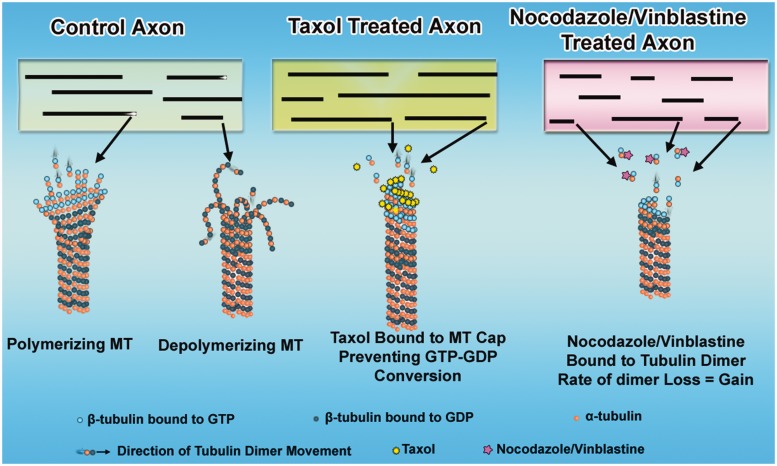
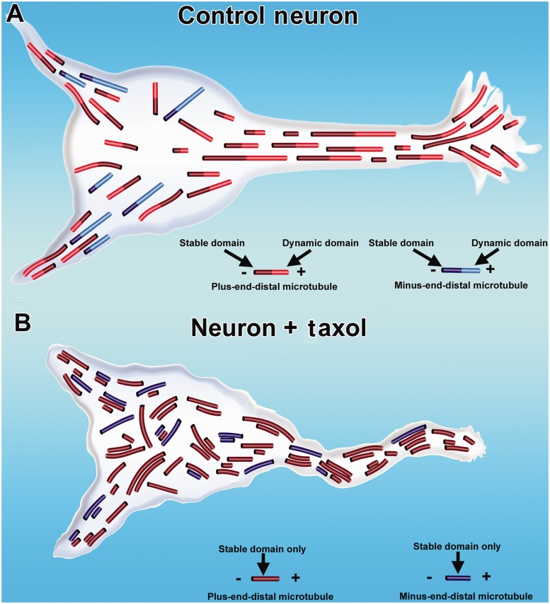
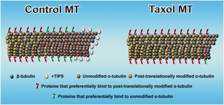
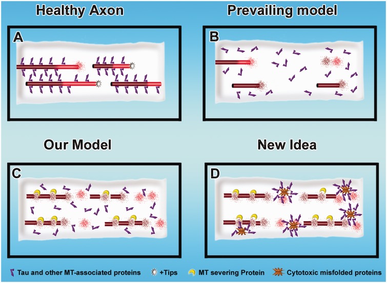
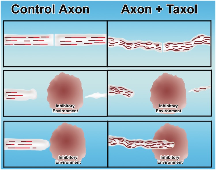
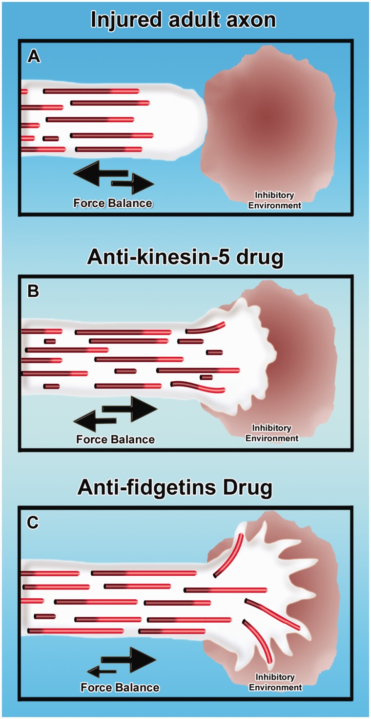
Contemporary research has revealed a great deal of information on the behaviours of microtubules that underlie critical events in the lives of neurons. Microtubules in the neuron undergo dynamic assembly and disassembly, bundling and splaying, severing, and rapid transport as well as integration with other cytoskeletal elements such as actin filaments. These various behaviours are regulated by signalling pathways that affect microtubule-related proteins such as molecular motor proteins and microtubule severing enzymes, as well as a variety of proteins that promote the assembly, stabilization and bundling of microtubules. In recent years, translational neuroscientists have earmarked microtubules as a promising target for therapy of injury and disease of the nervous system. Proof-of-principle has come mainly from studies using taxol and related drugs to pharmacologically stabilize microtubules in animal models of nerve injury and disease. However, concerns persist that the negative consequences of abnormal microtubule stabilization may outweigh the positive effects. Other potential approaches include microtubule-active drugs with somewhat different properties, but also expanding the therapeutic toolkit to include intervention at the level of microtubule regulatory proteins.

Keywords: axon; dendrite; microtubule; neuron; taxol.


Figures

Figure 1
Figure 1
Effects of microtubule-active drugs on microtubule dynamics. Shown are a control axon and axons during the first moments of exposure to microtubule-active drugs. In the control axon, the microtubules (MT) display dynamic instability at their plus ends. In taxol-treated axons, the microtubules are stabilized (no longer lose subunits) and no longer show dynamic instability at their plus ends. Hence, they continue to assemble. In axons treated with low concentrations of microtubule depolymerizing drugs, the microtubules become ‘kinetically stabilized,’ which means they lose and gain subunits at the same rate, resulting in no length change.
Figure 2
Figure 2
Effects of taxol on microtubule organization in axons and dendrites. In control neurons, microtubules are nearly uniformly plus-end-distal in the axon. In the dendrite, microtubules have a mixed orientation (A). Each microtubule consists of a stable domain toward the minus end of the microtubule, with most microtubules also consisting of a dynamic (labile) domain toward the plus end of the microtubule. Dendritic microtubules are less stable than axonal microtubules, as indicated by shorter stable domains on dendritic microtubules in the illustration. In neurons treated with taxol (B), the density of microtubules increases, the normal domain structure of individual microtubules is lost because the microtubules are stabilized all along their lengths, and flaws arise in the normal polarity patterns of the microtubules. Such abnormalities can lead to degeneration of axons and dendrites.
Figure 3
Figure 3
Effects of taxol on microtubule composition. Control microtubule (MT) shows more post-translationally modified subunits toward the minus end of the microtubule and more unmodified subunits toward the plus end. Different complements of microtubule-related proteins associate with regions of the microtubule that are richer in modified or unmodified subunits. At the plus end of the microtubule is an enrichment of +tips. In the case of microtubules stabilized with taxol, the subunits become predominantly modified all along the length of the microtubule, and hence the complement of microtubule-related proteins favours those that are associated with modified subunits. Also, in the presence of taxol, the +tips no longer appear at the plus end of the microtubule.
Figure 4
Figure 4
Mechanisms of microtubule loss during nerve degeneration. (A) A healthy axon. (B and C) Three mechanistic possibilities for microtubule loss during tau-based neurodegeneration. In the prevailing model (B), when tau detaches from them, the microtubules become less stable and depolymerize by their normal dynamic properties. In our model (C), when tau detaches from them, the microtubules become more sensitive to proteins such as katanin that actively promote microtubule loss (Sudo and Baas, 2011). (C) A ‘gain of function’ mechanism by which cytotoxic mutated proteins associated with neurodegeneration could promote microtubule loss by sequestering proteins such as tau.
Figure 5
Figure 5
Taxol-based strategy for augmenting regeneration of injured adult axons. (Left) Control situation (not treated with taxol) and situation with taxol treatment (right). (Top) Cut axon. Middle and bottom panels show the degeneration of the distal stump and the failure of the proximal cut end of the axon to regenerate through the inhibitory environment generated at the lesion site. In the control situation, the axon attempts to regenerate but fails as it encounters the inhibitory environment. In the taxol situation, the stabilized microtubules enable the axon to grow through the inhibitory environment.
Figure 6
Figure 6
New microtubule-based strategies for augmenting regeneration of injured adult axons. Injured adult axon does not regenerate through an inhibitory environment, with the balance of forces favouring retraction rather than growth (A). Treatment with anti-kinesin-5 drugs can shift the balance of forces toward axonal growth, notably increase the frequency of short microtubules in the axon undergoing transport, and enable the axon to enter the inhibitory environment (Lin et al., 2011b). Theoretical at this time is the idea that inhibiting fidgetins (fidgetin and/or fidgetin-like 2) may cause a consequential increase in the fraction of the microtubule mass that is dynamic/labile and restore the axon to a more juvenile state of growth, thus enabling it to grow faster and enter the inhibitory environment.

References

    1. Aguirre-Portoles C, Bird AW, Hyman A, Canamero M, Perez de Castro I, Malumbres M. Tpx2 controls spindle integrity, genome stability, and tumor development. Cancer Res. 2012;72:1518–28. - PubMed
    1. Akhmanova A, Steinmetz MO. Tracking the ends: a dynamic protein network controls the fate of microtubule tips. Nat Rev Mol Cell Biol. 2008;9:309–22. - PubMed
    1. Amos LA, Löwe J. How taxol stabilises microtubule structure. Chem Biol. 1999;6:R65–9. - PubMed
    1. Baas PW. Microtubules and neuronal polarity: lessons from mitosis. Neuron. 1999;22:23–31. - PubMed
    1. Baas PW, Ahmad FJ. The plus ends of stable microtubules are the exclusive nucleating structures for microtubules in the axon. J Cell Biol. 1992;116:1231–41. - PMC - PubMed

Publication types