. 2013 Aug;14(8):812-20.
doi: 10.1038/ni.2639. Epub 2013 Jun 30.

CD36 coordinates NLRP3 inflammasome activation by facilitating intracellular nucleation of soluble ligands into particulate ligands in sterile inflammation

Affiliations

CD36 coordinates NLRP3 inflammasome activation by facilitating intracellular nucleation of soluble ligands into particulate ligands in sterile inflammation

Frederick J Sheedy et al. Nat Immunol. 2013 Aug.

Abstract

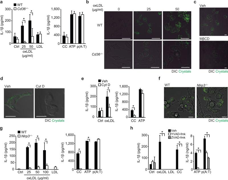
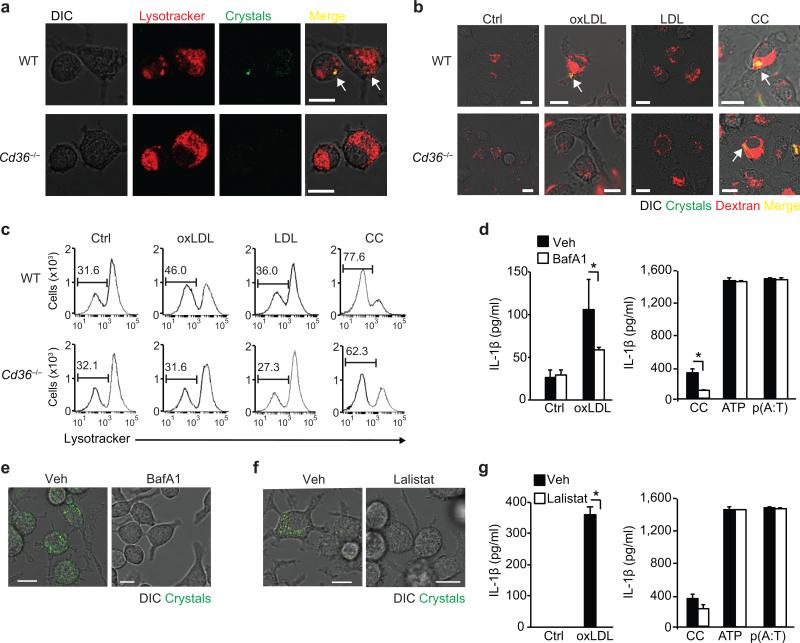
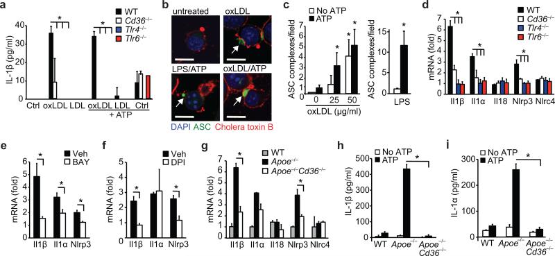
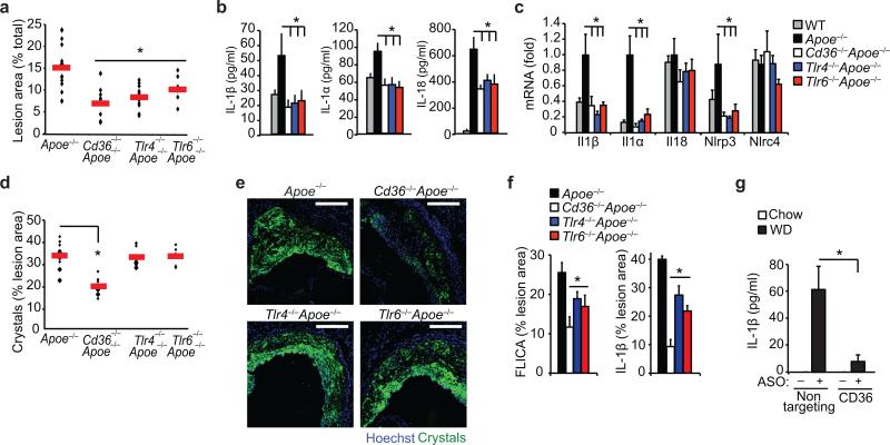
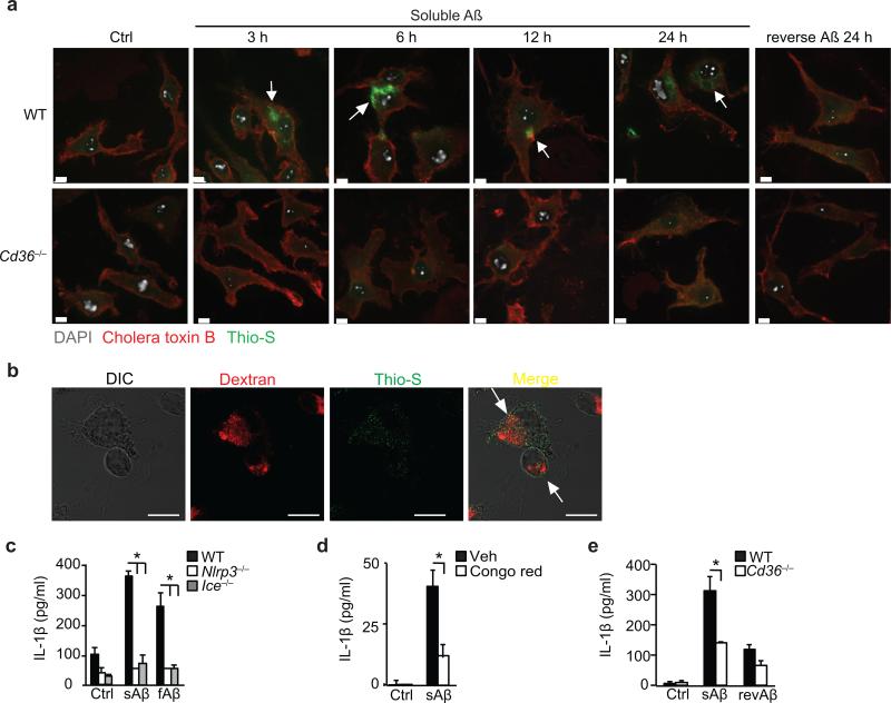
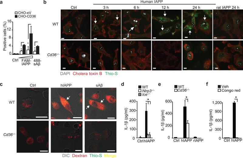
Particulate ligands, including cholesterol crystals and amyloid fibrils, induce production of interleukin 1β (IL-1β) dependent on the cytoplasmic sensor NLRP3 in atherosclerosis, Alzheimer's disease and diabetes. Soluble endogenous ligands, including oxidized low-density lipoprotein (LDL), amyloid-β and amylin peptides, accumulate in such diseases. Here we identify an endocytic pathway mediated by the pattern-recognition receptor CD36 that coordinated the intracellular conversion of those soluble ligands into crystals or fibrils, which resulted in lysosomal disruption and activation of the NLRP3 inflammasome. Consequently, macrophages that lacked CD36 failed to elicit IL-1β production in response to those ligands, and targeting CD36 in atherosclerotic mice resulted in lower serum concentrations of IL-1β and accumulation of cholesterol crystals in plaques. Collectively, our findings highlight the importance of CD36 in the accrual and nucleation of NLRP3 ligands from within the macrophage and position CD36 as a central regulator of inflammasome activation in sterile inflammation.


Figures

Figure 1
Figure 1. CD36-mediated uptake of oxLDL generates intracellular crystals and activates the NLRP3 inflammasome
(a) IL-1β ELISA of supernatants from LPS-primed wild-type (WT) or CD36-deficient (Cd36-/-) BMDM incubated for 24 h with PBS vehicle (Ctrl), oxLDL or unmodified LDL (left panel) alongside the following inflammasome activators (right panel), cholesterol crystals (CC, 1 mg/mL), ATP (5 μM, 1 h) and poly(dA:dT) [p(A:T), 100 ng/well, 6 h]. (b) Confocal reflection microscopy of WT or Cd36-/- peritoneal macrophages treated for 24 h with oxLDL as indicated. (c) Confocal reflection microscopy of peritoneal macrophages treated with oxLDL (50 μg/mL, 24 h) and subsequently treated with methyl-β-cyclodextrin (MβCD, 10 μM, 1 h) or vehicle (Veh). (d) Confocal reflection microscopy of oxLDL-treated BMDM (50 μg/mL, 24 h) following pre-treatment with cytochalasin D (Cyt D (1 μM, 1 h) or vehicle control (Veh). (e) IL-1β ELISA of supernatants from LPS-primed BMDM treated for 24 h with PBS vehicle (Ctrl), oxLDL (50 μg/mL) or cholesterol crystals and ATP as before. (f) Confocal reflection microscopy of WT or NLRP3-deficient (Nlrp3-/-) BMDM treated with oxLDL (50 μg/mL, 24 h). (g) IL-1β ELISA of supernatants from LPS-primed WT or Nlrp3-/- BMDM treated as in a). (h) IL-1β ELISA of supernatants from LPS-primed BMDM treated with the indicated inflammasome activators following 1 h pre-treatment with ZVAD-fmk (pan-caspase inhibitor, 20 μM) or YVAD-fmk (caspase-1 inhibitor, 20 μM). Data in a, e, g and h are mean ± s.d. of triplicate samples within a single experiment and are representative of three independent experiments. Images in b, c,d and f are representative of 3 independent experiments. Scale bar = 10 μm. *P<0.05.
Figure 2
Figure 2. CD36-mediated cholesterol crystal formation and NLRP3 activation occurs via a lysosomal pathway
(a) Combined confocal reflection microscopy of wild-type (WT) or CD36-deficient (Cd36–/–) BMDM incubated for 8 h with oxLDL (50 μg/mL) alongside Lysotracker-Red. (b) Combined confocal reflection microscopy of WT or Cd36–/– BMDM incubated for 24 h with PBS vehicle (Ctrl), oxLDL (50 μg/mL), unmodified LDL (50 μg/mL) or cholesterol crystals (CC, 1 mg/mL) alongside fluorescent Dextran-Red. (c) Flow cytometry measurements of Lysotracker Red-fluoresence in WT or Cd36–/– BMDM treated as in (b). (d,g) IL-β ELISA of supernatants from LPS-primed BMDM incubated the indicated inflammasome activators following pre-treatment with 500 nM bafilomycin A1 (BafA1) (d) or 20 nM Lalistat (g). (e,f) Confocal reflection microscopy of oxLDL-loaded BMDM (50 μg/mL, 24 h) following pre-treatment with BafA1 or Lalistat (as in d and g). Data in d and g are mean ± s.d. of triplicate samples within a single experiment and are representative of three independent experiments. Images in a-c, e and f are representative of three independent experiments. Scale bar = 10 μm. *P<0.05.
Figure 3
Figure 3. CD36 regulates priming of the NLRP3 inflammasome by oxLDL through TLR4-TLR6
(a) IL-1β ELISA of supernatants from wild-type (WT), CD36-deficient (Cd36–/–), TLR4-deficient (Tlr4–/–) or TLR6-deficient (Tlr6–/–) peritoneal macrophages primed with oxLDL, unmodified LDL (50 μg/mL, 18 h) or PBS vehicle (Ctrl) and subsequently treated with ATP (5 μM, 1 h). (b) Confocal microscopy of ASC-CFP expressing immortalized macrophages treated as indicated. (c) Quantification of ASC-oligomerization (speck formation) per 20x field for the indicated treatments. (d-f) Relative mRNA expression of the indicated gene in oxLDL-treated WT, Cd36–/–, Tlr4–/– or Tlr6–/– peritoneal macrophages (d) or from WT peritoneal macrophages following pre-treatment with BAY 11-7083 (BAY, an NF-κB inhibitor, 10 μM,) (e) or diphenylene iodonium (DPI, an NAPDH oxidase inhibitor, 1 μM) (f), measured by qRT-PCR. (g) Relative mRNA expression in in-vivo formed foam cell macrophages from mice of the indicated genotype fed a western diet and expressed relative to macrophages from chow-fed mice. (h-i) IL-1β (h) or IL-1α (i) production from in-vivo formed foam cells from mice of the indicated genotype treated ex-vivo with ATP (5 μM, 1 h). Data in a, h-i are mean ± s.d. of triplicate samples within a single experiment and are representative of three independent experiments. Data in c-f and g is mean ± s.e.m. of three independent experiments. Images in b are representative of three independent experiments. Scale bar =10 μm. *P<0.05.
Figure 4
Figure 4. Inflammasome activity is impaired in atherogenic mice deficient in CD36 and its signaling partners TLR4 and TLR6
Mice of the indicated genotype (Apoe–/–, Cd36–/–Apoe–/–, Tlr4–/– Apoe–/–, Tlr6–/–Apoe–/–, or control C57/BL6 Apoe+/+) were fed a western diet for 12 weeks, and atherosclerosis and inflammasome activity was assessed. (a) Lesion area in the aorta en face measured as a % of total aortic area. (b) Serum cytokine concentrations measured by Raybiotech Protein Array (IL1β, IL1α) or ELISA (IL18). (c) qRT-PCR analysis of aortic mRNA. (d-e) Plaque crystal content measured in serial sections throughout aortic root by combined confocal reflection microscopy illustrated in (e) and quantified in (d). (f) Plaque caspase-1 activity measured in serial sections throughout aortic root using FAM-YVAD-fmk FLICA probe fluorescence (left) or anti-IL-1β immunofluorescent staining (right) (n=8 sections/mouse) and expressed as % of total plaque area. (g) Serum IL-1β concentrations from Apoe–/– mice before (chow) or after western diet (WD) feeding for 4 wk during which mice were treated with a non-targeting or CD36 antisense oligonucleotide (ASO). Data is presented for n=15 mice (Apoe–/–), n=10 mice/group (Cd36–/–Apoe–/– and Tlr4–/–Apoe–/–) and n=7 mice (Tlr6–/–Apoe–/–) (a,d). Horizontal bars indicate the mean and symbols indicate individual mice. All other data is mean ± s.e.m. for n=4 mice/group (b-c), n=6 mice/group (f) and n=5 mice/group (g). Scale bar = 200 μm. *P<0.05.
Figure 5
Figure 5. CD36 uptake of soluble Aβ induces intracellular NLRP3-activating amyloid formation
(a) Confocal microscopy of wild-type (WT) or CD36-deficient (Cd36–/–) BMDM incubated with 10 μM soluble Aβ for the indicated times or control reverse peptide (reverse Aβ) for 24 h. After incubation cells were fixed and stained with Alexa-568 conjugated cholera toxin B and thioflavin-S (Thio-S) to visualize intracellular amyloid. (b) Confocal microscopy of BMDM incubated for 6 h with soluble Aβ (10 μM) and fluorescent Dextran as a lysosomal marker, then fixed and stained with thioflavin-S to visualize intracellular amyloid. (c) IL-1β ELISA of supernatants from LPS primed WT, NLRP3-deficient (Nlrp3–/–) or Caspase-1 deficient (Ice–/–) BMDM treated for 24h with DMSO-vehicle (Ctrl), soluble Aβ (sAβ, 10 μM) or fibrillar β-amyloid (fAβ, 1 μM). (d) IL-β ELISA of supernatants from LPS-primed BMDM pre-treated with Congo red (200 μM) for 1 h and incubated for 24h with DMSO vehicle (Ctrl) or sAβ (10 μM). (e) IL-1β ELISA of supernatants from LPS primed WT or Cd36–/– BMDM treated for 24 h with DMSO-vehicle (Ctrl), sAβ (10 μM) or reverse peptide (revAβ, 10 μM). Data in c-e are mean ± s.d. of triplicate samples within a single experiment and are representative of three independent experiments. Images in a-b are representative of three independent experiments. Scale bar = 10 μm. *P<0.05.
Figure 6
Figure 6. CD36 regulates NLRP3 activation by IAPP
(a) Binding of FAM-conjugated human IAPP, (FAM-IAPP; 5-10 μM) and HiLyte-488-conjugated soluble Aβ (488-sAβ) to control (CHO-eV) or CD36-expressing CHO (CHO-CD36) cells. (b) Confocal microscopy of WT or Cd36–/– BMDM incubated with 10 μM soluble human IAPP for the indicated times or control rat IAPP for 24 h. Cells were fixed and stained with Alexa-568 conjugated cholera toxin B and thioflavin-S (Thio-S) to visualize intracellular amyloid. (c) Confocal microscopy of WT or Cd36–/– BMDM incubated with human IAPP (hIAPP) or soluble Aβ (sAβ, 10 μM, 24 h) and fluorescent Dextran-Red, then fixed and stained with thioflavin-S. Arrows indicate thioflavin-S-reactive amyloid. (d) IL-1β ELISA of supernatants from LPS primed WT, NLRP3-deficient (Nlrp3–/–) or Caspase-1 deficient (Ice–/–) BMDM treated for 6 h with vehicle (Ctrl) or huIAPP (10 μM). (e) IL-1β ELISA of supernatants from LPS primed WT or Cd36–/– BMDM treated for 6h with vehicle (Ctrl), hIAPP (10 μM) or control rat IAPP (rIAPP, 10 μM). (f) IL-β ELISA of supernatants from LPS-primed BMDM pre-treated with Congo Red (200 μM, 1 h) and incubated for 6 h with vehicle (Ctrl) or hIAPP (10 μM). Data in a, d-f are mean ± s.d. of triplicate samples within a single experiment and are representative of three independent experiments. Images in b-c are representative of three independent experiments. Scale bar = 10 μm. *P<0.05.

Comment in

References

    1. Abela GS. Cholesterol crystals piercing the arterial plaque and intima trigger local and systemic inflammation. J. Clin. Lipidol. 2010;4:156–164. - PubMed
    1. Meyer-Luehmann M, et al. Rapid appearance and local toxicity of amyloid-beta plaques in a mouse model of Alzheimer's disease. Nature. 2008;451:720–724. - PMC - PubMed
    1. Westermark GT, Westermark P, Berne C, Korsgren O. Nordic Network for Clinical Islet T. Widespread amyloid deposition in transplanted human pancreatic islets. N. Engl. J. Med. 2008;359:977–979. - PubMed
    1. Duewell P, et al. NLRP3 inflammasomes are required for atherogenesis and activated by cholesterol crystals. Nature. 2010;464:1357–1361. - PMC - PubMed
    1. Halle A, et al. The NALP3 inflammasome is involved in the innate immune response to amyloid-beta. Nat. Immunol. 2008;9:857–865. - PMC - PubMed

Publication types

MeSH terms