Review
. 2013 Jul;12(7):526-42.
doi: 10.1038/nrd4003.

Strategies for optimizing the response of cancer and normal tissues to radiation

Affiliations
Review

Strategies for optimizing the response of cancer and normal tissues to radiation

Everett J Moding et al. Nat Rev Drug Discov. 2013 Jul.

Abstract

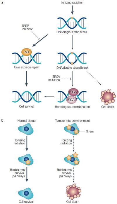
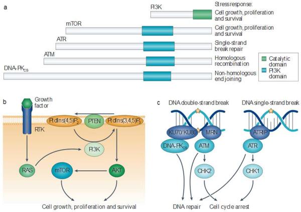
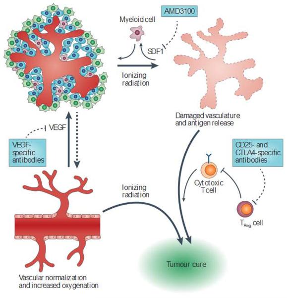
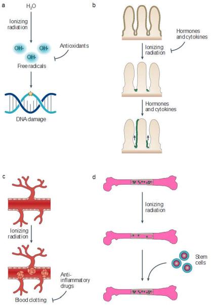
Approximately 50% of all patients with cancer receive radiation therapy at some point during the course of their treatment, and the majority of these patients are treated with curative intent. Despite recent advances in the planning of radiation treatment and the delivery of image-guided radiation therapy, acute toxicity and potential long-term side effects often limit the ability to deliver a sufficient dose of radiation to control tumours locally. In the past two decades, a better understanding of the hallmarks of cancer and the discovery of specific signalling pathways by which cells respond to radiation have provided new opportunities to design molecularly targeted therapies to increase the therapeutic window of radiation therapy. Here, we review efforts to develop approaches that could improve outcomes with radiation therapy by increasing the probability of tumour cure or by decreasing normal tissue toxicity.


Figures

Figure 1
Figure 1. Selectively targeting tumour cells through synthetic lethality
a | Ionizing radiation causes single-strand and double-strand DNA breaks. Some single-strand DNA breaks can be repaired by base excision repair. Poly(ADP-ribose) polymerase (PARP) inhibitors block base excision repair, causing some single-strend breaks to become double-strand breaks. In normal cells, BRCA (breast cancer susceptibility)-mediated homologous recombination can repair these breaks However, cancer cells with mutations in BRCA1 or BRCA2 are unable to repair this damage, allowing PARP inhibitors to radiosensitize these cellsvia synthetic lethality. b | Similarly synthetic lethality could potentially be used to sensitize cellsin the harsh tumour microenvironment Drugs that block stress survivel signalling pathways should preferentially kill tumour cells that are exposed to stresses such as hypoxia, oxidative stress or cell-cell contact abnormalities (right side) but not normal cells, which exist in a less harsh microenvironment (left side). These drugs have the potential to kill tumour cells in the absence of radiation, but radiation provides an additional stress that could enhance the probability of tumour eradication.
Figure 2
Figure 2. Inhibiting P13 Kand the P13 K-like protein kinase family
a | The phosphoinositide 3-kinase (H3K)-like protein kinase family includes several cellular kinases with catalytic domains that are homologous to PI3K(these are known as PI3K domains) This protein family coordinates diverse cellular responses that are crucial for the response to ionizing radiation. b | PI3K and mammalian target of rapamyc in (mTOR) are activated by numerous growth factors to stimulate cellular growth, survival and proliferation. Growth factors bind to receptor tyrosine kinases. Leading to downstream activation of PI3K by the RAS family of small GTFases. Active PI3K phosphorylates phosphatidylinositol-4,5-bisphophate (Ptdlns(4,5)P2) in the cellular membrane, converting it to the second messenger phosphatidylinositol-3,4,5-trisphophate (Ptdlns(3,4,5)P3). Ptdlns(3,4,5)P3 can activate the protein kinase AKT, leading to increased cell growth, proliferation and survivalby activating several proteins including mTOR. c | DNA-dependent protein kinase catalytic subunit (DNA-PKCCS), ataxia telangiectasia mutated (ATM) and ATR (ataxia telangiectasia and RAD3-related) respond to DNA damage by activating cell cycle arrest and engaging distinct DNA repair programmes. Because the catalytic domain is conserved across these diverse kinases, it is possible that a single drug could target several family members, thereby inhibiting some or all of these DNA repair and cell survival pathways. KU70 and/or KU80, the MRN complex(composed of MREll, RAD50 and Nijmegen breakage syndrome protein 1 (NBSl)) and ATR-interacting protein (ATRIP)sense DNA damage and promote the activation of DNA-PKCS, ATM and ATR. respectively. These kinases then phosphorylate several target sinside the cell, including checkpoint kinase | (CHK1) and CHK2.PTEN, phosphat ase and tensin homolog.
Figure 3
Figure 3. Enhancing tumour cure by modulating the tumour microenvironment
The tumour microenvironment can regulate the response of tumours to radiation by altering tumour oxygenation and vascular rebuilding, and by regulating the clearance of surviving cells by the immune system. Targeted drugs can alter the interaction between tumour cells and their microenvironment, which may enhance tumour cell killing and increase the probability of a cure. Anti-angiogenic therapies such as blockade of vascular endothelial growth factor (VEGF) can cause vascular normalization, leading to a window of increased tumour oxygenation. Myeloid cells are recruited to inradiated tumours by cytokines suchasstromal cell-derived factor 1 (SDF1). Blocking their recruitment may impair vascular regrowth. Irradiated tumours release antigens that can be recognized by the immune system, leading to the destruction of tumour cells by cytotoxic T cells. Regulatory T(TReg) cells can abrogate this response,so targeting these cells with CD25- and cytotoxic T lymphocyte antigen 4 (CTLA4)-specific antibodies may enhance tumour cure by radiation.
Figure 4
Figure 4. The tissue-dependent role of p53 in the response of cells to radiation
a | In unstressed cells, HDM2 ubiquitylates the tumour suppressor p53, leading to its degradation. Numerous cellular stresses increase the translation of TP53 mRNA and the phosphorylation of p53 protein, increasing its stability. p53 acts predominantly as a transcription factor that upregulates the expression of target genes to arrest cell growth or lead to cellular senescence and apoptosis, depending on the cellular context. Ribosomal protein |26 (RPL26) binds to TP53 mRNA and increases its translation. The p53 transcriptional targets GADD45 (growth arrest and DNA-damage-inducible protein 45) and p21 lead to cell cycle arrest, whereas BCL-2-associated X protein (BAX) and the BH3-only proteins PUMA (p53-upregulated modulator of apoptosis) and PMAIP1 (PMA-induced protein 1) can trigger apoptosis. b | The consequence Of p53 loss on the cellular radiation response varies depending on the type of tissue As a result, p53 inhibitors may be useful as both radiosensitizers and radioprotectors.
Figure 5
Figure 5. Strategies to protect and mitigate normal tissues from radiation damage
a |Antioxidants can scavenge free radicals that damage DNA b | Hormones and cytokines can be administered before or shortly after radiation to enhance cellular survival and facilitate tissue repopulation. c | Anti-inflammatory drugs can be used to reduce autoinflammatory responses and vascular damage that results in late radiation effects, d | Stem cell scan be used to reconstitute tissues that have been damaged by radiation. Amifostine, an antioxidant, is approved by the US Food and Drug Administration for use in combination with radiation therapy, and paliformin (a recombinant form of keratinocyte growth factor) has been approved for preventing mucositis from total-body irradiation. Several other hormones, cytokines and anti-inflammatory drugs have shown promise as mitigators of radiation injury in animal models. Although bone marrow transplantation has been established as a mitigator of the haematopoietic syndrome, substantial work will be necessary to enable the use of stem cells to regenerate solid tissues following exposure to radiation.

References

    1. Intensity Modulated Radiation Therapy Collaborative Working Group Intensity-modulated radiotherapy: current status and issues of interest. Int. J. Radiat. Oncol. Biol. Phys. 2001;51:880–914. - PubMed
    1. Lo SS, et al. Stereotactic body radiation therapy: a novel treatment modality. Nature Rev. Clin. Oncol. 2010;7:44–54. - PubMed
    2. This manuscript reviews prospective clinical trials-and current clinical use of stereotactic body radiation therapy.

    1. Timmerman R, et al. Excessive toxicity when treating central tumors in a phase II study of stereotactic body radiation therapy for medically inoperable early-stage lung cancer. J. Clin. Oncol. 2006;24:4835–4839. - PubMed
    1. Forquer JA, et al. Brachial plexopathy from stereotactic body radiotherapy in early-stage NSCLC: dose-limiting toxicity In apical tumor sites. Radiother. Oncol. 2009;93:408–413. - PubMed
    1. Sahgal A, Larson DA, Chang EL. Stereotactic body radiosurgery for spinal metastases: a critical review. Int. J. Radiat Oncol. Biol. Phys. 2008;71:652–665. - PubMed

Publication types

MeSH terms

Substances