. 2013 Jun 28;340(6140):1235970.
doi: 10.1126/science.1235970.

Control of ribosomal subunit rotation by elongation factor G

Affiliations

Control of ribosomal subunit rotation by elongation factor G

Arto Pulk et al. Science. .

Abstract

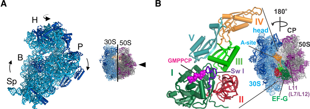
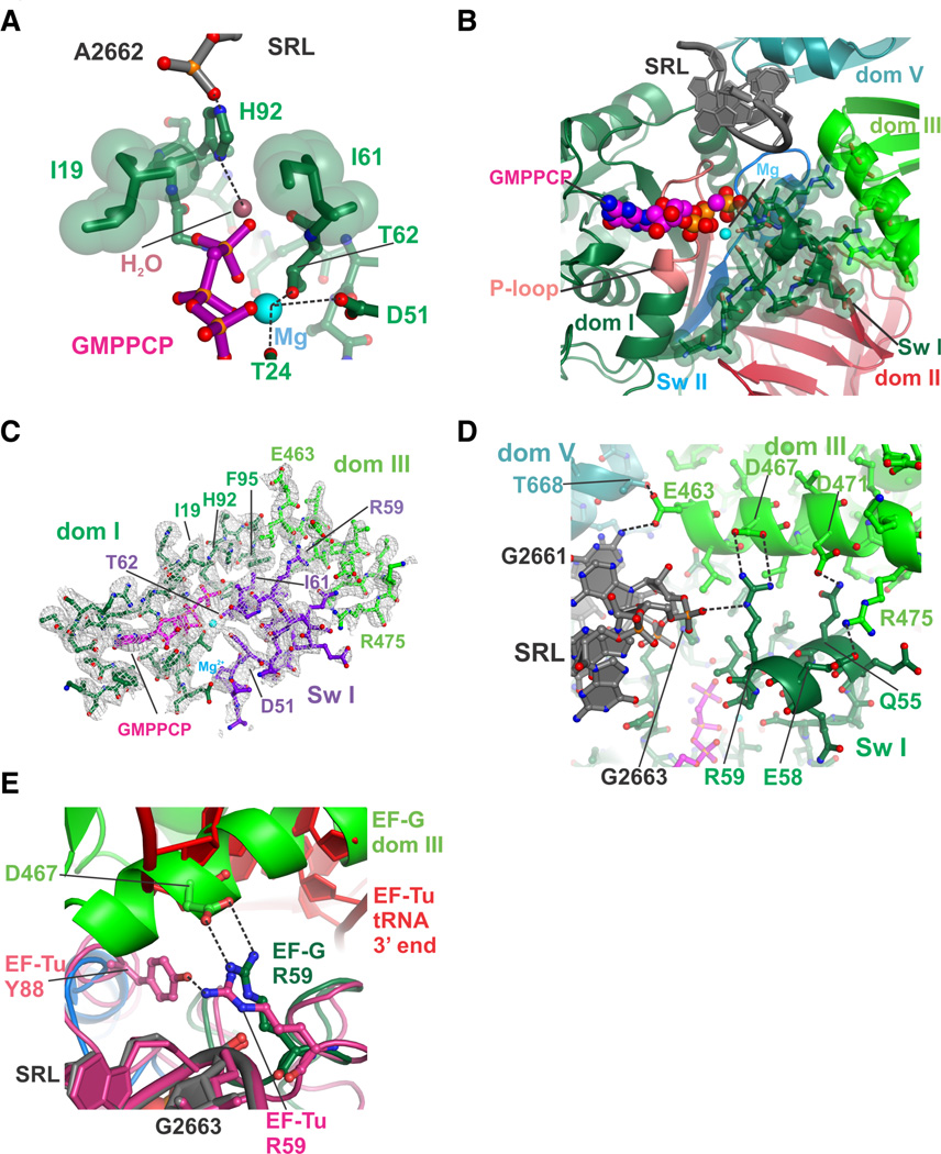
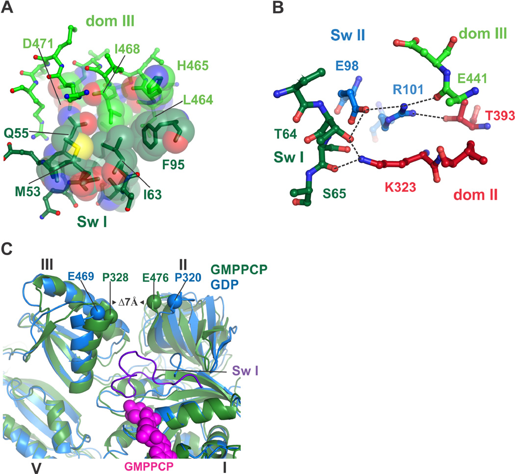
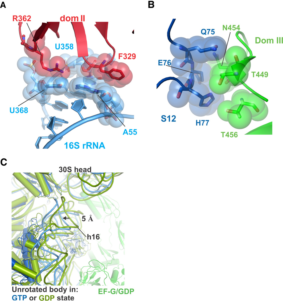
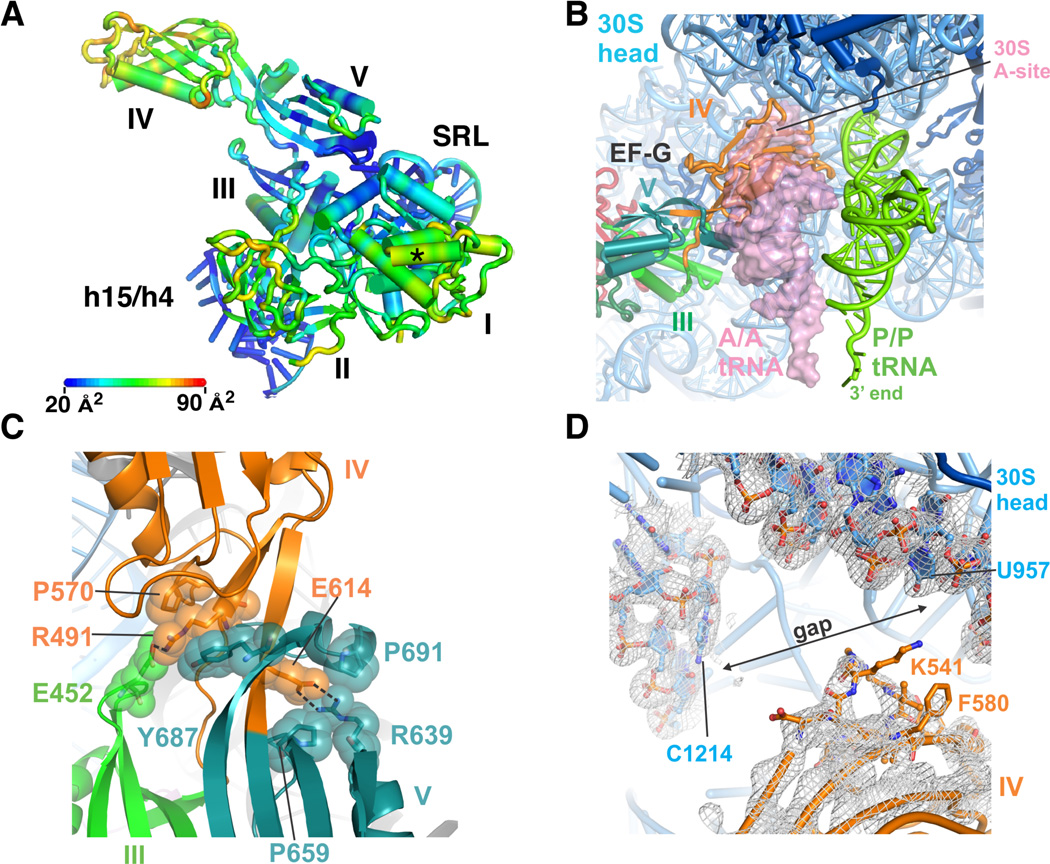
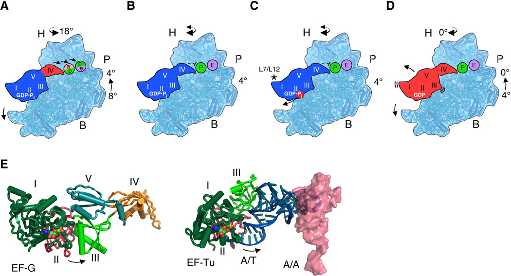
Protein synthesis by the ribosome requires the translocation of transfer RNAs and messenger RNA by one codon after each peptide bond is formed, a reaction that requires ribosomal subunit rotation and is catalyzed by the guanosine triphosphatase (GTPase) elongation factor G (EF-G). We determined 3 angstrom resolution x-ray crystal structures of EF-G complexed with a nonhydrolyzable guanosine 5'-triphosphate (GTP) analog and bound to the Escherichia coli ribosome in different states of ribosomal subunit rotation. The structures reveal that EF-G binding to the ribosome stabilizes switch regions in the GTPase active site, resulting in a compact EF-G conformation that favors an intermediate state of ribosomal subunit rotation. These structures suggest that EF-G controls the translocation reaction by cycles of conformational rigidity and relaxation before and after GTP hydrolysis.


Figures

Figure 1
Figure 1. Global structural rearrangements in the ribosome in EF-G/GMPPCP complexes
(A) Schematic illustrating two degrees of freedom in the 30S subunit within the 70S ribosome. 30S subunit rotation encompasses the body (B) and platform (P) domains, whereas the 30S subunit head domain (H) swivels around a nearly orthogonal rotational axis. (B) EF-G is shown bound to the 70S ribosome oriented 180° from the view shown to the left. Domains in EF-G are numbered: domain I (dark green), domain II (red), domain III (light green), domain IV (orange), and domain V (light blue). Switch I (sw I) (amino acids 38–64) in domain I is highlighted in purple spheres. The 30S subunit is in blue, and 50S subunit in grey and magenta.
Figure 2
Figure 2. Compact arrangement of EF-G domains I and III in the GTP state
(A) Position of the catalytic histidine H92. Gate residues Isoleucine 19 and Isoleucine 61 are shown with green spheres with van der Waals radii, along with the position of the proposed activated water and coordinated Mg2+ ion. (B) View of the GTPase active site, including swI, switch II (swII), P-loop, Sarcin-Ricin Loop in 23S rRNA (SRL) and the GTP nucleotide analog GMPPCP. Amino acids in swI that become ordered upon GTP binding and form salt bridges with EF-G domain III are shown as spheres with van der Waals radii. (C) Four-fold NCS averaged electron density for the GTPase domain of EF-G near the GTP binding pocket. Shown are EF-G swI, domain I, domain III, the GTP analog GMPPCP color-coded as in panel A, and a magnesium ion (light blue). Electron density (grey grid) is contoured at 1 standard deviation from the mean. (D) Closeup view of interactions between swI (dark green) and domain III (green) in the EF-G/GMPPCP complex. Contacts within hydrogen-bonding distance are indicated with dashed lines. (E) Essential salt bridge between EF-G residue R59 in swI and residue D467 in domain III. The corresponding interactions in the EF-Tu/aminoacyl-tRNA decoding complex are also shown. EF-G is color coded as in Figure 1B, while the EF-Tu/tRNA complex is colored magenta (protein, SRL) and red (tRNA).
Figure 3
Figure 3. A network of contacts between EF-G domains extends from the GTPase center towards the small ribosomal subunit
(A) A new hydrophobic core forms between domains I and III in EF-G in the GTP state. Positions in swI of domain I (dark green carbons) and domain III (green carbons) are shown. (B) Polar and ionic interactions formed between swI and swII and domains II and III in the GTP state, color-coded as in Fig. 1B. Dashes indicate atoms within hydrogen-bonding distance. (C) Closure of domains II and III due to binding of the GMPPCP form of EF-G to the ribosome (7 Å) is indicated. The structure of the ribosome with EFG/ GDP/fusidic acid is shown in blue (32). The ribosome in an intermediate rotation state with EF-G/GMPPCP is in green.
Figure 4
Figure 4. Contacts between EF-G and the 30S subunit are maintained during ribosomal subunit rotation
(A) Contacts between EF-G domain II (red) and 16S rRNA (light blue) near helices h5 (A55 or U358) and h15 (U368). (B) Contacts between EF-G domain III (green) and ribosomal protein S12 (blue), for the ribosome in an intermediate state of rotation. (C) Movement of the 30S subunit body domain away from the subunit interface in the unrotated state induced by EF-G binding in the GTP state (blue). For comparison, the structure of the ribosome with EF-G/GDP/fusidic acid in the unrotated state is shown in green and olive (32).
Figure 5
Figure 5. Conformational rigidity of EF-G/GMPPCP in the intermediate rotational state
(A) Atomic displacement parameters (B-factors) of EF-G/GMPPCP bound to the ribosome in an intermediate state of subunit rotation. Scale bar indicates the range of color-coded B-factors. Domains in EF-G and helices in 16S and 23S rRNA are indicated. The asterisk indicates the site of interaction seen between EF-G and ribosomal proteins L7/L12 in (32). (B) Position of EF-G domain IV (orange) in the ribosomal A site. The position of tRNAs in the A site (A/A tRNA, transparent pink surface) and P site (P/P tRNA, green) are derived from the superposition of the ribosome structure in (55) with the ribosome in an intermediate state of subunit rotation, using the 30S subunit platform as a frame of reference. (C) Salt bridges between EF-G domain III (green) residue E452, domain IV (orange) residues R491 or E614 and domain V (aqua) residue R639, buttressed by hydrophobic packing. (D) Lack of contacts between EF-G domain IV and the head domain of the 30S subunit of a representative ribosome in an intermediate state of rotation. (2Fobs–2Fcalc) difference electron density is shown at a contour of 1 standard deviation from the mean.
Figure 6
Figure 6. Model of EF-G controlled translocation of mRNA and tRNA
(A) EF-G in the activated GDP•Pi state requires movement of A-site tRNA out of the decoding site (pink) of the ribosomal 30S subunit platform into the P site (green), and P-site tRNA into the E site (purple) to accommodate EF-G domain IV, as indicated by arrows. The intermediate tRNA sites in the 30S subunit and the head rotation angle are based on (10). At this point, domains I-III and V of EF-G (blue) are rigidly bound to the intermediate rotation state of the ribosome, whereas domain IV (red) moves to occupy the A site. (B) Stable interactions between EF-G domain IV and the remainder of EF-G position domain IV to prevent back-translocation of P-site tRNA. The 30S subunit head domain may remain dynamic in this post-translocation state (46, 51, 56). (C) Phosphate release from the GTPase domain of EF-G, stimulated by proteins L7/L12 (asterisk), disrupts inter-domain contacts in EF-G, allowing the ribosome to revert to the unrotated state, (D), from which EF-G/GDP dissociates from the ribosome. (E) Opening of domains II and III in EF-G after GTP hydrolysis follows the same trajectory as tRNA release from EF-Tu during mRNA decoding. The position of A-site tRNA (pink, PDB entry 3I8G) (57) was compared to that for EF-Tu in an mRNA decoding complex with the GTP analog GMPPCP (6).

Comment in

References

    1. Bourne HR, Sanders DA, McCormick F. The GTPase superfamily: conserved structure and molecular mechanism. Nature. 1991 Jan 10;349:117. - PubMed
    1. Clementi N, Polacek N. Ribosome-associated GTPases: the role of RNA for GTPase activation. RNA Biol. 2010 Sep-Oct;7:521. - PubMed
    1. Liang H. Sordarin, an antifungal agent with a unique mode of action. Beilstein J Org Chem. 2008;4:31. - PMC - PubMed
    1. Sohmen D, Harms JM, Schlunzen F, Wilson DN. Enhanced SnapShot: Antibiotic inhibition of protein synthesis II. Cell. 2009 Oct 2;139:212. - PubMed
    1. Schmeing TM, Voorhees RM, Kelley AC, Gao YG, et al. The crystal structure of the ribosome bound to EF-Tu and aminoacyl-tRNA. Science. 2009 Oct 30;326:688. - PMC - PubMed

Publication types